Rev Bras Ter Intensiva. 2015;27(2):92-95
Should microcirculation monitoring be used
to guide luid resuscitation in severe sepsis and
septic shock?
COMMENTARY
Tissue hypoperfusion and subsequent limited oxygen transport are critical features conducting to organ failure during shock states. herefore, early identiication of tissue hypoperfusion and adequate resuscitation are key for improving the probability of survival after septic shock.(1,2) However, how to identify organ perfusion abnormalities at the bedside and select the type and amount of luids required to improve tissue hypoxia remain highly controversial. Traditionally, clinical signs, such as reduced blood pressure and urinary output, altered consciousness, and mottled skin, have been used to identify tissue perfusion abnormalities. Consequently, current hemodynamic monitoring during shock states mainly focuses on detection of pressure-derived hemodynamic variables related to systemic circulation. However, it has been largely recognized that monitoring these macro-hemodynamic variables is not suicient to rule out persistent abnormalities of tissue oxygenation. Indeed, the usefulness of resuscitation targets, such as global oxygen-derived parameters, has been strongly questioned,(3) and recent data have failed to demonstrate beneicial efects of using central venous oxygen saturation as a goal of resuscitation.(4-6)
Fluid resuscitation therapy primarily aims to optimize cardiac output on the assumption that increasing macro blood low can improve the convective transport of oxygen to the tissues and, therefore, maintain cellular respiration and support organ function.(7,8) hus, luid therapy targeting central venous pressure has been widely recommended to achieve adequate cardiac performance.(9) However, high positive luid balances have also been associated with unfavorable clinical outcomes.(10) In this sense, dynamic approaches to assessing volume responsiveness to luid administration seem to be superior to static variables.(11,12) Unfortunately, macro-hemodynamic optimization guided by either dynamic or static variables does not guarantee adequate tissue perfusion or adequate cellular respiration.
Oxygen transport to tissues is governed by convective and difusive components. he convective component is determined by the microcirculatory blood low itself, i.e., the number of red blood cells (RBCs) entering the microcirculation, and by oxygen content. In normal conditions, inlow and outlow pressures control the driving pressure at the microvascular level. hus, the convective transport is regulated upstream at the arteriolar level through microcirculatory inlow changes with subsequent micro-hematocrit Gustavo A. Ospina-Tascón1,2, Humberto
Madriñán-Navia1,2
1. Department of Intensive Care Medicine, Fundación Valle del Lili - Cali, Colombia. 2. Universidad ICESI - Cali, Colombia.
Conflicts of interest: None.
Submitted on April 4, 2015 Accepted on May 12, 2015
Corresponding author:
Gustavo A. Ospina-Tascón Intensive Care Unit Fundación Valle del Lili
Av. Simón Bolívar. Cra 98 # 18-49 Cali, Colombia
E-mail: [email protected]
Responsible editor: Jorge Ibrain Figueira Salluh
A ressuscitação volêmica na sepse grave e choque séptico deve ser
guiada pela microcirculação?
Should microcirculation monitoring be used to guide fluid resuscitation in severe sepsis and septic shock? 93
Rev Bras Ter Intensiva. 2015;27(2):92-95
modiications and is limited downstream by venous pressure. Meanwhile, the difusive component of oxygen transport is determined by the gradient between the capillary and mitochondrial oxygen partial pressures, the difusional distance and the area available for gas exchange, according to Fick´s law.
Unfortunately, current resuscitation procedures are based on the assumption that defects in oxygen transport arise from a lack of perfusion. hus, resuscitation eforts are mostly focused on the promotion of convective low based on the assumption that hypovolemia is the main limiting factor of blood low. However, a substantial contribution of oxygen tissue transport is determined by the capacity of difusion of oxygen from RBCs to the cells, and this can be quantiied by functional capillary density (FCD), i.e., the density of capillaries with lowing RBCs carrying oxygen. In normal conditions, the microvascular blood low is carefully matched to the metabolic demands of tissues. However, septic shock is characterized by decreased FCD in addition to increased heterogeneity of blood low, with zones of well-perfused vessels in close proximity to non-perfused capillaries.(13) he persistence of such alterations has been shown to be related to multiorgan dysfunction, even when global hemodynamics appear to be optimal.(14) Indeed, increases in cardiac output may be insuicient to correct tissue hypoxemia because microcirculation alterations might persist. hus, global hemodynamic targets should be integrated with functional microcirculatory goals, as this would optimize both convective and difusive components in order to maximize oxygen delivery to cells. Microcirculatory responsiveness should be deined as an increase in convective microvascular low in response to luid load in addition to a reduction of the heterogeneity of low, resulting in a more balanced distribution of oxygen to the tissues (Figure 1). Nevertheless, eforts to increase blood low by luid administration could be counterbalanced by a decrease in FCD resulting in a limited oxygen difusive capacity (Figure 2). hus, excessive luid administration could increase the distance between capillaries, reducing oxygen difusion to the tissues and inally to the mitochondria.
Orthogonal polarization spectral and sidestream dark ield imaging techniques have helped us gain a
better understanding of microcirculatory derangements during severe sepsis and septic shock at the bedside. In fact, deeper microvascular alterations, such as a reduced percentage of small perfused vessels (PPV), decreased FCD and increased heterogeneity of low, have been shown to be related to more severe organ dysfunctions and unfavorable outcomes.(13,15,16) Interestingly, reductions in FCD in sepsis are completely explained by decreased PPV, while RBC velocities are similar in both survivors and non-survivors.(16) hese indings suggest that variables that describe the difusional component of oxygen transport, i.e., FCD and heterogeneity of microvascular blood low, are more closely related to clinical outcomes than pure convective components, such as RBC velocity.(16) Remarkably, these derangements are amenable to correction over time and are dissociated from global hemodynamics. Consequently, micro-hemodynamics cannot be predicted by the typical systemic hemodynamic parameters, although more severe microcirculatory alterations coexist with high lactate levels and the requirement of higher doses of vasopressors.(17)
In a recent study, Ospina-Tascón et al.(18) explored the efect of luids on microcirculatory blood low using the sidestream dark ield imaging technique in severe sepsis and septic shock. hey found that early, but not late luid challenge can increase the PPV, with subsequent improvement in FCD and decreased heterogeneity of blood low. Analogous to macro-hemodynamic luid responsiveness predictors, the prior status of the microcirculation is strongly related to the response to volume expansion.(18,19) Indeed, a recent study(20) demonstrated that the efects of luid load on the microcirculation are dependent on both basal microvascular perfusion and the magnitude of the increase in cardiac output.
94 Ospina-Tascón GA, Madriñán-Navia H
Rev Bras Ter Intensiva. 2015;27(2):92-95
REFERENCES
1. Vincent JL, De Backer D. Circulatory shock. N Engl J Med. 2013; 369(18):1726-34.
2. Cecconi M, De Backer D, Antonelli M, Beale R, Bakker J, Hofer C, et al. Consensus on circulatory shock and hemodynamic monitoring. Task force of the European Society of Intensive Care Medicine. Intensive Care Med. 2014;40(12):1795-815.
3. Bellomo R, Reade MC, Warrillow SJ. The pursuit of a high central venous oxygen saturation in sepsis: growing concerns. Crit Care. 2008;12(2):130. 4. ProCESS Investigators, Yealy DM, Kellum JA, Huang DT, Barnato AE,
Weissfeld LA, Pike F, et al. A randomized trial of protocol-based care for early septic shock. N Engl J Med. 2014;370(18):1683-93.
5. Peake SL, Bailey M, Bellomo R, Cameron PA, Cross A, Delaney A, Finfer S, Higgins A, Jones DA, Myburgh JA, Syres GA, Webb SA, Williams P; ARISE Investigators, for the Australian and New Zealand Intensive Care Society Clinical Trials Group. Australasian resuscitation of sepsis evaluation (ARISE): A multi-centre, prospective, inception cohort study. Resuscitation. 2009;80(7):811-8.
6. Mouncey PR, Osborn TM, Power GS, Harrison DA, Sadique MZ, Grieve RD, Jahan R, Harvey SE, Bell D, Bion JF, Coats TJ, Singer M, Young JD, Rowan KM; ProMISe Trial Investigators. Trial of early, goal-directed resuscitation for septic shock. N Engl J Med. 2015;372(14):1301-11.
7. Santry HP, Alam HB. Fluid resuscitation: past, present, and the future. Shock. 2010;33(3):229-41. Review.
8. Vincent JL, Weil MH. Fluid challenge revisited. Crit Care Med. 2006;34(5):1333-7.
9. Dellinger RP, Levy MM, Rhodes A, Annane D, Gerlach H, Opal SM, Sevransky JE, Sprung CL, Douglas IS, Jaeschke R, Osborn TM, Nunnally ME, Townsend SR, Reinhart K, Kleinpell RM, Angus DC, Deutschman CS, Machado FR, Rubenfeld GD, Webb S, Beale RJ, Vincent JL, Moreno R; Surviving Sepsis Campaign Guidelines Committee including The Pediatric Subgroup. Surviving Sepsis Campaign: international guidelines for management of severe sepsis and septic shock, 2012. Intensive Care Med. 2013;39(2):165-228.
10. Boyd JH, Forbes J, Nakada TA, Walley KR, Russell JA. Fluid resuscitation in septic shock: a positive fluid balance and elevated central venous pressure are associated with increased mortality. Crit Care Med. 2011;39(2):259-65. 11. Pinsky MR, Payen D. Functional hemodynamic monitoring. Crit Care.
2005;9(6):566-72.
12. Teboul JL, Monnet X. Detecting volume responsiveness and unresponsiveness in intensive care unit patients: two different problems, only one solution. Crit Care. 2009;13(4):175.
13. De Backer D, Ospina-Tascon G, Salgado D, Favory R, Creteur J, Vincent JL. Monitoring the microcirculation in the critically ill patient: current methods and future approaches. Intensive Care Med. 2010;36(11):1813-25. 14. Doerschug KC, Delsing AS, Schmidt GA, Haynes WG. Impairments in
microvascular reactivity are related to organ failure in human sepsis. Am J Physiol Heart Circ Physiol. 2007;293(2):H1065-71.
15. Trzeciak S, Dellinger RP, Parrillo JE, Guglielmi M, Bajaj J, Abate NL, Arnold RC, Colilla S, Zanotti S, Hollenberg SM; Microcirculatory Alterations in Resuscitation and Shock Investigators. Early microcirculatory perfusion derangements in patients with severe sepsis and septic shock: relationship to hemodynamics, oxygen transport, and survival. Ann Emerg Med. 2007;49(1):88-98, 98.e1-2.
Figure 1 - Effects of adequate fluid administration on microvascular blood flow.
Progressive increasing of convective flow after fluid loading during microcirculatory conditions of pure
convective flow derangements (A1, A2) and combined with increased heterogeneity of blood flow (B1, B2).
An optimal fluid administration corrects convective and heterogeneity blood flow disturbances (C). Note that
the number of vessels with adequate flow per tissue area represents the functional capillary density (FCD),
i.e., the major determinant of the diffusive component of oxygen transport to tissues. Black arrows pointing
down represent the magnitude of the blood flow.
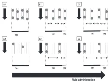
Figure 2 - Effects of fluid overload on microvascular blood flow. Inadequate fluid overload conducts to progressive decrease in functional capillary density in cases of homogeneous (A1, B1,
C1) or heterogeneous (A2, B2, C2) microcirculatory blood flow. Progressive increase in distance between
capillaries impairs oxygen diffusive capacities to tissues despite apparently normal convective flow (A1, B1,
C1) or in cases of apparent corrected convective component with persistence of increased heterogeneity
(B2, C2). TA1 depicts the original tissue area. TA2 depicts the increased tissue area due to edema. Black
Should microcirculation monitoring be used to guide fluid resuscitation in severe sepsis and septic shock? 95
Rev Bras Ter Intensiva. 2015;27(2):92-95
16. Edul VS, Enrico C, Laviolle B, Vazquez AR, Ince C, Dubin A. Quantitative assessment of the microcirculation in healthy volunteers and in patients with septic shock. Crit Care Med. 2012;40(5):1443-8.
17. Hernandez G, Boerma EC, Dubin A, Bruhn A, Koopmans M, Edul VK, et al. Severe abnormalities in microvascular perfused vessel density are associated to organ dysfunctions and mortality and can be predicted by hyperlactatemia and norepinephrine requirements in septic shock patients. J Crit Care. 2013;28(4):538.e9-14.
18. Ospina-Tascon G, Neves AP, Occhipinti G, Donadello K, Büchele G, Simion D, et al. Effects of fluids on microvascular perfusion in patients with severe sepsis. Intensive Care Med. 2010;36(6):949-55.
19. Pranskunas A, Koopmans M, Koetsier PM, Pilvinis V, Boerma EC. Microcirculatory blood flow as a tool to select ICU patients eligible for fluid therapy. Intensive Care Med. 2013;39(4):612-9.