Transitory increased blood pressure
after upper airway surgery for snoring
and sleep apnea correlates with the
apnea-hypopnea respiratory
disturbance index
1Programa de Pós-Graduação em Ciências Fisiológicas, Centro Biomédico,
Universidade Federal do Espírito Santo, Vitória, ES, Brasil
2Service d’ORL, Biochimie, Explorations Cardiovasculaires and Pneumologie,
Hôpital Saint-Antoine, Paris, France
3Departamento de Ciências Fisiológicas, Escola Superior de Ciências da
Santa Casa de Misericórdia de Vitória (EMESCAM), Vitória, ES, Brasil
4Departamento de Área Aplicada à Saúde, Faculdade Salesiana de Vitória,
Vitória, ES, Brasil M.T.M. Araújo4,
M. Ouayoun2,
J.M. Poirier2,
M.M. Bayle2,
E.C. Vasquez1,3
and B. Fleury2
Abstract
A transitory increase in blood pressure (BP) is observed following upper airway surgery for obstructive sleep apnea syndrome but the mechanisms implicated are not yet well understood. The objective of the present study was to evaluate changes in BP and heart rate (HR) and putative factors after uvulopalatopharyngoplasty and septoplasty in normotensive snorers. Patients (N = 10) were instrumented for 24-h ambulatory BP monitoring, nocturnal respiratory monitoring and urinary catecholamine level evaluation one day before surgery and on the day of surgery. The influence of postsurgery pain was prevented by analgesic therapy as confirmed using a visual analog scale of pain. Compared with preoperative values, there was a significant (P < 0.05) increase in nighttime but not daytime systolic BP (119 ± 5 vs 107 ± 3 mmHg), diastolic BP (72 ± 4 vs 67 ± 2 mmHg), HR (67 ± 4 vs 57 ± 2 bpm), respiratory disturbance index (RDI) characterized by apnea-hypopnea (30 ± 10 vs 13 ± 4 events/h of sleep) and norepinephrine levels (22.0 ± 4.7 vs 11.0 ± 1.3 µg l-1 12 h-1) after surgery. A positive
correlation was found between individual variations of BP and indi-vidual variations of RDI (r = 0.81, P < 0.01) but not between BP or RDI and catecholamines. The visual analog scale of pain showed similar stress levels on the day before and after surgery (6.0 ± 0.8 vs 5.0 ± 0.9 cm, respectively). These data strongly suggest that the cardiovascular changes observed in patients who underwent uvulopalatopharyngo-plasty and septouvulopalatopharyngo-plasty were due to the increased postoperative RDI. Correspondence
E.C. Vasquez
Programa de Pós-Graduação em Ciências Fisiológicas
Centro Biomédico, UFES Av. Marechal Campos, 1468 29042-755Vitória, ES Brasil
E-mail: [email protected] Research supported by CAPES, CNPq and ResMed (Sydney, Australia).
Received June 9, 2003 Accepted October 16, 2003
Key words
•Snorers
•Obstructive sleep apnea
syndrome
•Uvulopalatopharyngoplasty •Septoplasty
Introduction
Among the postoperative complications described in patients with obstructive sleep apnea syndrome (OSAS) undergoing upper airway surgical procedures, transient sys-temic hypertension has been reported (1-3). Recently, Riley et al. (1) reported that among 210 OSAS patients examined, 70% required postoperative antihypertensive medications during their hospital stay (1,4). This increase in blood pressure (BP) could in turn promote postoperative complications such as bleed-ing, stroke or myocardial ischemia in those patients with systemic hypertension and/or cardiovascular diseases (1). The mechan-isms for transient elevation of BP in this critical situation are not understood. It is possible that increases in upper airway resis-tance due to postoperative airway edema or induced by oral breathing may decrease the upper airway area, thereby causing an in-crease in the frequency of the obstructive respiratory events during sleep (5-8). These repetitive episodes of apnea often coincide with marked hypoxia, hypercapnia, and arousals during sleep, all of which are known to increase sympathetic nervous outflow (9-11). These repetitive acute changes in sym-pathetic activity may be involved in the patho-genesis of cardiovascular disturbances, mainly BP elevation (12). Moreover, there is evidence that changes in sympathetic activ-ity may be implicated in the development of sustained hypertension in these patients (13-16). Additionally, preoperative anxiety and fear and postoperative pain can induce a psychological stress that in turn can alter the homeostasis of BP, heart rate (HR) and neu-ral activity (17-19). Severe pain has been frequently reported after upper airway sur-gery for OSAS, leading to the use of newer approaches for postoperative analgesia, such as the combination of a nonopioid analgesic with nonsteroidal anti-inflammatory drugs (20,21), but their efficacy after upper airway surgery has not been evaluated.
The main objective of the present study was to evaluate the changes in BP and in putative factors causing its elevation, par-ticularly postoperative pain, in a group of snorers subjected to upper airway surgery.
Patients and Methods
The protocol was approved by the Hospi-tal Ethics Committee and informed consent was obtained from each patient. The study was performed on 10 normotensive (systolic BP: 125 ± 3 mmHg; diastolic BP: 81 ± 3 mmHg) patients with a mean age of 46 ± 1 years and a mean body mass index (BMI) of 23 ± 1 kg/m2. All patients were snorers and
most of them had mild OSAS. The mean respiratory disturbance index (RDI) of the group was 13 ± 4 events/h of recording (range: 2 to 30 events/h). We excluded from the study those patients who were: a) older than 60 years or younger than 18 years, b) had a body weight more than 1.6 times the ideal body weight (22) and/or had a BMI higher than 30 kg/m2, c) had moderate to
severe respiratory disease, and d) had car-diac arrhythmia and systolic BP ≥160 and/or diastolic BP ≥95 mmHg.
Nocturnal respiration monitoring
All studies were carried out at the Hôpital Saint-Antoine, Paris, France. Nocturnal res-piration monitoring studies, including meas-urements of snoring and body movements, were carried out by a continuously recording technique (PolyMesam®, Taema, France)
from 10:00 pm to 6:00 am on the preopera-tive day, on the day of surgery and one year after surgery. Nocturnal respiratory events were assessed by measuring naso-buccal air-flow using a dual thermistor. Chest and ab-dominal wall respiratory movements were measured using noncalibrated inductive res-piratory plethysmography, oxyhemoglobin saturation (SaO2) was analyzed by finger
intensity was measured using a microphone placed at the level of the neck, body position was monitored through a sensor positioned on the chest, and cardiac rhythm was moni-tored by electrocardiogram. Throughout the study, the movements of the subject were recorded with a wrist actigraph, which con-tained a motion detector and a system to record and store the number of wrist move-ments. Wrist movement intervals higher than 10 s were considered and computed every hour from 11:00 pm to 6:00 am. Every pe-riod without wrist activity was considered to be a period of sleep (23). RDI was indicated by the number of recognized apneic and hypopneic episodes recorded per hour of recording. It means that all respiratory dis-turbances during the evaluation period were added and divided by the number of hours of evaluation time. An event of obstructive ap-nea was defined as an 80% or greater de-crease in airflow amplitude through the nose and mouth lasting more than 10 s in the presence of visible thoracic and abdominal respiratory efforts. An event of hypopnea was defined as a 10-s or longer decrease in oronasal airflow amplitude and the ampli-tude of thoracic and abdominal respiratory efforts to values less than 50% of those observed during normal breathing prior to the event. Desaturation events were recog-nized when oxygen saturation was 4% be-low baseline saturation, but desaturation was not a criterion for scoring either apnea or hypopnea.
Ambulatory blood pressure monitoring
Fully automatic systolic and diastolic BP and HR profiles were recorded continuously and noninvasively for 24 h using a system for ambulatory monitoring of BP (SpaceLabs 90207, SpaceLabs Inc., Redmond, Wash-ington, DC, USA). Oscillometric units were used at 15-min sampling intervals through-out the recording period one day before sur-gery and on the day of sursur-gery and the
proce-dure was repeated again one year later. The readings were edited and averaged automati-cally by an interface (SpaceLabs ABP90209), set to discard systolic BP readings higher than 260 and lower than 70 mmHg, diastolic readings higher than 150 and lower than 40 mmHg, pulse pressure readings higher than 150 and lower than 20 mmHg, and readings with the diastolic BP higher than the systolic BP. The interface was also set to define the daytime and nighttime periods as the inter-vals between 7:00 am and 9:00 pm. On average, 95.8% BP readings per patient sat-isfied the editing criterion (range 91-100). At least two valid ambulatory BP readings per hour for the entire 24-h period were obtained for each patient. The presence of the “dip” in the BP recording observed dur-ing nocturnal sleep was investigated. A sub-ject was considered a “nondipper” if his/her systolic and diastolic BP did not decrease by 10% during sleep compared to the mean values observed during the daytime.
Urinary catecholamines
Samples for determination of urinary norepinephrine and epinephrine were col-lected from 8:00 pm to 8:00 am during the three recording occasions (preoperative, post-operative and one year after surgery). Urine specimens for each sample were collected into polyethylene containers, acidified with 6 N HCl as preservative, kept at +4ºC over-night and stored at -20ºC until the time for assay. Urinary catecholamines were deter-mined by high performance liquid chroma-tography with electrochemical detection (24,25).
Anesthetic and surgical procedures
All patients received 100 mg hydroxy-zine dihydrochloride (orally) one hour prior to surgery. General anesthesia was induced with propofol (Diprivan®), the trachea was
isoflurane and atracurium dibesylate (Tracu-rium®). Injections of sufentanil were
per-formed as decided by the anesthesiologist responsible for the patient. Uvulopalato-pharyngoplasty (UPPP) was performed as originally described by Fujita et al. (26). Bilateral tonsillectomy was then performed followed by septoplasty in all patients. Pa-tients were monitored in the recovery room postoperatively. Antibiotics were used and postoperative pain was controlled with a combination of ketoprofen (Profenid, 50 mg,
iv; Specia Laboratory, Paris, France) and propacetamol hydrochloride (Prodafalgan, 2 g, iv; UPSA Laboratory, Rueil Malmaison, France), an acetaminophen pro-drug, diluted in 100 ml dextrose.
Evaluation of postoperative pain
The first pain medication with ketopro-fen (50 mg, iv) plus propacetamol (2 g, iv) was administered at the time of extubation in the operating room and the second was ad-ministered 4 h later. After this period, the drug was administered every 6 h for 28 h.
A subjective criterion was used to evalu-ate the postoperative pain scores and the efficacy of the analgesic combination. A 10-cm long ruler with endpoints marked as “no pain” to “severe pain” was used as a visual analog scale (VAS) to assess the pain inten-sity described by the patient (27-29). After the patient had pointed at the pain classifica-tion on the VAS the corresponding score was measured by the investigator. The first pain evaluation was carried out in the recov-ery room, when the patient was able to per-form VAS evaluation before returning to his room (control period). The other measure-ments were made 30 min before and 3 h after the administration of the analgesic combina-tion over a period of 28 h. The locacombina-tion and kind of pain reported by the patient were also recorded using a validated French version of the McGill Pain Questionnaire (Question-naire Douleur de Saint-Antoine, QDSA; 30).
The psychological stress, especially anxi-ety, was also evaluated using a VAS that permitted the patient to self-report the level of anxiety, which ranged from “no anxiety” to “severe anxiety”. This evaluation was per-formed in the recovery room concomitantly with pain evaluation. In addition, the devel-opment of sensory and affective components of the anxiety state was assessed using the QDSA. We also wanted to determine the subjective impression of sleep since there is no objective sleep, and for this purpose we used the VAS changing the endpoints of the long ruler from “no sleep” to “good sleep” to assess the sleep score described by the pa-tient.
Statistical analysis
Results are reported as means ± SEM. One-way analysis of variance (ANOVA) for repeated measures followed by a protected
post hoc test (Fisher least significant differ-ence procedure) was used for comparisons of the mean values of the parameters re-corded on the three different occasions (31). Linear regression analysis was employed to obtain the correlation between changes in BP and RDI and between changes in BP and catecholamines. The level of significance was set at P < 0.05 for all comparisons.
Results
Wrist activity and disordered sleep breathing
No differences in mean wrist motion du-ration were observed between the preopera-tive night (5.2 ± 3.1 movements/h) and the night following surgery (5.4 ± 3.0 move-ments/h), suggesting that there were similar sleep times. No significant changes were observed between the two nights in terms of subjective sleep quality evaluation meas-ured by the VAS (6.0 ± 0.8 and 5.0 ± 0.9 cm, respectively).
ob-served during the night following surgery when compared with the preoperative night (30 ± 10 vs 13 ± 4 events/h, respectively; P < 0.05). One year later the values were similar to those observed on the preoperative day. These results were accompanied by a ten-dency (P > 0.05) for the mean nocturnal SaO2 to decrease during the night after
sur-gery (7.0 ± 5.0% below baseline saturation) compared to the preoperative values (4.3 ± 3.3% below the baseline saturation), and to those observed one year later (4.9 ± 4.2% below the baseline saturation).
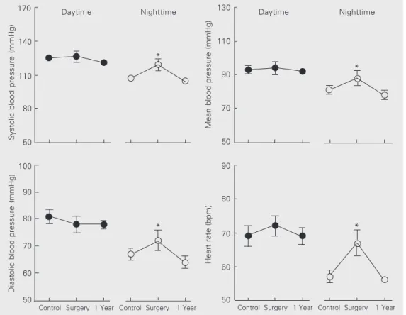
Cardiovascular changes
Figure 1 shows average BP and HR val-ues continuously recorded over a period of 24 h. Data showed a significant (P < 0.05) increase in nighttime values of mean systolic (119 ± 5 vs 107 ± 3 mmHg) and diastolic BP (72 ± 4 vs 67 ± 2 mmHg), mean BP (88 ± 4 vs
81 ± 3 mmHg), and HR (67 ± 4 vs 57 ± 2
bpm) compared to preoperative control val-ues. One year later, when the patients were monitored again, these values were normal-ized (104 ± 3, 64 ± 2, 78 ± 3 mmHg and 56 ± 1 bpm, respectively). Nocturnal dipping in BP was observed in 9 of the 10 patients on the preoperative day and one year later but only in one patient on the day of surgery.
Pre- and postoperative correlation analy-sis was performed to obtain a first assess-ment of the degree of relationship between BP values and other dependent variables. Figure 2 shows that both systolic and dia-stolic BP were significantly correlated with RDI (r = 0.81 and r = 0.74, respectively; P < 0.01).
Urinary catecholamines
The 12-h catecholamine urinary excre-tion rates showed a significant (P < 0.05) increase of norepinephrine during the night immediately following surgery (22.0 ± 4.7
Systolic blood pressure (mmHg)
170
140
110
80
50
Diastolic blood pressure (mmHg)
100
90
80
70
60
50
Mean blood pressure (mmHg)
130
110
90
70
50
Daytime Nighttime Daytime Nighttime
Heart rate (bpm)
90
80
70
60
50
Control Surgery 1 Year Control Surgery 1 Year Control Surgery 1 Year Control Surgery 1 Year
*
*
* *
µg l-1 12 h-1) as compared to values observed
during the preoperative night (11.0 ± 1.3 µg l-1 12 h-1) and were normalized one year later
(12.0 ± 2.3 µg l-1 12 h-1). Similar results were
observed for epinephrine excretion rates (3.1 ± 0.9, 2.0 ± 0.2, 1.7 ± 0.4 µg l-1 12 h-1,
respectively). Data analysis of absolute val-ues showed that urinary catecholamine ex-cretion rates did not correlate with BP (sys-tolic: r = 0.33 and dias(sys-tolic: r = 0.19) nor with RDI desaturation index (r = 0.29) and similar negative correlations were observed when values were determined as changes (∆ values).
Stress and pain evaluation
No differences in anxiety scores quanti-fied by the VAS were observed among the five postoperative periods (4, 10, 16, 22 and 28 h) compared to the same periods of time on the preoperative day. In addition, all scores were below the limit values of the test (5 cm), which means that these patients were
not abnormally anxious during the preopera-tive period. The evaluation of time course of VAS pain intensity scores for patients re-ceiving the analgesic combination showed no significant differences among the mean values measured in the five postoperative periods in the recovery room (4 h: 5.0 ± 0.7, 10 h: 3.7 ± 0.7, 16 h: 4.0 ± 0.5, 22 h: 3.8 ± 0.5 and 28 h: 2.2 ± 0.4 cm) when compared to the preoperative control values (5.1 ± 0.7 cm).
Discussion
The present study confirms that upper airway surgery (UPPP) associated with septoplasty is followed by a significant el-evation of RDI. This phenomenon was ac-companied by an elevation of systemic BP and HR in normotensive snorers or mild OSAS patients during the first night postsur-gery. Since postoperative pain was controlled by an analgesic, the elevation of BP and HR seems to be a direct consequence of surgi-cally induced disordered breathing.
An increase in RDI and oxyhemoglobin desaturation (5-9) has been reported fre-quently after upper airway surgery in OSAS patients. As expected, in our study the mean RDI value increased significantly during the night after UPPP plus septoplasty surgery both in snorers and mild OSAS patients com-pared to the values observed on the preop-erative day. We speculate that postoppreop-erative airway edema associated with upper airway occlusion and the mouth breathing due to nasal packing after septoplasty could worsen the unstable respiratory status in these pa-tients (8).
Esclamado et al. (2) retrospectively re-viewed 135 patients with OSAS undergoing UPPP and associated procedures and noted that 13% showed complications such as air-way obstruction after extubation or hemor-rhage. On the other hand, Riley et al. (1) reported that among 210 patients analyzed after a surgical procedure performed for the
Changes in systolic blood
pressure (mmHg)
40
30
20
10
0
-10
30
10 20
0
-10
Changes in diastolic blood
pressure (mmHg)
-1.00 -0.50 0.00 0.50 1.00 1.50 Changes in RDI (events/h of recording)
y = 19.1387x + 6.9792 r = 0.81
y = 13.0742x + 3.1137 r = 0.74
-1.00 -0.50 0.00 0.50 1.00 1.50 Changes in RDI (events/h of recording) Figure 2. Regression analysis
correction of OSAS, 53% required intraop-erative and 70% postopintraop-erative antihyperten-sive medications. Among these patients, only 31% had a history of hypertension. The sur-gical procedure included an UPPP for the majority of these patients (77%), but it was generally associated with a mandibular os-teotomy with genioglossus advancement and/ or hyoid myotomy and suspension (68%). In our study the surgical procedure always con-sisted of UPPP and septoplasty. The nasal surgery was required to correct a significant nasal septum deformity in all patients and was conducted at the same time of the UPPP to limit the number of procedures requiring general anesthesia. There was an acute sig-nificant increase of BP in 80% of these normotensive patients during the night im-mediately following UPPP surgery as com-pared to the preoperative night. Even a mod-erate acute elevation in BP involves a higher risk of complications, especially postopera-tive bleeding, for any given value even if these pressures have been long-standing (2,3). This increase in BP could in turn promote another kind of postoperative complications such as stroke or myocardial ischemia in patients with ischemic cardiovascular dis-ease.
It is interesting to observe that in our study the normotensive patients undergoing surgical UPPP plus septoplasty had only noc-turnal BP increases, e.g., during the night after surgery they presented no nocturnal “dipping”. Perhaps the most critical aspect of the transient postoperative hypertension occurring after UPPP surgery is to evaluate their consequences during a period while the patients are sleeping. We chose to study only normotensive subjects in order to obtain an uncomplicated effect on BP caused by sur-gery and sleep disordered breathing in simple snorers or in patients with very mild sleep apnea. We speculate that the immediate con-sequences of surgery for BP could be more severe and require treatment in borderline or hypertensive patients, as reported by Riley et
al. (1). The acute and paradoxical creation of repetitive upper airway occlusions due to the surgical procedure allowed us to obtain data that permitted the understanding of the vas-cular consequences of apnea for normoten-sive snorers.
intratho-racic pressures generated by the obstructed inspiratory efforts (9,33).
Although other investigators have pro-posed pain as a cause for postoperative hy-pertension associated with increased circu-lating catecholamines, our results show that the combination of propacetamol and keto-profen was effective for pre- and postopera-tive analgesia, reducing peripheral nocicep-tion and hence reducing the pain and inflam-matory response to surgical trauma (20,21). In addition, because this analgesic combina-tion does not cause adverse effects such as those produced by opioids (respiratory de-pression, sedation, urinary retention, nausea and vomiting) it did not contribute to the repetitive surgically induced disordered breathing (20).
The transient BP elevation observed im-mediately after a common upper airway sur-gery in normotensive snorers with or
with-out mild OSAS seems to be associated with repetitive increases of sleep disordered breathing created by the surgical procedure that could also increase the sympathetic ac-tivity and the observed changes in BP. Fu-ture experiments should test the hypothesis that the transient elevation of BP and HR following surgery could be prevented using a continuous positive airway pressure asso-ciated with a face mask (34).
Acknowledgments
We thank Mr. Jean Valty and Mr. Dussaule Jean Claude for thoughtful com-ments, and Marie France Roussignol and Marie José Bellegarde for biochemical anal-ysis, all of them from the Cardiovascular and Pneumology Service of the Otorhinolaryn-gology Department at the Saint-Antoine Hospital, Paris, France.
References
1. Riley RW, Powell NB, Guilleminault C, Pelayo R, Troell RJ & Li KK (1997). Obstructive sleep apnea surgery: risk management and complications. Otolaryngology - Head and Neck Surgery, 117: 648-652.
2. Esclamado RM, Glenn MG, McCulloch TM & Cummings CW (1989). Perioperative complications and risk factors in the surgical treat-ment of obstructive sleep apnea syndrome. Laryngoscope, 99: 1125-1129.
3. Haavisto L & Suonpaa J (1994). Complications of uvulopalatopharyn-goplasty. Clinical Otolaryngology, 19: 243-247.
4. Laslett L (1995). Hypertension. Preoperative assessment and peri-operative management. Western Journal of Medicine, 162: 215-219.
5. Sanders MH, Johnson JT, Keller FA & Seger L (1988). The acute effects of uvulopalatopharyngoplasty on breathing during sleep in sleep apnea patients. Sleep, 11: 75-89.
6. Gabrielczyk MR (1988). Acute airway obstruction after uvulopalato-pharyngoplasty for obstructive sleep apnea syndrome. Anesthesiol-ogy, 69: 941-943.
7. Burgess LPA, Derderian SS, Morin GV, Gonzalez C & Zajtchuk JT (1992). Postoperative risk following uvulopalatopharyngoplasty for obstructive sleep apnea. Otolaryngology - Head and Neck Surgery, 106: 81-86.
8. Rombaux P, Liistro G, Hamoir M, Eloy P, Bertrand B & Collard P (1998). Nocturnal oxymetry in patients with total nasal packing.
Acta Otorhinolaryngologica Belgica, 52: 223-228.
9. Morgan BJ (1996). Acute and chronic cardiovascular responses to sleep disordered breathing. Sleep, 19: S206-S209.
10. Morgan BJ, Crabtree DC, Palta M & Skatrud JB (1995). Combined hypoxia and hypercapnia evokes long-lasting sympathetic activa-tion in humans. Journal of Applied Physiology, 79: 205-213. 11. Narkiewicz K & Somers VK (1997). The sympathetic nervous
sys-tem and obstructive sleep apnea: implications for hypertension.
Journal of Hypertension, 15: 1613-1619.
12. Coy TV, Dimsdale JE, Israel S-A & Clausen J (1996). Sleep apnoea and sympathetic nervous system activity: a review. Journal of Sleep Research, 5: 42-50.
13. Fletcher EC, Miller J, Schaaf JW & Fletcher JG (1987). Urinary catecholamines before and after tracheotomy in patients with ob-structive sleep apnea and hypertension. Sleep, 10: 35-44. 14. Dimsdale JE, Coy TV, Ziegler MG, Ancoli-Israel S & Clausen J
(1995). The effect of sleep apnea on plasma and urinary catechol-amines. Sleep, 18: 377-381.
15. Baylor P, Mouton A, Shamoon HH & Goebel P (1995). Increased norepinephrine variability in patients with sleep apnea syndrome.
American Journal of Medicine, 99: 611-615.
16. Marrone O, Riccobono L, Salvaggio A, Mirabella A, Bonanno A & Bonsignore MR (1993). Catecholamines and blood pressure in ob-structive sleep apnea syndrome. Chest, 103: 722-727.
17. Heller PH, Perry F, Narfeh K, Gordon NC, Wachter-Shikura N & Levine J (1984). Cardiovascular autonomic response during preop-erative stress and postoppreop-erative pain. Pain, 18: 33-40.
19. McEwen BC & Stellar E (1993). Stress and the individual. Mechan-isms leading to disease. Archives of Internal Medicine, 153: 2093-2101.
20. Code W (1993). NSAIDs and balanced analgesia. Canadian Journal of Anaesthesia, 40: 401-405.
21. Fletcher D, Nègre I, Barbin C, François A, Carreres C, Falgueirettes C, Barboteu A & Samii K (1997). Postoperative analgesia with iv
propacetamol and ketoprofen combination after disc surgery. Cana-dian Journal of Anaesthesia, 44: 479-485.
22. Metropolitan Life Foundation (1983). 1983 metropolitan height and weight tables. Statistical Bulletin (Metropolitan Life Foundation), 64: 1.
23. Maus TL, McLaren JW, Shepard JW & Brubaker RF (1996). The effects of sleep on circulating catecholamines and aqueous flow in human subjects. Experimental Eye Research, 62: 351-358. 24. Goldstein DS, Feuerstein G, Izzo JL, Kopin IJ & Keiser HR (1981).
Validity and reliability of liquid chromatography with electrochemi-cal detection for measuring plasma levels of norepinephrine and epinephrine in men. Life Sciences, 28: 467-475.
25. Orsulak PJ, Kizuka P, Grab E & Schildkraut J (1983). Determination of urinary normetanephrine and metanephrine by radial compres-sion LC/EC. Clinical Chemistry, 29: 305-309.
26. Fujita AS, Conway W, Zorick F & Roth T (1981). Surgical correction of anatomic abnormalities in obstructive sleep apnea syndrome: uvulopalatopharyngoplasty. Otolaryngology - Head and Neck
Sur-gery, 89: 923-934.
27. Wewers ME & Lowe NK (1990). A critical review of visual analogue scales in the measurements of clinical phenomena. Research in Nursing and Health, 13: 227-236.
28. McCormack HM, Horne DJ & Sheather S (1988). Clinical applica-tions of visual analogue scales: a critical review. Psychological Medicine, 18: 1007-1019.
29. Huskisson EC (1983). Visual analogue scales. In: Melzack R (Editor),
Pain Measurement and Assessment. Raven, New York, 33-37. 30. Boureau F & Paquette C (1988). Translated versus reconstructed
McGill Pain Questionnaire: a comparative study of two French forms. In: Dubner R, Gebbart GF & Bond MR (Editors), Proceedings of the 5th World Congress on Pain. Elsevier, New York, 395-402. 31. Snedecor GW & Cochran WG (1989). Statistical Methods. Iowa
University Press, Ames, IA, USA.
32. Fletcher EC, Lesske J, Behm R, Miller III CC, Stauss H & Unger T (1992). Carotid chemoreceptors, systemic blood pressure, and chronic episodic hypoxia mimicking sleep apnea. Journal of Applied Physiology, 72: 1978-1984.
33. Guilleminault C & Stoohs R (1995). Arousal, increased respiratory efforts, blood pressure and obstructive sleep apnea. Journal of Sleep Research, 4: S117-S124.