The efficacy of antihypertensive drugs in chronic
intermittent hypoxia conditions
Lucilia N. Diogo * and Emília C. Monteiro
Centro de Estudos de Doenças Crónicas, CEDOC, NOVA Medical School/Faculdade de Ciências Médicas, Universidade Nova de Lisboa, Lisboa, Portugal
Edited by:
Rodrigo Iturriaga, Pontificia Universidad Católica de Chile, Chile Reviewed by:
Camillo Di Giulio, University of Chieti, Italy
Harold D. Schultz, University of Nebraska Medical Center, USA *Correspondence:
Lucilia N. Diogo, Faculdade de Ciências Médicas, Universidade Nova de Lisboa, Campo Mártires da Pátria, 130, 1169-056 Lisboa, Portugal
e-mail: [email protected]
Sleep apnea/hypopnea disorders include centrally originated diseases and obstructive sleep apnea (OSA). This last condition is renowned as a frequent secondary cause of hypertension (HT). The mechanisms involved in the pathogenesis of HT can be summarized in relation to two main pathways: sympathetic nervous system stimulation mediated mainly by activation of carotid body (CB) chemoreflexes and/or asphyxia, and, by no means the least important, the systemic effects of chronic intermittent hypoxia (CIH). The use of animal models has revealed that CIH is the critical stimulus underlying sympathetic activity and hypertension, and that this effect requires the presence of functional arterial chemoreceptors, which are hyperactive in CIH. These models of CIH mimic the HT observed in humans and allow the study of CIH independently without the mechanical obstruction component. The effect of continuous positive airway pressure (CPAP), the gold standard treatment for OSA patients, to reduce blood pressure seems to be modest and concomitant antihypertensive therapy is still required. We focus this review on the efficacy of pharmacological interventions to revert HT associated with CIH conditions in both animal models and humans. First, we explore the experimental animal models, developed to mimic HT related to CIH, which have been used to investigate the effect of antihypertensive drugs (AHDs). Second, we review what is known about drug efficacy to reverse HT induced by CIH in animals. Moreover, findings in humans with OSA are cited to demonstrate the lack of strong evidence for the establishment of a first-line antihypertensive regimen for these patients. Indeed, specific therapeutic guidelines for the pharmacological treatment of HT in these patients are still lacking. Finally, we discuss the future perspectives concerning the non-pharmacological and pharmacological management of this particular type of HT.
Keywords: antihypertensive drugs, blood pressure, chronic intermittent hypoxia, hypertension, obstructive sleep apnea
CHRONIC INTERMITTENT HYPOXIA-RELATED DISORDERS Is is well established that intermittent hypoxia (IH) affects control of breathing, the autonomic nervous system and the cardiovas-cular system (Foster et al., 2007). Chronic intermittent hypoxia (CIH) is a feature that is present in interstitial lung disease
(Fletcher et al., 1992a) and sleep-disordered breathing (SDB), and
it has also been shown to occur in patients with hepatopulmonary syndrome (Tanné et al., 2005; Ogata et al., 2006; Palma et al., 2008). Since several years ago, there has been growing interest concerning CIH due to the high relevance of the part assumed to be played by sleep-related breathing disorders in chronic diseases. Sleep apnea/hypopnea disorders include centrally originated diseases and obstructive sleep apnea (OSA). Central sleep apnea (CSA) is characterized by a lack of drive to breathe during sleep, resulting in insufficient or absent ventilation and compro-mised gas exchange (Eckert et al., 2007). In CSA, the cessation of respiration during sleep is not associated with ventilatory effort and there is sleep fragmentation due to arousals associ-ated with reflexes activassoci-ated by the ensuing hypoxemia (Paiva and
Attarian, 2014). The major manifestations of CSA include high
altitude-induced periodic breathing, idiopathic CSA, narcotic-induced central apnea, obesity hypoventilation syndrome, and Cheyne-Stokes breathing in heart failure (Eckert et al., 2007). While the precipitating mechanisms involved in the several types of CSA may diverge, unstable ventilatory drive during sleep is the principal underlying feature (Eckert et al., 2007). CSA is diagnosed in approximately 5% of the patients who undergo a polysomnographic study (Khan and Franco, 2014). On the other hand, OSA is briefly characterized by repetitive episodes of airflow cessation (apnea) or airflow reduction (hypopnea) caused by an obstructed or collapsed upper airway during sleep. Unlike CSA, obstruction occurs in OSA despite the central drive to breathe and inspiratory muscle activity (Levitzky, 2008). An apprecia-ble number of factors are known to be linked to upper-airway collapse, namely reduced airway dilator muscle activity during sleep, upper-airway anatomy, obesity, decreased end-expiratory lung volume, ventilatory control instability, and rostral fluid shifts
(Kapur, 2010). The repetitive episodes of apnea and hypopnea
arousals and significant changes in sleep architecture. OSA is affecting a growing proportion of the common population, and the estimated prevalence in the 1990s was 9% for women and 24% for men among middle-aged adults (Young et al., 1993). In addi-tion, CSA can occur concomitantly with OSA. This last condiaddi-tion, recently labeled complex sleep syndrome, is observed in approxi-mately 15% of the patients following treatment with continuous positive airway pressure (CPAP) (Paiva and Attarian, 2014). In a few words, complex sleep syndrome is a form of SDB in which CSA persists or emerges when obstructive events have disap-peared using a positive pressure device (Khan and Franco, 2014). In clinical practice, when a few central apneas are observed in polysomnograms of patients with OSA, they are normally ignored because we do not presently understand their potencial clinical relevance.
Nowadays, it is well known that the outcomes of these sleep-related breathing disorders can lead to vascular diseases, con-tributing to a considerable increase in overall cardiovascular risk. The desaturation-reoxygenation sequence, a typical pattern cou-pled with the majority of respiratory events, is thought to be responsible for most of the associated cardiovascular morbid-ity (Lévy et al., 2012). Although OSA has been associated with several cardiovascular conditions, it has been more closely etio-logically connected to systemic HT (Kapa et al., 2008), and the link between HT and OSA is now widely accepted and supported by different findings. Most episodes of OSA are coupled with sleep disruption, whichper seincreases sympathetic nerve activity and blood pressure (Morgan et al., 1996). In addition, the occurrence of arousals appears to enhance the pressor effects of asphyxia dur-ing OSA (Morgan et al., 1998), contributing synergistically to blood pressure increase. In any case, studies in both animals and humans underline the major role of hypoxia itself in promoting an increase in blood pressure (Brooks et al., 1997b; Tamisier et al.,
2011).
Regarding CSA, this SDB, like OSA, is strongly linked to car-diac disease and cardiovascular outcomes (Brenner et al., 2008). Indeed, the majority of patients with CSA have underlying car-diovascular disease, primarily heart failure, which is considered the most common risk factor for CSA, followed by atrial fibril-lation (Bradley and Phillipson, 1992). Moreover, like OSA, CSA has been implicated in heart failure pathophysiology (Mehra, 2014) and occurs in 30–50% of patients with left ventricular dys-function and heart failure caused by HT, cardiomyopathy and ischemic heart disease (Bradley and Floras, 2003). Thus, CSA has significant co-morbidity with many cardiac conditions, which clearly contributes to an increase in the associated mortality and morbidity.
Besides systemic HT, chronic intermittent alveolar and sys-temic arterial hypoxia-hypercapnia can cause pulmonary HT (PH). SDB has also been found to be associated with PH, being considered one of the potential etiologies of PH (Galie et al., 2009). During episodes of OSA, the subsequent oscillations in PaO2 lead to a cyclical pattern of vasoconstrictions and relax-ations in the pulmonary circulation responsible for the marked fluctuations observed in pulmonary arterial pressure (Dempsey
et al., 2010). The perpetuation of this pattern leads to fixed
ele-vations in pulmonary pressure (Dempsey et al., 2010). Some
data suggest that even slight changes in pulmonary function, in the absence of lung disease, are able to induce PH in patients with OSA. Furthermore, it is important to bear in mind that PH could also be a cause of abnormal arterial blood gases dur-ing wakefulness (Dempsey et al., 2010) and that OSA itself can lead to PH (Sajkov and McEvoy, 2009). The major conse-quence of the increased pulmonary artery pressure, together with increased blood viscosity (a consequence of the renal release of erythropoietin subsequent to hypoxemia), is the occurrence of right ventricle hypertrophy leading tocor pulmonale(Levitzky, 2008). The prevalence of this chronic cardiopulmonary condition among patients with SDB is estimated to range from 17 to 52%
(Minic et al., 2014), and 20–30% of untreated OSA patients
suf-fer from PH (Dumitrascu et al., 2013). Even if PH in this group of patients is typically not severe (Badesch et al., 2010), OSA patients with PH have a higher mortality rate than OSA patients without
PH (Minai et al., 2009). A recent meta-analysis shows that CPAP
is associated with a mild but statistically significant reduction in pulmonary artery pressure in OSA patients (Sun et al., 2014). This decrease might translate into a better outcome in patients with PH secondary to OSA. However, more studies are needed to confirm this assumption.
Taking into account its high prevalence and its associated adverse impact on cardiovascular, metabolic and other health outcomes, this review focuses on OSA and systemic HT.
OSA AND HT: HOW RELEVANT IS THIS LINKAGE?
Since 2003, OSA has formally been recognized as a frequent and important secondary cause of HT and is one of the first causes to be screened mainly in patients with a suggestive pheno-type, refractory HT and a non-dipping profile (Chobanian et al.,
2003; Mancia et al., 2007). More recently, OSA has been
iden-tified as an independent risk factor for HT (Lavie et al., 2000;
Peppard et al., 2000; Marin et al., 2012), as one of the major
clin-ical conditions that favors poorly controlled HT (Oliveras and
Schmieder, 2013), and as the most common condition
associ-ated with resistant HT (Pedrosa et al., 2011). OSA and HT are two prevailing risk factors for several cardiovascular events (Wang
and Vasan, 2005; Baguet et al., 2009). Due to their high
preva-lence and cardiovascular morbidity (Wolf et al., 2007; Malhotra
and Loscalzo, 2009), OSA and HT are now acknowledged as
public health problems. Epidemiological data show that the esti-mated overall prevalence of HT among patients with OSA is approximately 50% and an estimated 30–40% of hypertensive patients are diagnosed with OSA (Calhoun, 2010), confirming the bidirectional relationship between OSA and HT. Moreover, OSA and HT are chronic diseases mostly diagnosed in active adults and because of the associations between OSA and obe-sity and advancing age, the public health burden of OSA related to cardiovascular disease is expected to rise in the coming years
(Dempsey et al., 2010). The use of both antihypertensive drugs
(AHDs) and CPAP in these patients is for life and consequently treatment is associated with a high impact both in terms of costs and in patients’ quality of life. Indeed, OSA generates an impres-sive economic burden, including medical costs, when compared to other equally relevant chronic diseases (Kapur, 2010; Badran
OSA AND HT: WHAT IS THE PROBLEM?
CPAP is considered the gold standard treatment for mild, mod-erate and severe OSA due to its remarkable ability in providing pneumatic splitting of the upper airway and effectiveness in reducing the apnea-hypopnea index (AHI), symptoms, and car-diovascular morbidity and mortality (Hla et al., 2002; Pepperell et al., 2002; Wolf et al., 2007; Epstein et al., 2009; Mannarino
et al., 2012). Besides preventing hypoxemia, sleep disturbance
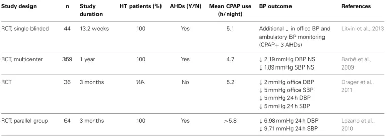
and apnea episodes, CPAP reduces sympathetic activity, systemic inflammation and oxidative stress (Yorgun et al., 2014). However, the results found for the effectiveness of CPAP on blood pres-sure (BP) control are still controversial.Table 1summarizes the results of original studies in which the effect of CPAP on BP has been analyzed. Whereas some studies and meta-analyses (Bakker
et al., 2014; Varounis et al., 2014) have reported modest effects for
CPAP in lowering BP, others tend to support the beneficial effect of CPAP treatment on BP reduction and attenuating the risk of developing HT. In any case, although the lowering effect of CPAP on BP is relevant in terms of overall cardiovascular risk reduction, this effect is very limited when compared to the performance of AHDs in patients with essential HT (Pépin et al., 2009). Thus, treating HT in patients with sleep apnea is proving to be a difficult task and there is consensus that the use of AHDs is mandatory. In spite of this, data on AHDs regimens in patients with OSA are scarce and there is a lack of specific therapeutic guidelines for the pharmacological treatment of HT in these patients. Furthermore, the effects of AH agents on OSA patients are not consistent (Parati
et al., 2012) and there are no data on the efficacy of specific AHDs
regimens when associated with CPAP.
A new treatment for OSA patients is the oral appli-ance/mandibular advancement device (Guralnick and Bakris, 2012). Oral appliance therapy is an important alternative to CPAP for some patients with mild to moderate OSA (Iftikhar et al., 2013). Despite a recent study (Andrén et al., 2013) and a recent meta-analysis (Iftikhar et al., 2013) which have shown some beneficial effects of this device in reducing blood pressure mea-surements, larger and longer randomized control trials are needed to confirm the effects of oral appliance therapy on BP control.
Clearly, more studies are required to identify first-line AHDs regimens for optimal BP control in this particular group of hypertensive patients (Tsioufis et al., 2010; Parati et al., 2013). Moreover, HT related to OSA needs to be managed as a specific entity and an earlier diagnosis of this type of HT seems to be as relevant as the selection of AHDs regimens. This work provides, for the first time, a systematic review on the efficacy of AHDs in HT related to OSA.
WHAT MODELS ARE AVAILABLE TO STUDY HT RELATED TO OSA?
Due to the high complexity and heterogeneity associated with OSA, considerable variability can be observed between reports addressed at the study of this disease. In addition, the scarcity of opportunities for patient investigation, in particular at the cellular level, has compromised progress in understanding the patho-physiology of OSA and the development of novel and specific treatments for this disorder. To overcome some of these limita-tions, several animal models and more recently, a model of OSA
in healthy human volunteers (Tamisier et al., 2009, 2011) have been developed. Animal models, especially of IH, mimic OSA more easily than human models. The small size of rodents allows more rapid and intense changes in SaO2whereas humans require longer periods of hypoxia to induce arterial oxyhaemoglobin desaturation (Foster et al., 2007). The combination of these two approaches is certain to contribute to the consolidation of preven-tion strategies and the development of more suitable treatments for OSA patients.
ANIMAL MODELS
The major advantage of the use of animal models is that they allow single components of the disease to be evaluated, accu-rately controlling the triggering events in terms of both severity and duration, and providing homogeneous populations (Lévy
et al., 2012). These models also provide an excellent opportunity
to explore the underlying mechanistic pathways of HT related to OSA and their consequences under controlled conditions. Moreover, animal models have enabled the study of parameters that have proved difficult to assess in humans, particularly due to the need for organ harvesting to explore the mechanisms under-lying the consequences of IH at the molecular level (Dematteis
et al., 2009). Thus, studies with animal models are good tools for
overcoming some confounding factors present in human studies (e.g., the presence of comorbidities, disease duration, and behav-ioral and environmental variables) (Badran et al., 2014), and for providing more specific information concerning the efficacy of drugs to be tested.
In 2009, Dematteis et al. used the terminology homologous (sharing the cause or pathophysiology of the human disease), predictive (responding to treatment similarly to the human dis-ease) and isomorphic (displaying symptoms similar to those of the human disease although their cause and pathophysiology may differ) to categorize sleep apnea models (Dematteis et al., 2009). According to these categories, most sleep apnea models are only partially isomorphic, focusing on a specific aspect of the human disease. As a matter of fact, none of the currently available ani-mal models reproduce all aspects of human sleep apnea and they present some important limitations. Nonetheless, the ani-mal models of sleep apnea have brought out most of the available knowledge in this field and furthermore, almost all cardiovascu-lar diseases known to be present in patients with OSA have been replicated in these models (Dumitrascu et al., 2013).
The effective use of animals to study sleep apnea implies recognition of the natural similarities and differences between animals and humans to ensure the reliability of the experimen-tal results. For instance, as rodents are nocturnal animals, the stimulus must be applied during the sleep-dominant phase of the diurnal cycle. Moreover, in humans the circadian distribu-tion of sleep tends to be consolidated and normally monophasic, with a daily sleep duration of 7–8 h, whereas it is polyphasic, relatively fragmented and with a duration of 12–15 h in rodents
(Toth and Bhargava, 2013). Another issue is related to the fact
Table 1 | CPAP effect on blood pressure.
Study design n Study
duration
HT patients (%) AHDs (Y/N) Mean CPAP use (h/night)
BP outcome References
RCT; parallel group; blinded endpoint
194 12 weeks 100 Yes 5 ↓3.1 mmHg MBP
↓3.2 mmHg DBP
↓3.1 mmHg SBP (NS)
Martínez-García et al., 2013
RCT; parallel group 118 4 weeks 10 Yes 4.9 ↓3.3 mmHg 24 h MBP Pepperell et al.,
2002
Case -controlled study
48 4 weeks 79 Yes 5.1 ↓5.2 mmHg DBP
↓3.8 mmHg SBP (NS)
Zhao et al., 2012
Prospective randomized trial
32 9 weeks 66 Yes 5.5 ↓ ±10 mmHg MBP
↓ ±10 mmHg DBP
↓ ±10 mmHg SBP
(During both day and night-time)
Becker et al., 2003
RCT; multicenter; parallel group
723 4 years 51.5 Yes 5.0 NS on new-onset HT Barbé et al.,
2012
Prospective, single-center, long-term follow-up
91 5 years 100 Yes NA NS on 24 h BP, SBP and DBP Kasiakogias
et al., 2013
RCT; parallel group 40 6 months 100 Yes 6.01 ↓Awake SBP (6.5 mmHg)
and DBP (4.5 mmHg) NS nocturnal SBP and DBP
Pedrosa et al., 2013
Retrospective chart review study
98 1 year 100 Yes 6.3 ↓5.6 mmHg MBP (resistant
HT group)
↓0.8 mmHg MBP (controlled
BP group)
Dernaika et al., 2009
RCT; crossover 28 8 weeks 100 Yes 4.8 ↓2.1 mmHg 24 h MBP (CPAP
group)
↓9.1 mmHg 24 h MBP
(valsartan group)
Pépin et al., 2009
Prospective cohort study
86 6 months 55 Yes 4.8 ↓4.92 mmHg 24 h MBP Robinson et al.,
2008
Observational study 24 12 weeks 0 No NA ↓5.3 mmHg 24 h MBP Yorgun et al.,
2014
Prospective cohort study
196 6 months 85 Yes NA ↓2.7 mmHg DBP
↓2.1 mmHg SBP
Börgel et al., 2004
RCT; multicenter; double-blinded
340 12 weeks 100 No 4.5 ↓1.5 mmHg MBP
↓1.3 mmHg mean DBP
↓2.1 mmHg mean SBP
Durán-Cantolla et al., 2010
RCT; multicenter; parallel group
44 6 weeks NA Yes 5.0 NS on 24 h SBP and DBP Barbé et al.,
2001
RCT; crossover study; sham placebo
35 10 weeks 100 Yes 5.2 NS on overall 24 h MBP Robinson et al.,
2006
Observational, monocentric; cohort study
495 3.4 years 40.4 Yes NA ↓Occurrence of systemic
arterial HT
Bottini et al., 2012
Table 1 | Continued
Study design n Study
duration
HT patients (%) AHDs (Y/N) Mean CPAP use (h/night)
BP outcome References
RCT; single-blinded 44 13.2 weeks 100 Yes 5.1 Additional↓in office BP and
ambulatory BP monitoring
(CPAP+3 AHDs)
Litvin et al., 2013
RCT, multicenter 359 1 year 100 Yes 4.7 ↓2.19 mmHg DBP NS
↓1.89 mmHg SBP NS
Barbé et al., 2009
RCT 36 3 months NA No 5.2 ↓2 mmHg office DBP
↓5 mmHg office SBP
↓5 mmHg 24 h DBP
↓5 mmHg 24 h SBP
Drager et al., 2011
RCT; parallel group 64 3 months 100 Yes >5.8 ↓6.98 mmHg 24 h DBP
↓9.71 mmHg 24 h SBP
Lozano et al., 2010
AHDs, antihypertensive drugs; BP, blood pressure; CPAP, continuous positive airway pressure; DBP, diastolic blood pressure; HT (%), percentage of hypertensive patients; MBP, mean blood pressure; NA, information not available; NS, no significant effect; RCT, randomized controlled trials; SBP, systolic blood pressure;↓, decrease.
additional care must be taken to minimize external factors (e.g., light exposure, photoperiod, noise, disruptions in the home environment, and post-surgical care in studies, for instance requiring implantation of telemetric devices) able to influence sleep in animals used in experimental research (reviewed in
Toth and Bhargava, 2013).
The experimental animal models developed to mimic OSA have recently been reviewed (Dematteis et al., 2009; Golbidi et al., 2012; Davis and O’Donnell, 2013; Toth and Bhargava, 2013) and assembled taking into account the main injuries trig-gered by OSA. Despite attempts to use large animals (e.g., dogs, lambs, and pigs) to simulate upper airway obstruction, most research on the cardiovascular consequences of OSA has been performed in rodents. Alternative models (e.g., cell cultures incubated in specific devices that perform oxygen fluctuations mimicking sleep apnea-related IH), mainly relevant to signal-ing investigation (Kumar et al., 2003; Gozal et al., 2005; Ryan
et al., 2005), represent a complementary approach to the most
widely used sleep models. However, in spite of the recommen-dations to refine, reduce and replace (the 3Rs programme), these alternative models cannot replace animal models in the study of HT.
The natural models of sleep apnea include the English bull-dog, the historic natural model of spontaneous obstruction
(Hendricks et al., 1987), the sleep-related central apnea models
[e.g., Sprague-Dawley rats (Carley et al., 2000), spontaneously hypertensive (SH) rats (Carley et al., 1998), C57BL/6J (Julien
et al., 2003; Liu et al., 2010)], and the Zucker obese rat in which
apnea is obesity-related (Ray et al., 2007; Lee et al., 2008; Iwasaki
et al., 2012). The experimentally-induced models (e.g., the sleep
deprivation model, induced airway obstruction and the CIH model) are the most widely used. Due to model limitations and lack of extensive study, we only briefly describe the induced airway obstruction model and the sleep deprivation model. Special focus will be given to the CIH model, based on the assumption that IH is the most effective paradigm to induce HT related to OSA and
probably the most relevant stimulus regarding the cardiovascular sequelae of OSA.
Induced airway obstruction model
Briefly, the airway obstruction model involves surgical interven-tion (an endotracheal tube), which is an invasive procedure, or alternatively the use of a specific chamber with a latex neck collar that induces recurrent airway obstruction. This latter procedure, developed byFarré et al. (2007), is associated with high levels of stress due to the restriction of animal movement. In both approaches, the degree of obstruction is adjustable (Golbidi et al., 2012) and in the case of induction of obstruction through endo-tracheal tube, the PaCO2can be adjusted to mimic human sleep apnea (Golbidi et al., 2012). Many experiments using this method have not monitored the sleep state of the animals, but more recent studies have incorporated sophisticated apparatus that is able to detect sleep-awake states and allow close coordination between the initiation of airway obstruction and sleep onset (Schneider
et al., 2000).
This model allows the study of the potential consequences of strenuous breathing against an obstructed airway and can be used to study the cardiovascular consequences and risk factors of OSA (e.g., systemic inflammation and coagulation), and to investigate the mechanisms that underlie OSA (Salejee et al., 1993; Nácher et al., 2007, 2009; Almendros et al., 2008, 2011; Othman et al., 2010). However, to the best of our knowledge, no study has yet shown that this obstruction model is able to mimic HT related to OSA. Furthermore, when testing AHDs, it became crucial to ensure the selection of a stress-free paradigm as it has been shown that any source of external stress on rodents can significantly increase heart rate and blood pressure (Brown et al., 2000; Kramer
et al., 2000; Balcombe et al., 2004; Bonnichsen et al., 2005) and
Sleep deprivation model
In the last few years, several approaches have been used to trigger sleep deprivation in different animals, the rat being the animal of choice to date (Colavito et al., 2013). In the “multiple platform technique,” the animal is aroused from sleep when the charac-teristic loss of muscle tone that accompanies paradoxical sleep causes it to fall off the platform (Suchecki and Tufik, 2000). The “gentle handling” procedure, by far the most popular method, is based on direct interaction with the experimenter, who actively keeps the animal awake through the use of external stimulation (e.g., mild noises, tapping or gentle shaking of the cage, or by direct contact with the animal either using a soft brush or by hand), or by the introduction of novel objects or nesting material in the cages, which typically leads to active exploratory behavior
(Colavito et al., 2013).
These models are most often used to evaluate the neurophys-iological aspects of OSA (Van Dongen et al., 2003; Haack and Mullington, 2005; McKenna et al., 2007; Ward et al., 2009; Nair
et al., 2011) due to the high similarity between the structures of
the nervous systems of rodents and humans (Badran et al., 2014), and to illustrate some mechanistic pathways induced by this trig-ger (McGuire et al., 2008; Tartar et al., 2010; Liu et al., 2011;
Perry et al., 2011). Nevertheless, some studies have also aimed to
evaluate the cardiovascular outcomes induced by this OSA fea-ture and have suggested that sleep fragmentation may have a far more important role in cardiovascular changes observed in OSA patients (Golbidi et al., 2012). Even so, sleep deprivation studies have produced mixed results regarding BP outcomes.
In 1997, Brooks et al. suggested that sleep fragmentation, trig-gered by auditory stimulus, induced only acute changes in BP and did not affect daytime BP (Brooks et al., 1997a,b). In the same way, Bao et al.’s results showed that sleep fragmentation in rats, using acoustic stimuli for 35 days, did not elicit an increase in BP, probably due to some adaptation behavior (Bao et al., 1997). However, more recent studies have shown that sleep depri-vation leads to increased plasma concentrations of epinephrine and norepinephrine (Andersen et al., 2005), ET-1/2 levels (Palma
et al., 2002), and increased heart rate and systolic blood
pres-sure (Andersen et al., 2004; Perry et al., 2007). In addition, sleep fragmentation enhances plasma inflammatory cytokines (e.g., TNF-α, IL-6, IL-1α, and IL-1β), leading to increased oxida-tive stress and inflammation (Yehuda et al., 2009). These results add further evidence demonstrating that sleep deprivation may lead to serious cardiovascular consequences and may aggravate hypertensive features. However, despite the potential of the sleep deprivation model to induce HT related to OSA, it does not exactly mimic sleep fragmentation and presents one major short-coming regarding the evaluation of AHDs efficacy that should be taken into account. Sleep deprivation is a stressful method and it is still unclear whether the method is itself a stressful stimulus
(Palma et al., 2002). Thus, in conclusion, sleep deprivation
mod-els are useful tools for unveiling various aspects of sleep function, studying the effects of sleep loss on subsequent brain function at the molecular, cellular and physiological levels, and evaluating cognitive impairment, but should be used with caution when-ever stress can act as a confounding factor and compromise data interpretation.
CIH model
IH is now established as the dominant model of sleep apnea. Generally, this model makes use of specific ventilated chambers in which the animals are housed and cyclically exposed either to normoxia/hypoxia or room air to mimic the most relevant consequences of OSA. Hypoxic conditions can also be achieved by surgical intervention (an endotracheal tube) or by the use of a mask, which involves animal restraint and consequently high levels of stress (Golbidi et al., 2012). In either case, animals breathe nitrogen-enriched air alternating with oxygen or normal air (Dematteis et al., 2009). Thus, as with O2, nitrogen plays an important role in this model as the flushing of the chambers with this gas allows the gradual lowering of O2. The duration of the hypoxic and normoxic phases of the IH cycle, as well as the slopes of FiO2, decrease and increase, and are dependent on cage/chamber size and the gas flows and mixtures (Dematteis
et al., 2009).
The standard animal model of OSA was that described in the landmark study of Fletcher and Bao (1996). Despite the presence of some drawbacks, this model has successfully been employed to study the changes in systemic arterial pressure and the impact of IH on a wide range of cardiovascular outcomes. One of the major limitations pointed to in this model is the absence of recurrent upper airway obstruction, abolishing the acute hemodynamic changes due to the negative intrathoracic pressure (Badran et al., 2014). Marked negative intrathoracic pressure induces acute hemodynamic changes that are probably the starting point for chronic cardiovascular diseases (Bonsignore
et al., 1994). Despite the absence of upper airway occlusion, some
respiratory efforts (intermittent tachypnea) occur, correspond-ing to a fluctuatcorrespond-ing hyperventilation that follows the IH cycles
(Dematteis et al., 2009). However, this disadvantage allows the
evaluation of CIH effects, namely chronic blood gas exchanges, without the interference of the mechanical aspects of OSA.
This model also fails to reproduce the transient hypercapnia, or at least eucapnia, which occurs in humans determined by air-way occlusion. The first question concerning this issue should be: is PaCO2relevant in humans? Hypercapnia is not a standard parameter analyzed in polysomnographic recordings in patients and therefore there is no consensus on the impact of PaCO2 in arterial blood pressure in patients with OSA. In clinical stud-ies of patients with moderate OSA, the changes in PaCO2 have seemed to be irrelevant (Epstein et al., 2001) or have shown a slight increase (Tilkian et al., 1976) during the apneic events. However, a PaCO2increase may contribute to the severity of the cardiovascular consequences of OSA (Cooper et al., 2005). The results shown by Fletcher et al. in rats suggest that the exposure to hypercapnia during IH is not a critical factor as the effect of IH on diurnal BP is similar, independently of the lower or higher levels of CO2(Fletcher et al., 1995). Moreover, Bao et al. found that eucapnic IH in rats is a more powerful stimulus for induc-ing acute BP increase than hypocapnic IH (Bao et al., 1997). Similarly, Lesske et al. showed comparable changes in BP between two groups submitted to IH with or without hypercapnia (Lesske
et al., 1997). On the other hand, based on the results of
in BP (Kanagy, 2009). Concretely, eucapnic hypoxia induces a faster and greater increase than hypocapnic hypoxia (Kanagy, 2009), through mechanisms that presently remain unknown. Moreover, the greatest increases in BP have been observed in stud-ies in which hypocapnia was prevented by CO2 administration
(Morgan, 2009). Likewise, Tamisier et al., in a study performed
in humans, reported that hypercapnic hypoxia leads to greater sympathetic activation than hypocapnic hypoxia (Tamisier et al., 2009). In line with these findings, the presence of hypocap-nic or eucaphypocap-nic hypoxia conditions leads to an underestimated increase in BP that must be taken into account. In conclusion, although some data suggest that PaCO2may influence physiolog-ical responses to IH, further studies are needed to evaluate the combined effect of IH and hypercapnia. Another drawback that could be attributed to the IH paradigm is the fact that it is not accompanied by sleep fragmentation and does not incorporate monitoring of sleep.
Each group of researchers has applied its own specific paradigm and these discrepancies may compromise the straight-forward comparison of the results. The several paradigms of CIH, which simulate the cyclical pattern of hypoxia experienced by patients with OSA, diverge in some respects, namely in the animal species involved, e.g., Sprague-Dawley rats (Fletcher et al., 1995; Kanagy et al., 2001; Tahawi et al., 2001; Allahdadi et al., 2005;
Chen et al., 2005; Phillips et al., 2005; Lai et al., 2006), Wistar
rats (Dunleavy et al., 2005; Lefebvre et al., 2006), C57BL/6J mice
(Julien et al., 2003), and CF-1 mice (Rosa et al., 2011), the
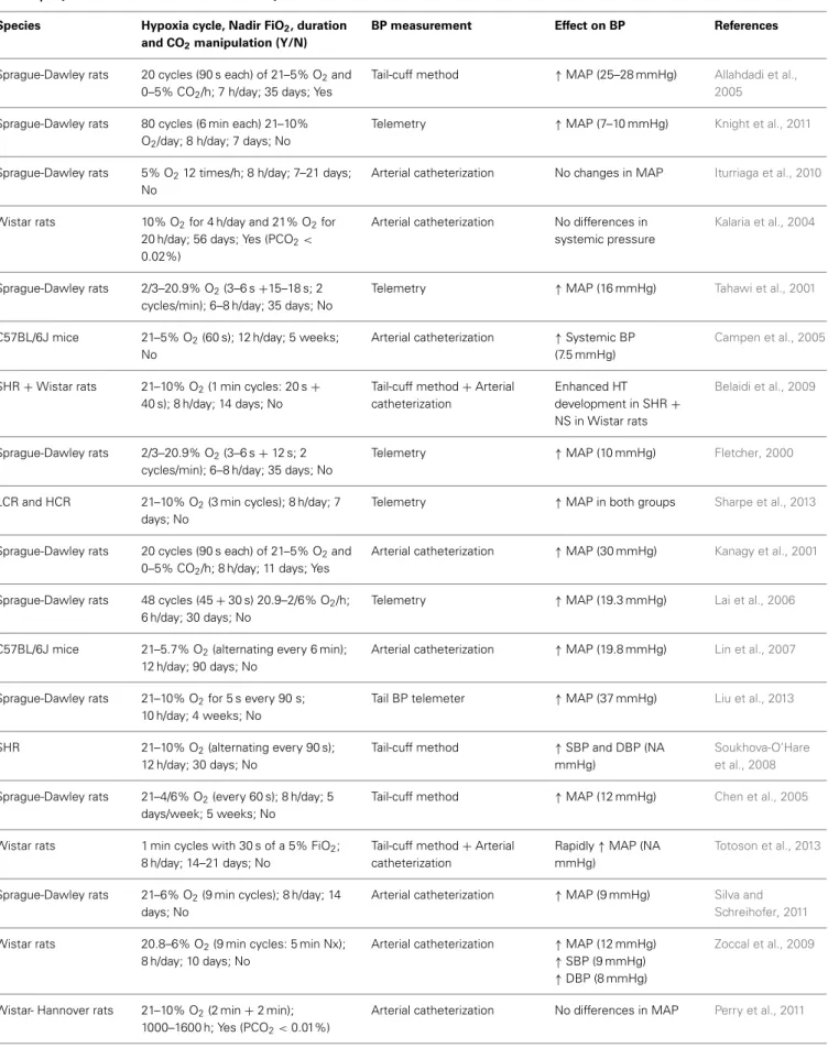
sever-ity of hypoxia, the number of hypoxic episodesperhour of sleep, the number of days of hypoxic exposure (exposure duration), and CO2manipulation.Table 2summarizes the variability observed in the CIH models.
These models typically create moderate to severe oxygen desaturation, thereby mimicking severe forms of OSA and may therefore not be applicable to mild and moderate clinical OSA
(Dematteis et al., 2009). CIH models with cycles of FiO2of 5% or
less usually mimic severe forms of OSA in humans and produce maximal changes in BP and heart rate (Dematteis et al., 2008). However, higher FiO2 (8–10%) has been used in rodent models of CIH (Soukhova-O’Hare et al., 2008; Knight et al., 2011; Perry
et al., 2011; Bathina et al., 2013).
The duration and frequency of hypoxic/normoxic periods are adjustable; usually, the higher the frequency the shorter the IH cycles (Golbidi et al., 2012). There is a sizeable discrepancy regarding the duration of IH cycles, ranging from 120 cycles/h (30 s cycle;Fletcher et al., 1992a; Julien et al., 2003; Dematteis
et al., 2008), 80 cycles/h (6 min cycle; Knight et al., 2011), 60
cycles/h (1 min cycle;Campen et al., 2005), and when the cham-bers are larger, longer cycles are often used, reducing the number of cycles/h (Zoccal et al., 2007, 2008; Silva and Schreihofer, 2011) of daytime exposure, from 4 h/day (Kalaria et al., 2004), 6 h/day
(Lai et al., 2006), 7 h/day (Fletcher et al., 1992a), 8 h/day (Chen
et al., 2005; Belaidi et al., 2009; Zoccal et al., 2008, 2009; Knight et al., 2011; Silva and Schreihofer, 2011; Dyavanapalli et al., 2014;
Schulz et al., 2014), 10 h/day (Liu et al., 2013) to 12 h/day (Lin
et al., 2007). The exposure duration of 8 h/day seems to be that
on which there is the greatest consensus (seeTable 2). The dura-tion of exposure seems to affect the study outcomes more than
the hypoxic nadir or the rate of hypoxic cycling (Davis and
O’Donnell, 2013).
An advantage of CIH models is they allow exposures that can be extended over months, enabling the investigation of chronic consequences that might occur in humans (Toth and Bhargava, 2013). The number of days necessary to induce an increase in BP seems to be dependent on the CIH paradigm. Some authors suggest that the BP increase triggered by CIH represents a time-dependent effect (Prabhakar et al., 2001; Hui et al., 2003;
Dematteis et al., 2008; Zoccal et al., 2009). Moreover, both the
time and severity of hypoxia have been shown to play an impor-tant role in the cardiovascular response (Li et al., 2007; Perry
et al., 2007). It has recently been shown that a period of 14 days
is not long enough to induce structural changes in cardiovascular structures, but these are already apparent after 35 days of incuba-tion (Dematteis et al., 2008). Moreover, Iturriaga et al. report that the exposure of rats to CIH for 14 days enhanced the ventilatory response to hypoxia and produced a significant shift in heart rate variability, but these cardiorespiratory alterations occurred with-out noticeable changes in mean arterial BP until 21 days of CIH exposure (Iturriaga et al., 2010). Whereas some short-term pro-tocols (7–14 days) cause a significant increase in BP (Belaidi et al., 2009; Knight et al., 2011; Silva and Schreihofer, 2011; Bathina
et al., 2013), others show an increase in BP that occurs only after
long-term exposure (35 days) to CIH (Prabhakar et al., 2001,
2005; Chen et al., 2005; Zoccal et al., 2009) (seeTable 2). Finally,
most IH paradigms in rodents do not include CO2 supplementa-tion (Fletcher et al., 1999; Lin et al., 2007; Iturriaga et al., 2010;
Perry et al., 2011; Bathina et al., 2013). In fact, only some authors
have manipulated the CO2levels (Ooi et al., 2000; Kantores et al.,
2006; Dyavanapalli et al., 2014) and fixed the values along the
protocol (seeTable 2).
Independently of the paradigm used to induce HT related to OSA, previous reviews are unanimous in reporting the develop-ment of mild HT, despite the divergent changes in arterial blood gases (Kanagy, 2009) (seeTable 2). The exceptions found in this review (Kalaria et al., 2004; Belaidi et al., 2009; Iturriaga et al.,
2010; Perry et al., 2011) are all related to the method used for
BP measurement. It is apparent that arterial catheterization is not an accurate method of measuring BP in CIH models. The meth-ods most often used for BP measurement (for a review, seeKurtz
et al., 2005) in IH models (seeTable 2) are the tail-cuff method
(Allahdadi et al., 2005; Chen et al., 2005; Soukhova-O’Hare et al.,
2008; Belaidi et al., 2009; Totoson et al., 2013), radiotelemetry
(Fletcher, 2000; Tahawi et al., 2001; Lai et al., 2006; Knight et al.,
2011; Bathina et al., 2013; Sharpe et al., 2013; Dyavanapalli et al.,
2014; Schulz et al., 2014), and arterial catheterization (Kanagy
et al., 2001; Kalaria et al., 2004; Campen et al., 2005; Lin et al., 2007; Belaidi et al., 2009; Zoccal et al., 2009; Iturriaga et al., 2010; Perry et al., 2011; Silva and Schreihofer, 2011; Totoson et al.,
2013).
HUMANS
Table 2 | Reports on the effects of CIH on blood pressure.
Species Hypoxia cycle, Nadir FiO2, duration and CO2manipulation (Y/N)
BP measurement Effect on BP References
Sprague-Dawley rats 20 cycles (90 s each) of 21–5% O2and
0–5% CO2/h; 7 h/day; 35 days; Yes
Tail-cuff method ↑MAP (25–28 mmHg) Allahdadi et al.,
2005
Sprague-Dawley rats 80 cycles (6 min each) 21–10%
O2/day; 8 h/day; 7 days; No
Telemetry ↑MAP (7–10 mmHg) Knight et al., 2011
Sprague-Dawley rats 5% O212 times/h; 8 h/day; 7–21 days;
No
Arterial catheterization No changes in MAP Iturriaga et al., 2010
Wistar rats 10% O2for 4 h/day and 21% O2for
20 h/day; 56 days; Yes (PCO2< 0.02%)
Arterial catheterization No differences in
systemic pressure
Kalaria et al., 2004
Sprague-Dawley rats 2/3–20.9% O2(3–6 s+15–18 s; 2
cycles/min); 6–8 h/day; 35 days; No
Telemetry ↑MAP (16 mmHg) Tahawi et al., 2001
C57BL/6J mice 21–5% O2(60 s); 12 h/day; 5 weeks;
No
Arterial catheterization ↑Systemic BP
(7.5 mmHg)
Campen et al., 2005
SHR+Wistar rats 21–10% O2(1 min cycles: 20 s+
40 s); 8 h/day; 14 days; No
Tail-cuff method+Arterial catheterization
Enhanced HT
development in SHR+
NS in Wistar rats
Belaidi et al., 2009
Sprague-Dawley rats 2/3–20.9% O2(3–6 s+12 s; 2
cycles/min); 6–8 h/day; 35 days; No
Telemetry ↑MAP (10 mmHg) Fletcher, 2000
LCR and HCR 21–10% O2(3 min cycles); 8 h/day; 7
days; No
Telemetry ↑MAP in both groups Sharpe et al., 2013
Sprague-Dawley rats 20 cycles (90 s each) of 21–5% O2and
0–5% CO2/h; 8 h/day; 11 days; Yes
Arterial catheterization ↑MAP (30 mmHg) Kanagy et al., 2001
Sprague-Dawley rats 48 cycles (45+30 s) 20.9–2/6% O2/h;
6 h/day; 30 days; No
Telemetry ↑MAP (19.3 mmHg) Lai et al., 2006
C57BL/6J mice 21–5.7% O2(alternating every 6 min);
12 h/day; 90 days; No
Arterial catheterization ↑MAP (19.8 mmHg) Lin et al., 2007
Sprague-Dawley rats 21–10% O2for 5 s every 90 s;
10 h/day; 4 weeks; No
Tail BP telemeter ↑MAP (37 mmHg) Liu et al., 2013
SHR 21–10% O2(alternating every 90 s);
12 h/day; 30 days; No
Tail-cuff method ↑SBP and DBP (NA
mmHg)
Soukhova-O’Hare et al., 2008
Sprague-Dawley rats 21–4/6% O2(every 60 s); 8 h/day; 5
days/week; 5 weeks; No
Tail-cuff method ↑MAP (12 mmHg) Chen et al., 2005
Wistar rats 1 min cycles with 30 s of a 5% FiO2;
8 h/day; 14–21 days; No
Tail-cuff method+Arterial catheterization
Rapidly↑MAP (NA
mmHg)
Totoson et al., 2013
Sprague-Dawley rats 21–6% O2(9 min cycles); 8 h/day; 14
days; No
Arterial catheterization ↑MAP (9 mmHg) Silva and
Schreihofer, 2011
Wistar rats 20.8–6% O2(9 min cycles: 5 min Nx);
8 h/day; 10 days; No
Arterial catheterization ↑MAP (12 mmHg)
↑SBP (9 mmHg)
↑DBP (8 mmHg)
Zoccal et al., 2009
Wistar- Hannover rats 21–10% O2(2 min+2 min);
1000–1600 h; Yes (PCO2<0.01%)
Arterial catheterization No differences in MAP Perry et al., 2011
Table 2 | Continued
Species Hypoxia cycle, Nadir FiO2, duration and CO2manipulation (Y/N)
BP measurement Effect on BP References
Sprague-Dawley rats 10 cycles (6 min each) of 21–6% O2
and 0–5% CO2/h; 8 h/day; 28 days; Yes
Telemetry ↑SBP (39 mmHg)
↑DBP (33 mmHg)
Dyavanapalli et al., 2014
C57BL/6J mice 21–7% O2(120 s each cycle); 5
days/week; 8 h/day; 6 weeks; No
Telemetry Significant↑MAP Schulz et al., 2014
Sprague-Dawley rats 21–10% O2(cycle duration: NA);
8 h/day; 7 days; No
Telemetry ↑MAP that persisted
after CIH exposure
Bathina et al., 2013
BP, blood pressure; CIH, chronic intermittent hypoxia; DBP, diastolic blood pressure; h, hour; HCR, high aerobic capacity rats; HT, hypertension; LCR, low aerobic capacity rats; MAP, mean arterial pressure; NA, information not available; NS, no significant effect; Nx, normoxia; min, minutes; s, seconds; SBP, systolic blood pressure; SHR, spontaneously hypertensive rats;↑, increase.
short-term IH models, generally the exposure time (20–60 min) and the duration of the hypoxia or voluntary apnea period (30 s) are very limited. The protocols of Cutler et al. and Tamisier et al. are good examples of short-term models (Cutler et al., 2004;
Tamisier et al., 2009). In contrast, Foster et al. made use of a
chronic model, exposing healthy human volunteers to an hour of IH (5 min hypoxia alternating with 5 min normoxia) daily for 2 weeks (Foster et al., 2005). As in the animal models of IH, only some studies have controlled the level of CO2(Foster et al., 2005), whereas others have not (Tamisier et al., 2009). Regardless of the protocol followed, exposing humans to CIH implies careful supervision.
In 2001, Xie et al. exposed nine healthy human subjects dur-ing wakefulness to 20 min of isocapnic hypoxia (arterial O2 saturation, 77–87%) and 20 min of normoxic hypercapnia (end-tidal PCO2, 15.3–8.6 Torr above eupnea) on two separate days. The subjects breathed through a leak-free nasal mask and the neurocirculatory and ventilatory responses to these two stimuli were further evaluated (Xie et al., 2001). These authors found that hypoxia induced a sympathetic activation that outlasted the chemical stimulus, whereas hypercapnia evoked a short-lived sympathetic activation (Xie et al., 2001). Years later, in a study performed with a larger sample (n=31), Cutler et al. used a model of IH induced by voluntary apnea (30 s of hypoxic apnea every 1 min—simulating an AHI of 60/h—for 20 min) to deter-mine if the cessation of breathing is important in prolonged sym-pathetic activation (Cutler et al., 2004). This study also included two other groups that were exposed to intermittent hypercap-nic hypoxia and to intermittent isocaphypercap-nic/hypoxia, respectively
(Cutler et al., 2004). Their results support the hypothesis that
short-term exposure to intermittent hypoxic apnea results in sus-tained elevation of post-ganglionic muscle sympathetic nerve activity and that hypoxia is the primary mediator of this response
(Cutler et al., 2004). The data reported by Leuenberger et al.
one year later were in line with these results (Leuenberger et al., 2005). They also found, in a study that enrolled 26 patients, a sustained sympathetic activation and also a transient elevation of BP following 30 min of voluntary end-expiratory apneas primed with a hypoxic gas mixture and lasting for 20 s in each minute
(Leuenberger et al., 2005).
Foster et al. carried out three main studies in healthy human volunteers. The first aimed to determine the ventilatory, cardio-vascular and cerebral tissue oxygen response to two protocols of
IH (Foster et al., 2005). This study involved 18 patients randomly
assigned to short-duration IH (1 h of 12% O2separated by 5 min of normoxia) or long-duration IH (30 min of 12% O2). Both groups had 10 exposures over 12 days. Their findings show a rise in mean arterial blood pressure (MAP) that occurs throughout the daily exposure to short-duration IH but not during expo-sure to long-duration IH; moreover, they demonstrate that the vascular processes required to control blood flow and O2 sup-ply to cerebral tissue in a healthy human are delayed following exposure to 12 days of isocapnic IH (Foster et al., 2005). In 2009, the same group reinforced the enrollment of IH on the patho-genesis of cardiovascular and cerebrovascular disease in patients with OSA (Foster et al., 2009). They exposed 10 healthy subjects to IH (2 min of hypoxia: nadir PET,O2=45.0 mmHg, alternat-ing with 2 min of normoxia: peak PET,O2=88.0 mmHg for 6 h) for 4 consecutive days and concluded that IH alters BP (MAP increased by 4 mmHg) and induces an increase in cerebral vascu-lar resistance (Foster et al., 2009). More recently, Foster et al. have assessed the role of the type I angiotensin II receptor in mediat-ing an increase in arterial pressure associated with a smediat-ingle 6-h IH exposure (Foster et al., 2010). For that, they exposed nine healthy subjects to sham IH, IH with placebo medication, and IH with the type I angiotensin II receptor antagonist (losartan). Their find-ings demonstrate a significant increase in arterial pressure after exposure to isocapnic IH (Foster et al., 2010). Furthermore, since this increase is prevented by the blockade of AT1receptors, these results suggest an important role for the rennin-angiotensin-aldosterone system (RAAS) in the pathophysiology of HT related to OSA (Foster et al., 2010).
delivered O2 for 15 s every 2 min during sleep while subjects breathed 13% O2in a hypoxic tent to create 30 cycles/h of cyclic desaturation-reoxygenation (SpO2range: 95–85%), and exposed subjects overnight for 8–9 h/day for 2 or 4 weeks (Tamisier et al., 2009). Among other results, they show that waking normoxic arterial pressure increased significantly at 2 weeks for systolic and for diastolic at 4 weeks, that patients developed a sustained BP increase during the day and exhibited a steeper BP decrease at night compared to baseline BP values, and finally, that this model produces clinically relevant fluctuations in SaO2(Tamisier et al., 2009). Although undoubtedly relevant, the authors recognize the presence of several respects in which their model does not mimic sleep apnea, e.g., no negative intrathoracic pressure development, higher percentage of sleep time at<90% SaO2and poikilocapnia
(Tamisier et al., 2009). However, some of these limitations can
be overcome to achieve a pattern of IH more akin to OSA fea-tures. This model was further used by the same group in 2011 to shed light on the profile of the BP increase previously described to determine if it is sustained and to explore potential under-lying physiological mechanisms. The authors found that only 2 weeks of severe IH exposure produces a sustained daytime BP increase in the setting of sympathetic activation and blunted vas-cular sympathic baroreflex gain in healthy volunteers (Tamisier
et al., 2011).
In conclusion, to date, only a small number of studies have been conducted using healthy human models of IH and these have primarily been aimed at elucidating the role of IH in sus-tained sympathetic activation and cerebrovascular regulation. Only a few studies have evaluated BP outcomes (Foster et al.,
2009, 2010; Tamisier et al., 2009, 2011) and none of these models
have truly been used to assess the efficacy of AHDs in the treat-ment of HT related to OSA. In fact, in the later work of Foster et al., losartan (the angiotensin II AT1receptor antagonist) was used only to demonstrate a mechanistic pathway rather than to evaluate its efficacy (Foster et al., 2010). Thus, future research in this field is clearly needed.
WHAT ARE THE MECHANISMS INVOLVED IN THE PATHOGENESIS OF HT RELATED TO OSA?
Fletcher et al. were pioneers in demonstrating the hyperten-sive effect of CIH (Fletcher et al., 1992a) and the role of the sympathetic nervous system, peripheral receptors and rennin-angiotensin system in this response (Fletcher et al., 1992b, 1999,
2002; Fletcher, 2000). This group also showed that surgical
dener-vation of peripheral chemoreceptors, adrenal demedullation and chemical denervation of the peripheral nervous system prevented the increase in BP in response to CIH stimulus (Fletcher et al.,
1992b; Bao et al., 1997). After Fletcher et al.’s first work, many
reports enabled confirmation of the relationship between IH and BP increases and contributed to elucidating the underlying mech-anisms. Kanagy et al. reported increased plasma endothelin-1 levels in rats exposed for 11 days to CIH, which also demon-strated an appreciable increase in MAP (Kanagy et al., 2001). In 2006, Lai et al. suggested that chronic IH-induced sustained HT was associated with the facilitation of cardiovascular sympa-thetic outflow followed by decreases in baroreflex sensitivity in conscious rats (Lai et al., 2006). Along the same line, the work
undertaken by Zoccal et al. provided strong evidence to sup-port the idea that rats submitted to CIH show an increase in sympathetic activity, which seems to be essential in the mainte-nance of high BP values in the CIH model (Zoccal et al., 2007). Another group revealed that although elevated sympathetic ner-vous system activity (SNA) may contribute to CIH-induced HT, reduced adrenergic vascular reactivity buffers the cardiovascular impact of exaggerated acute raises in SNA (Silva and Schreihofer, 2011). Data attained by Knight et al. indicated that CIH induces an increase in FosB/FosB in autonomic nuclei and suggested that activator protein-1 (AP-1) transcriptional regulation may contribute to stable adaptative changes that support chroni-cally elevated BP (Knight et al., 2011). Also in 2011, Liu et al. demonstrated that CIH activates the HIF-1α/endothelin system, through CIH-NADPH oxidase-mediated ROS production, and this enhances the development of resistant vasoconstriction and elevates BP in rats (Liu et al., 2011). The study undertaken by Bathina et al. revealed that the knockdown of tyrosine hydrox-ylase in the nucleus of the solitary (NTS) tract reduces the CIH-induced persistent increase in MAP, suggesting that nora-drenergic A2 neurons in nucleus tractus solitarius play a role in the cardiovascular responses to CIH (Bathina et al., 2013). More recently, Schulz et al. have shown that NADPH oxidase 2 (NOX2) knockout blocks the development of HT induced by CIH, suggesting that this type of HT is mediated by reactive oxy-gen species (ROS) derived from the activation of NOX2 within cells located outside the cardiovascular system (Schulz et al.,
2014).
The mechanisms involved in the genesis of HT related to OSA have recently been reviewed (Lavie and Lavie, 2009; Bosc et al., 2010; Sunderram and Androulakis, 2012; Zhang and Si, 2012;
Lévy et al., 2013) and broadly include the following:
sympa-thetic nervous system stimulation mediated mainly by the activa-tion of carotid body chemoreflexes, decreased vascular responses to nitric oxide, increased plasma concentrations of endothe-lin, and elevation of proinflammatory cytokines (TNF-α, IL-6, VEGF). While for some of these mechanisms (e.g., activation of the RAAS, endothelial dysfunction, systemic inflammation, metabolic anomalies, and genetic contribution) the relationship with OSA and subsequent cardiovascular morbidity remain par-tially unclear and there is a need to gather more evidence, for others (e.g., the increase in sympathetic activity and acute effects of negative intrathoracic pressure), there seems to be more agree-ment on the linkage and it is well-docuagree-mented (Parati et al., 2013). In fact, based on data attained from patients with OSA, it is widely accepted that sympathetic activation, inflammation and oxidative stress play major roles in the pathophysiology of this particular type of HT. In addition, the use of animal models has revealed that CIH is the critical stimulus underlying sympa-thetic activity and HT, and that this effect requires the presence of functional arterial chemoreceptors (Fletcher, 2000). However, it should be also mentioned that HT related to OSA probably results not only from increased carotid chemoreflex but also from decreased baroreceptor activity (Dumitrascu et al., 2013). It is also important to highlight the potential role of obesity as an interme-diate factor in the pathway of HT related to OSA (Young et al.,
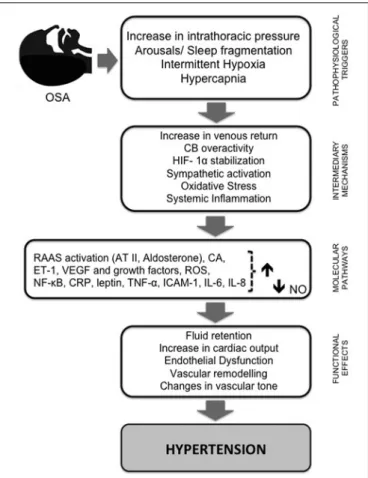
The mechanisms involved in the pathogenesis of HT can be summarized in relation to two main pathways: sympathetic ner-vous system stimulation mediated mainly by activation of carotid body (CB) chemoreflexes and the systemic effects of CIH, mainly due to the activation of NOX2 and subsequent ROS produc-tion. Figure 1 illustrates the hypothesized pathways by which intermittent hypoxia leads to HT.
WHAT IS ALREADY KNOWN CONCERNING THE EFFICACY OF AHDs?
HUMANS
Despite the considerable number of studies involving OSA patients, only a few have investigated the efficacy of different AHDs and in general, they tend to be individual drug studies. Moreover, most of the studies only take into account the number of drugs taken by patients to adjust this variable and are difficult to interpret as most of the patients were already under AHDs reg-imens. This lack of information could be attributed to the large number of possible different AHDs regimens observed in OSA patients.Table 3summarizes the most relevant studies that have investigated the efficacy of AHDs in OSA patients.
In a study undertaken by Pelttari et al., the AH effects of four different AHDs (atenolol: a beta-blocker; isradipine: a calcium channel blocker; hydrochlorothiazide: a diuretic; spirapril: an angiotension-converting enzyme inhibitor) in obese patients with OSA and HT were compared using ambulatory blood pressure monitoring (ABPM) (Pelttari et al., 1998). This study revealed that although daytime HT was quite easily controlled by the single use of these drugs (especially with atenolol and isradip-ine; diuretics did not significantly lower BP) none of the AHDs were able to produce a significant decrease in nocturnal BP
(Pelttari et al., 1998). Mayer et al. carried out another
compara-tive study between cilazapril (an angiotension-converting enzyme inhibitor) and metoprolol (a beta-blocker) (Mayer et al., 1990). Their findings showed that despite the short period of therapy (1 week), both metoprolol and cilazapril lowered nighttime BP in OSA patients (Mayer et al., 1990).
A multiple crossover study examined the BP-lowering effect of the five major AHDs classes (atenolol: beta-blocker; amlodipine: calcium channel blocker; enalapril: angiotension-converting enzyme inhibitor; hydrochlorothiazide: diuretic; losartan: angiotensin receptor blocker) and showed that atenolol induced the most pronounced effect in lowering BP (Kraiczi
et al., 2000). Atenolol was more efficient in reducing mean
night-time diastolic and systolic BP (measured by ABPM) compared to amlodipine, enalapril, hydrochlorothiazide, and losartan (Kraiczi
et al., 2000). Salo et al. investigated the effects of four AHDs
(atenolol; isradipine: a calcium channel blocker; hydrochloroth-iazide; spirapril: an angiotension-converting enzyme inhibitor) on cardiovascular autonomic control and reactivity in HT OSA patients (Salo et al., 1999). This group reported that of the four drugs, only atenolol effected BP variability (Salo et al., 1999). Thus, the results of these two pilot studies are in line with those arguing the involvement of the sympathetic system in the patho-physiology of HT related to OSA, suggesting that beta-blockers, in particular atenolol, may have beneficial effects beyond BP reduc-tion in patients with OSA. However, both studies presented low
FIGURE 1 | Schematic diagram summarizing the pathways by which intermittent hypoxia leads to hypertension.Repetitive obstructive apneas or hypopneas lead to increased intrathoracic pressure, sleep fragmentation, recurrent hypercapnia, and intermittent hypoxia (IH). This last phenomenon plays a pivotal role in triggering several intermediary mechanisms and molecular pathways that contribute to the initiation and progression of cardiac and vascular pathology. First, IH enhances sympathetic nervous system activity, leading to vasoconstriction and systemic hypertension through RAAS activation, and an increase in catecholamine secretion and plasma level of vasoconstrictive ET-1. Episodic hypoxia also favors the stabilization of HIF-1αand the production of ROS, which is followed by increased expression of NF-κB and decreased NO bioavailability, the most important vasodilatory molecule synthesized by the endothelium. AT II and ET-1 both seem to be implicated in vascular remodeling and ROS formation, which is increased through the activation of vascular NADPH oxidase and xanthine oxidase. ROS molecules induce a cascade of inflammatory pathways linked to an overexpression of adhesion molecules and pro-inflammatory cytokines, and oxidative stress may trigger sympathetic hyperactivation andvice versa. ROS production is required for HIF-1αinduction and HIF-1αinduction is required for ROS production. In addition, HIF-1αpromotes the expression of ET-1 and transcriptional activation of VEGF and other growth factors. Activation of NF-κB also seems to be central in inflammation induced by IH due to its regulatory role in the production of pro-inflammatory mediators (e.g., TNF-α, IL-6, IL-8, ICAM-1, and CRP). These signaling pathway proteins, combined with RAAS, decreased expression of eNOS, and increased ROS production and stabilization of HIF-1, participate in the molecular mechanisms underlying the endothelial dysfunction induced by IH. Together, these mechanisms progress to fluid retention, changes in cardiac output and vascular tone, and vascular remodeling, leading to systemic HT, one of the major
consequences of OSA. AT II, angiotensin II; CA, catecholamine levels; CRP, C- reactive protein; CB, carotid body; ET-1, endothelin 1; HIF-1α,
hypoxia-inducible factorα; IH, intermittent hypoxia; IL, interleukin; ICAM-1,
FIGURE 1 | Continued
intercellular adhesion molecule; NO, nitric oxide; eNOS, endothelial nitric oxide synthase; NF-κB, nuclear factor-κ-light chain enhancer of activated B cells; RAAS, renin-angiotensin-aldosterone system; ROS, reactive oxygen species; TNF-α, tumor necrosis factorα; VEGF, vascular endothelial growth factor.
levels of causation, which could have limited the ability to detect differences between classes.
Nevertheless, it has been advanced that angiotension-converting enzyme inhibitors (ACEi) treatment could exacerbate OSA by inducing upper airway inflammation (Cicolin et al., 2006). The comparison between chronic treatments of ACEi and angiotensin AT1 receptor antagonists in terms of AH efficacy and levels of inflammatory markers has never been performed either in humans with OSA or in animal models. More recently, other study compared the effect of doxazosin (anα1- adrenergic recep-tor antagonist) and enalapril (an angiotensin-converting enzyme inhibitor) on nocturnal BP control and concluded that the for-mer has a proportionally poorer effect than the latter (Zou et al., 2010). In 1994, Grote et al. performed a study aimed at assessing the effectiveness of cilazapril (an angiotension-converting enzyme inhibitor) in managing high BP in patients with OSA. Although the study comprised a small sample size, the results suggested that cilazapril is effective in reducing BP in all sleep stages (Grote et al., 1994). In another small study, Heitmann et al. evaluated the effect of nebivolol (a third generation beta-blocker) on BP reduction and sleep apnea activity in HT patients with mild to moder-ate OSA in comparison with valsartan (an angiotensin receptor blocker) and concluded that the effect of these AHDs were similar
(Heitmann et al., 2010). Despite the same limitations, these
stud-ies highlight the role of the renin-angiotensin-aldosterone system (RAAS) in the pathophysiology of HT related to OSA.
In two past studies (Lozano et al., 2010; Litvin et al., 2013), patients either received CPAP in combination with AHDs or alternatively, the pharmacological treatment alone, allowing the evaluation of the effects of CPAP and AHDs independently or in conjunction. In the study undertaken by Lozano et al., patients were under an AHDs regimen with at least three drugs at adequate doses, including a diuretic (Lozano et al., 2010). The authors noted a significant decrease in the mean 24-h diastolic BP in patients who received CPAP in addition to conventional treat-ment, suggesting that resistant HT treated with both CPAP and AHDs provides greater BP reduction than AHDs alone (Lozano
et al., 2010). However, in patients who used CPAP less than the
average (5.6±1.52 h/night) and for those treated with conven-tional treatment alone, there was no significant difference in the 24-h ambulatory BP values (Lozano et al., 2010). These find-ings are in line with those reported by Litvin et al., attained with patients who received stepped dose titration of AHDs treatment (valsartan 160 mg+amlodipine 5–10 mg+hydrochlorothiazide 25 mg) for 3 months before CPAP was added (Litvin et al., 2013). These findings seem to suggest that the best strategy to treat HT related to OSA involves the combination of OSA treatment with CPAP and the use of AHDs. This combination is likely to be more effective in lowering both daytime and nighttime BP
than either treatment alone (Phillips and O’Driscoll, 2013). In addition, Pépin et al. explored RAAS inhibition using losartan in a crossover randomized control trial. In this study, the authors compared the efficacy of CPAP and valsartan in reducing BP in HT patients with OSA never treated for either condition (Pépin
et al., 2009). They reported that although the BP decrease was
significant with CPAP treatment, valsartan induced a four-fold higher decrease in mean 24-h BP than CPAP in this specific sample (Pépin et al., 2009).
In an earlier report, 74 of the 393 OSA patients using AH med-ications on a regular basis for more than 6 months were deemed to have been treated “ineffectively” (Lavie and Hoffstein, 2001), but the characterization of these medications was not reported. The same limitation is found in the study of Deleanu et al., which aimed to study the effect of medication-controlled HT on OSA patients (Deleanu et al., 2014). The authors suggested that con-trolled BP abates sleepiness and reduces remaining symptoms (e.g., headaches, impotence and morning fatigue). These findings could be much more interesting if the regimens responsible for these effects were revealed.
In a very recent study, Kario et al. aimed to evaluate the effects of bedtime dosing of vasodilating (nifedipine, a calcium channel blocker) vs. sympatholytic (carvedilol, a non-selectiveβ -blocker/α1-blocker) AH agents on the sleep BP profile in HT OSA patients (Kario et al., 2014). For this, they made use of a new BP monitoring method, the trigger sleep BP monitoring (TSP) method, which is based on the automated fixed-interval measure-ment function with an additional oxygen-triggered function that initiates BP measurement when oxygen desaturation falls below a set variable threshold continuously monitored by pulseoxime-try (Kario et al., 2014). The BP lowering effects of nifedipine on the mean and minimum sleep systolic BP were stronger than those of carvedilol; moreover, sleep systolic BP surge (the differ-ence between the hypoxia peak systolic BP—SBP—measured by the oxygen-triggered function and SBP within 30 min before and after the peak SBP) was only significantly reduced by carvedilol
(Kario et al., 2014). Thus, both drugs are effective in decreasing
sleep BP (Kario et al., 2014) but the effect of carvedilol seems to be related more specifically to the hypoxia stimuli than nifedipine. Finally, Cichelero et al. recently published the protocol of their randomized double-blind clinical trial, which seeks to compare the efficacy of chlorthalidone (a diuretic) with amiloride (also a diuretic) vs. amlodipine (a calcium channel blocker) as a first drug option in patients older than 40 years of age with stage I HT and moderate OSA (Cichelero et al., 2014). The findings of this study have not yet been reported.
In summary, individual drug studies find that the blockade ofβ1-adrenergic receptors (e.g., atenolol and nebivolol) and the renin-angiotensin-aldosterone (RAA) pathway, including both ACEi and angiotensin AT1 receptor antagonists, might be help-ful. Spironolactone (a mineralocorticoid receptor antagonist) has been proposed has a very useful tool in cases of resistant HT
(Ziegler et al., 2011a,b), a very prevalent condition in OSA
M
onteiro
Antih
y
pertensiv
e
d
rugs
in
CIH
c
onditions
Study design n CPAP (Y/N) AHDs; dosage (mg/day) BP measurement BP outcome References
RCT;
double-blinded; balanced incomplete block design (6 w each drug+3 w washout)
40 No Atenolol (50); amlodipine (5); enalapril (20);
hydrochlorothiazide (25); losartan (50)
Office BP 24 h ABPM
↓in office SBP and daytime ABPM NS for all drugs; Atenolol↓night-time 24 h SBP and DBP more effectively than amlodipine, enalapril or losartan
Kraiczi et al., 2000
RCT;
double-blinded; crossover schedule
(8 w each drug+2–3 w washout
15 No Atenolol (50); isradipine (2.5): hydrochlorothiazide (25); spirapril (6)
Office BP Slight↓BP for all drugs;
Only atenolol affected BP variability
Salo et al., 1999
RCT;
double-blinded; crossover (8 w each drug+2–3 w washout
18 NA Atenolol (50); isradipine (2.5); hydrochlorothiazide (25); spirapril (6)
24 h ABPM ↓mean 24 h SBP (except for HCTZ) ↓mean 24 h DBP (for all drugs)
NS↓mean night-time SBP and DBP (for all drugs)
Pelttari et al., 1998
RCT
(3 months each treatment)
75 Yes Treatment with at least 3 drugs at adequate doses, including a diuretic
24 h ABPM CPAP+AHDs regimen:↓4.9 mmHg 24 h DBP; AHDs regimen alone: NS
Lozano et al., 2010
RCT; single-blinded (3 w each regimen)
44 Yes Valsartan (160)+amlodipine (5–10)+hydrochlorothiazide (25)
Office BP 24 h ABPM
AHDs alone:↓office and 24 h SBP and DBP
Additional↓in office BP and ambulatory BP monitoring (CPAP+3 AHDs)
Litvin et al., 2013
RCT; crossover
(8 w each treatment+4 w washout)
23 Yes Valsartan (160) Office BP 24 h ABPM
CPAP:↓2.1 mmHg 24 h MBP and↓1.3 mmHg night-time MBP (NS)
VAL:↓9.1 mmHg 24 h MBP and↓6.1 mmHg night-time MBP
Pépin et al., 2009
RCT (8 w)
12 No Spironolactone (25–50) added to current medication (mean number of AHDs: 4.3 (SD=1.1)
Office BP 24 h ABPM
↓17 mmHg 24 h SBP ↓10 mmHg 24 h DBP
Gaddam et al., 2010
RCT; double-blinded (8 days)
12 NA Metoprolol (100); cilazapril (2.5)
Office BP 24 h ABPM
MET:↓13 mmHg 24 h SBP and↓5 mmHg 24 h DBP CIL:↓13 mmHg 24 h SBP and↓17 mmHg 24 h DBP
Mayer et al., 1990
RCT;
double-blinded; crossover (2 w each treatment+3 w washout)
16 No Doxazosin (4–8); enalapril (10–20)
24 h ABPM DOX:↓4.1 mmHg 24 h SBP and↓5.1 mmHg 24 h DBP EN:↓12.6 mmHg 24 h SBP and↓8.9 mmHg 24 h DBP 24 h MBP: no differences between groups
Zou et al., 2010
RCT;
double-blinded; parallel group; single center
(6 w)
31 No Nebivolol (5); valsartan (80) Office BP NEB:↓14.6 mmHg SBP and↓8.6 mmHg DBP VAL:↓11.6 mmHg SBP and↓8.9 mmHg DBP No differences between treatments
Heitmann et al., 2010
ontiersin.or
g
September
20
1
4
|
V
olume
5
|
Article
361
|