Neuropsychological dysfunction related
to earlier occupational exposure to
mercury vapor
1Instituto de Psicologia e Núcleo de Neurociências e Comportamento,
2Departamento de Medicina Legal, Ética Médica e Medicina Social e do Trabalho, 3Departamento de Psiquiatria, Faculdade de Medicina, Universidade de São Paulo,
São Paulo, SP, Brasil E.C. Zachi1,
D.F. Ventura1,
M.A.M. Faria2
and A. Taub3
Abstract
We assessed the neuropsychological test performances of 26 patients (mean age = 41.5 ± 6.1 years; mean years of education = 9.8 ± 1.8; 20 males) diagnosed with chronic occupational mercurialism who were former workers at a fluorescent lamp factory. They had been exposed to elemental mercury for an average of 10.2 ± 3.8 years and had been away from this work for 6 ± 4.7 years. Mean urinary mercury concentrations 1 year after cessation of work were 1.8 ± 0.9 µg/g creatinine. Twenty control subjects matched for age, gender, and education (18 males) were used for comparison. Neuropsychological assessment included attention, inhibitory control, verbal and visual memory, verbal fluency, manual dexterity, visual-spatial function, executive function, and semantic knowledge tests. The Beck Depres-sion Inventory and the State and Trait Inventory were used to assess depression and anxiety symptoms, respectively. The raw score for the group exposed to mercury indicated slower information processing speed, inferior performance in psychomotor speed, verbal spontane-ous recall memory, and manual dexterity of the dominant hand and non-dominant hand (P < 0.05). In addition, the patients showed increased depression and anxiety symptoms (P < 0.001). A statisti-cally significant correlation (Pearson) was demonstrable between mean urinary mercury and anxiety trait (r = 0.75, P = 0.03). The neuropsychological performances of the former workers suggest that occupational exposure to elemental mercury has long-term effects on information processing and psychomotor function, with increased depression and anxiety also possibly reflecting the psychosocial context.
Correspondence
E.C. Zachi
Instituto de Psicologia, USP Av. Prof. Mello Moraes, 1721 Bloco A, Sala D-09 05508-900 São Paulo, SP Brasil
E-mail: [email protected] Presented at the Symposium on Sensory and Neuropsychological Losses Due to Mercury Intoxication and to Other Neurodegenerative Processes. Studies in Humans and in Animal Models. Águas de Lindóia, SP, Brazil, August 25-29, 2004. Research supported by CAPES/ PROCAD (No. 0019/01-1), CNPq (No. 55.1639/2002-4), and FAPESP (Temático 02/12733-8) to D.F. Ventura. E.C. Zachi was the recipient of a Master’s fellowship from FAPESP (No. 03/03427-3) and D.F. Ventura was the recipient of a CNPq research fellowship.
Received March 6, 2006 Accepted January 19, 2007
Key words
•Mercury, occupational
exposure
•Cognitive assessment •Neuropsychological
dysfunction
•Neuropsychological tests
Introduction
The nervous system is considered to be the main organ affected by elemental mer-cury vapor (1-4), with accumulation in the nervous cells appearing to persist through-out life (5). Nevertheless, mercury continues
to be used for various industrial processes and products such as lamps, thermometers, barometers, etc.
Hg3+) vapor (6) during work, such as motor impairment, salivation, insomnia, memory losses, gingivitis (5), and erethism(7), which consists of changes of personality and be-havior such as excitability and excessive shyness (8). It is a central nervous system syndrome (9) involving sensory losses that affect visual function (10-12), emotional dis-turbances (13) and personality changes in-cluding irritability, agitation, mood lability, shyness, and depression (5,8,14-16). Symp-toms of mercurialism can be found in indi-viduals with a history of chronic exposure to mercury vapor years after the exposure has ceased. In this case, urinary mercury levels will show normal values because urine ex-cretion of large volumes of mercury occurs only during and shortly after exposure (5,7). A number of studies have shown that cognitive dysfunctions develop in individu-als exposed to mercury vapor. Some studies with industry workers currently exposed to mercury vapor have found deficits in short-term memory (17,18), mental arithmetic, switching attention, and reaction time (19), psychomotor function (19,20), logic memo-ry, and manual dexterity (21). However, not all studies find changes in the same param-eters (13,22,23) and this disagreement could not be explained by the exposure levels (23). Among the effects of mercury intoxication, motor impairment is apparently the most consistently reported symptom (9,23).
Neuropsychological evaluation was per-formed in most studies of intoxicated pa-tients during the period of exposure to el-emental mercury. Only a few have shown results of assessments after the cessation of the chronic exposure to mercury vapor (14,15,24-27). Some of these studies did not find significant differences between exposed and controls in any of the cognitive or motor functions assessed (25,27) while others have suggested that neuronal dysfunction is likely to be persistent even years after the end of chronic exposure (5,7,8,14,15,26). The like-lihood of reversibility of the
neuropsycho-logical deficit is therefore controversial. The purpose of the present study was to determine if there are long-term effects of exposure to elemental mercury vapor on cognitive and motor functions, as well as symptoms of depression and anxiety.
Material and Methods
Participants
Thirty-four former workers at a fluores-cent lamp plant were recruited from the out-patient population (total mercury-intoxicated patients available = 44) of the Hospital das Clínicas (HC), Faculdade de Medicina, Uni-versidade de São Paulo (FMUSP), São Paulo, SP, Brazil, where they had been treated. They had been exposed to elemental mer-cury vapor for 4 to 20 years (10.2 ± 3.8 years) and the period after cessation of expo-sure ranged from 1 to 18 years (6 ± 4.7 years). They were away from work because of health problems related to chronic occu-pational mercurialism and, in some cases, due to repetitive work. The inclusion crite-rion was a diagnosis of chronic occupational mercurialismand exclusion criteria were his-tory of alcoholism, drug abuse, cerebrovas-cular or endocrine disease, head injury, or chelation treatment. The diagnosis of mer-curialism was based on clinical examina-tion, neurological evaluaexamina-tion, exposure his-tory, and increased levels of mercury in urine during the time of exposure.
an epileptic episode history, and 3 refused to participate. The demographic characteris-tics of the participants are summarized in Table 1.
No significant differences were found between the two groups regarding age or years of formal education.
Urinary mercury samples were analyzed by cold vapor atomic absorption spectro-photometry. Metal concentrations were de-termined using a calibration curve consist-ing of 5 standards. In order to ensure accu-rate measurements, the calibration curves were checked by the Levey-Jennings method (28) as a quality control procedure. The data on urinary elemental mercury levels (U-Hg; µg/g creatinine, Cr) were collected from the medical records of HC-USP. The urinary mercury levels during exposure were avail-able for only 18 patients (46.8 ± 22.1 µg/g Cr). However, a high urinary mercury con-centration could be estimated for the whole group because the former workers came from the same lamp production line.
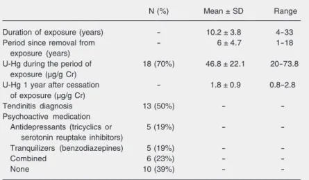
Mercurialism can be found in individu-als with normal U-Hg because the kidneys eliminate large volumes of mercury only during and shortly after the period of expo-sure (5,7). The mean U-Hg concentrations in the exposed group were normal 1 year after removal from exposure (1.8 ± 0.8 µg/g Cr). Thirteen patients were diagnosed with ten-dinitis (affecting one or both arms) related to repetitive work in manufacturing lamps. Twenty of them were taking medication for the treatment of depression and/or anxiety (Table 2).
Procedures
Each participant underwent neuropsycho-logical assessment during a session of about 1.5 h of duration, which included a brief clinical interview and neuropsychological testing. The anamnesis included questions concerning the subjects’ demographic data, job history, life-style habits, and clinical
data. The patients were also asked about their occupational histories at the fluores-cent lamp plant.
The neuropsychological battery included tests of attention, short term memory, and mental control (WMS Digit Span subtest) (29), inhibitory control (Stroop Interference
Table 2.Data related to the 26 mercury-exposed participants.
N (%) Mean ± SD Range
Duration of exposure (years) - 10.2 ± 3.8 4-33
Period since removal from - 6 ± 4.7 1-18
exposure (years)
U-Hg during the period of 18 (70%) 46.8 ± 22.1 20-73.8
exposure (µg/g Cr)
U-Hg 1 year after cessation - 1.8 ± 0.9 0.8-2.8
of exposure (µg/g Cr)
Tendinitis diagnosis 13 (50%) -
-Psychoactive medication
Antidepressants (tricyclics or 5 (19%) -
-serotonin reuptake inhibitors)
Tranquilizers (benzodiazepines) 5 (19%) -
-Combined 6 (23%) -
-None 10 (39%) -
-Data are reported as number of subjects with percent in parentheses, mean ± SD, and range. U-Hg = urinary mercury levels; Cr = creatinine.
Table 1. Demographic characteristics of subjects exposed to mercury.
Exposed group (N = 26) Control group (N = 20)
N(%) Mean ± SD Median N(%) Mean ± SD Median
Age (years) 41.5 ± 6.1 41.5 42.7 ± 8.2 42
Educational level (years) 9.8 ± 1.8 9.5 9.8 ± 2.2 11
Gender
Male 20 (77%) - - 18 (90%) -
-Female 6 (23%) - - 2 (10%) -
-Work
Employed 0 (0%) - - 20 (100%) -
-Away from work due 26 (100%) - - 0 (0%) -
-to mercury in-toxication Alcohol consumption
Drinkers 14 (54%) - - 15 (75%) -
-Non-drinkers 12 (46%) - - 5 (25%) -
-Smokers
Current 2 (8%) - - 4 (20%) -
-Ex-smokers 2 (8%) - - 0 (0%) -
-Non-smokers 22 (84%) - - 16 (80%) -
-Data are reported as number of subjects with percent in parentheses, mean ± SD, and median. There were no statistical differences between groups in terms of age and
Test) (30), verbal memory (Buschke Selec-tive Reminding Test, SRT) (30), visual mem-ory (WMS Visual Reproduction subtest) (29), manual dexterity (Grooved Pegboard; La-fayette Instrument), verbal fluency (FAS) (30), visuomotor ability (WAIS-R Block Design subtest) (31), executive function (Wisconsin Card Sorting Test) (32), and ver-bal knowledge (WAIS-R Vocabulary sub-test) (31). After this neuropsychological as-sessment the subjects were instructed to com-plete the Beck Depression Inventory (BDI) (33) and the State-Trait Anxiety Inventory (STAI) (34).
The study was approved by the Ethics Committee of the Psychology Institute, Uni-versity of São Paulo, and all subjects gave written informed consent prior to testing.
Statistical analysis
Data with non-normal distribution (years of education, cognitive and motor tests, and mood inventory scores) were log-trans-formed to achieve normalization. Independ-ent t-tests were applied to the continuous demographic variables (age and years of education) to assess differences between groups.
Group comparisons were made using the general linear model, which combines anal-ysis of variance and regression procedures and allows the assessment of continuous and categorical variables (covariates/confound-ers) simultaneously. Dependent variables were the test scores. Assessment of potential confounding variables was taken into ac-count. Age, raw score in the Vocabulary test as a measure of pre-morbid general ability, and drinking habits are potential confound-ers frequently considered in neuropsycho-logical studies. BDI scores were also used as covariates because depression is reported to compromise cognitive functions (35,36). Since anxiety showed a high correlation with depression, STAI scores were not consid-ered in order to avoid redundancies. Since
antidepressants (tricyclic and serotonin re-uptake inhibitors) and tranquilizers (benzo-diazepines) can impair cognitive and/or psy-chomotor skills (37,38), the use (yes/no) of one or both medications was included in the analysis. A diagnosis of tendinitis (yes/no) was considered to be a potential confounder in the assessment of the tests that require manual motor performances (Visual Repro-duction, Grooved Pegboard, and Block De-sign).
Pearson’s correlation coefficients were calculated to detect possible associations between exposure to mercury variables (urine Hg0 levels, duration of exposure and the time since the cessation of exposure) and neuropsychological performance within the exposed group.
Statistical analyses were performed us-ing the MINITAB 14.0 software (39).
Results
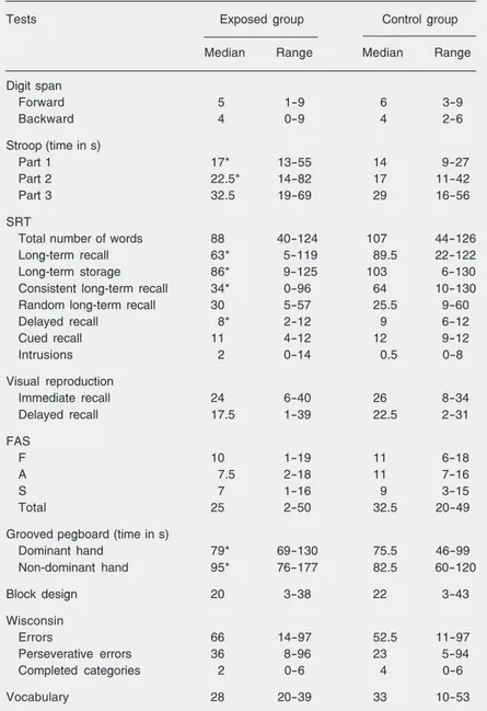
The group exposed to mercury had sig-nificantly different scores compared to the control group on variables related to the SRT, Stroop Test, and Grooved Pegboard after accounting for potential confounders (P < 0.05). The medians and ranges of the score tests, F-ratios, and P values are re-ported in Table 3. The exposed participants a) took a longer mean time in naming colors in the Stroop Test parts 1 (P = 0.03) and 2 (P = 0.01), but not in part 3 (P = 0.09), b) showed lower scores for verbal memory on the SRT term recall (P = 0.03), long-term storage (P = 0.00), consistent long-long-term recall (P = 0.01), and delayed recall (P = 0.00), c) showed longer mean times in manual dexterity with the dominant hand (P = 0.00) and the non-dominant hand (P = 0.00) in the Grooved Pegboard test. Exposed participants also had higher scores in the depression (BDI) and anxiety (STAI) inventories (P = 0.00; Figure 1).
with the control group, the former workers showed poorer results in the Stroop Test parts 1 and 2, that mainly require informa-tion processing and psychomotor speed, but not in part 3, a measure of inhibitory control. Indeed, their low SRT scores in long-term recall and consistent long-term recall sug-gest impairment of primary verbal memory spontaneous recall. In contrast, the cued re-call scores were not statistire-cally different between groups, indicating the integrity of verbal memory storage ability in the ex-posed group, but a difficulty in spontane-ously recalling the stored words. Although the groups differed significantly in long-term storage, this sub-item only reflects the sum of words recalled in two consecutive trials without being reminded during testing. The associations between the exposure indicators and the patient’s neuropsycho-logical performances calculated by Pearson correlation coefficients are presented in Table 4. The urinary mercury concentration meas-ured 1 year after the end of exposure was significantly correlated only with the STAI score (r = 0.75; P = 0.03). The concentra-tions of mercury in urine at the time of exposure, duration of exposure and the pe-riod of removal from exposure were not correlated with any test or inventory score (Table 4).
Discussion
The present study focused on determin-ing if neuropsychological and motor impair-ment could be detected in a group of workers formerly employed by a fluorescent lamp plant. They had been away from the source of exposure for an average of 6 years (1 to 18 years) and had a diagnosis of chronic occu-pational mercurialism.
The exposed subjects showed reduced performance in specific neuropsychological functions and motor skills. There were slowed information processing speeds (Stroop test parts 1 and 2) and impairment in verbal
memory (SRT), hand-eye coordination, and manual dexterity (Grooved Pegboard). We also found symptoms of depression (BDI) and anxiety (STAI) in these patients. When analyzing the verbal memory performance of the exposed subjects it is important to emphasize that although they failed
long-Table 3. Comparison of test scores between subjects exposed to mercury and
con-trols.
Tests Exposed group Control group
Median Range Median Range
Digit span
Forward 5 1-9 6 3-9
Backward 4 0-9 4 2-6
Stroop(time in s)
Part 1 17* 13-55 14 9-27
Part 2 22.5* 14-82 17 11-42
Part 3 32.5 19-69 29 16-56
SRT
Total number of words 88 40-124 107 44-126
Long-term recall 63* 5-119 89.5 22-122
Long-term storage 86* 9-125 103 6-130
Consistent long-term recall 34* 0-96 64 10-130
Random long-term recall 30 5-57 25.5 9-60
Delayed recall 8* 2-12 9 6-12
Cued recall 11 4-12 12 9-12
Intrusions 2 0-14 0.5 0-8
Visual reproduction
Immediate recall 24 6-40 26 8-34
Delayed recall 17.5 1-39 22.5 2-31
FAS
F 10 1-19 11 6-18
A 7.5 2-18 11 7-16
S 7 1-16 9 3-15
Total 25 2-50 32.5 20-49
Grooved pegboard (time in s)
Dominant hand 79* 69-130 75.5 46-99
Non-dominant hand 95* 76-177 82.5 60-120
Block design 20 3-38 22 3-43
Wisconsin
Errors 66 14-97 52.5 11-97
Perseverative errors 36 8-96 23 5-94
Completed categories 2 0-6 4 0-6
Vocabulary 28 20-39 33 10-53
Data are reported as median and range. SRT = Buschke Selective Reminding Test; FAS = verbal fluency.
ability are related to information processing deficit. High BDI and STAI scores were expected, since depression and anxiety are symptoms of chronic mercurialism. Never-theless, the possibility that these symptoms are associated with psychosocial problems related to being unemployed could not be ruled out. In this case, depression and anxi-ety would represent a reaction to the psycho-social context.
Few investigations have focused on the question of cognitive impairment associa-tion with past exposure to mercury vapor (14,15,24-27). Our findings in terms of pre-served functions were consistent with previ-ous studies that failed to detect impairment of immediate memory (24-27), vocabulary (24-26), and viso-spatial ability (14,15,24). Our results agree with the slowed infor-mation processing speed (test of color card reading) and motor deficits found in ex-miners (U-Hg = 3.2 ± 4.1 ng/g) who were previously exposed to high levels of mer-cury vapor for 15.5 years on average (SD = 8.7) (14,15). However, we did not detect visuo-spatial or short-term memory deficits. A possible explanation is the fact that the previous study examined ex-miners of a mercury mine 18 years after the end of expo-sure and certainly included individuals who were older and had been exposed to higher mercury vapor concentrations than in the present study. Indeed, a correlation was ob-served between the number of years after the cessation of exposure and better performance in the short-memory test (Digit Span) (15).
Mathiesen et al. (24) also found motor, psychomotor, visuomotor, and attention defi-cits in former workers of a chloralkali plant (mean U-Hg = 1.8 ± 1.3 nmol/mmol Cr) who had been exposed to mercury vapor for 7.9 ± 6.8 years (time since the end of exposure = 12.7 ± 11.7 years). In contrast to our results, scores in a visual memory ability test were also decreased, particularly in individuals with high cumulative exposure levels (U-Hg
≥3000 nmol/L). On the other hand,
Bast-Table 4.Pearson correlation coefficients between exposure variables and tests and
inventory scores among former workers.
Variable Domain r P
Duration of exposure All tests and inventories
-Period since removal from exposure All tests and inventories
-U-Hg during the period of exposure All tests and inventories
-U-Hg 1 year after removal of exposure Anxiety trait (STAI) 0.75 0.03
Other tests and inventories
-U-Hg = urinary mercury levels. See Procedures in Material and Methods. -, no
correlation.
Figure 1.Box plots representing
the performances (median, first and third quartiles, and 95 and 95 percentiles) of exposed and control participants on the Beck Depression Inventory (BDI) and State-Trait Anxiety Inventory (STAI) (P = 0.00). U-Hg = uri-nary mercury levels. P < 0.05 compared to control (independ-ent t-test).
Pettersen et al. (27) did not find visual memory impairment in former chloralkali workers (mean U-Hg = 1.65 nmol/mmol Cr, range 0.2-5.2 nmol/mmol Cr) exposed to mercury for 13.1 years on average (range 2.8-34.5) and removed from exposure for 4.8 years (range 4.2-10.0). The majority of these participants had been examined during the period of exposure in a previous study (22) that reported impairment of immediate visual memory, suggesting recovery of func-tion.
Our findings of verbal memory impair-ment support previous studies that showed a significantly worse performance in a list learning test (26) and in a word pair test (individuals with the highest intensity of exposure showed a deficit) (24) in former employees of a chloralkali plant. In contrast, Letz et al. (25) did not detect statistical dif-ferences in verbal memory between former workers of a heavy industrial plant and con-trols. However, these discordant findings may reflect the time since the cessation of exposure (30 years or more).
Motor dysfunction associated with a his-tory of past exposure to mercury vapor has been well described. In the present study, there was a significant difference between exposed participants and controls in the Grooved Pegboard test that required manual dexterity and hand-eye coordination. Indeed, the general linear model analysis was done considering a diagnosis of tendinitis to be a confounder, and this variable was not sig-nificantly associated with motor perfor-mance. Previous studies conducted after the cessation of exposure to mercury detected impairment of motor coordination (14,15), manual dexterity (14,15,24,26), and reac-tion time (14,15,24). Letz et al. (25) reported a significant association between hand-eye coordination and cumulative mercury expo-sure in former workers examined 30 years after the end of exposure.
The present study found no association between the impairment detected in the
pa-tients and duration of exposure to mercury or length of the period away from exposure. Because of the variability among the work-ers’ job categories in the plant, the degree of exposure was also variable, and therefore the failure to detect significant relationships is not surprising. Similarly, urinary mercury levels during the time of exposure were not observed to be associated with any test or inventory score. It is possible that effects of U-Hg on cognitive and motor functions were not detected because of the small sample size. However, urinary mercury concentra-tion measured 1 year after the end of expo-sure correlated positively with the anxiety trait (STAI). Mercury intoxication has been reported to provoke changes in personality traits as a symptom of erethism (3,13). Alter-ations have been reported even at very low exposure concentrations, such as higher trait anxiety scores in women exposed to elemen-tal mercury from denelemen-tal amalgams compared to women with no dental fillings, attributed to a possible dysfunctional norepinephrine metabolism (40).
The limitations of the present investiga-tion are mainly related to the demographic characteristics of the groups studied. A larger sample size might have helped detect other possible cognitive dysfunctions related to mercury exposure. Another problem is the variable duration of exposure and time after the end of exposure. Indeed, there were changes in type of occupation during em-ployment, with different mercury vapor lev-els in the air in different occupations.
sam-ple is recommended to confirm whether or not the neuropsychological and motor defi-cits observed persist for many years.
Acknowledgments
Appreciation is expressed to Marcelo
Fernandes Costa, Ph.D. (Universidade de São Paulo), for critical discussions of this work and for methodological support during the study. We are also grateful to the neuro-psychologist Shirley S. Lacerda (Hospital Albert Einstein)for assistance with data dis-cussion and interpretation.
References
1. Echeverria D, Aposhian HV, Woods JS, Heyer NJ, Aposhian MM, Bittner AC Jr, et al. Neurobehavioral effects from exposure to dental amalgam Hg(o): new distinctions between recent exposure and Hg
body burden. FASEB J 1998; 12: 971-980.
2. Anonymous. Mercury toxicity. Agency for toxic substance and
dis-ease registry. Am Fam Physician 1992; 46: 1731-1741.
3. Langworth S, Almkvist O, Soderman E, Wikstrom BO. Effects of occupational exposure to mercury vapour on the central nervous
system. Br J Ind Med 1992; 49: 545-555.
4. Vassallo DV, Massaroni L, Oliveira EM, Rossoni LV, do Amaral SM, Vassallo PF. Acute toxic actions of mercury on the cardiovascular
system. Arq Bras Cardiol 1996; 67: 39-45.
5. Faria MA. Chronic occupational metallic mercurialism. Rev Saúde
Pública 2003; 37: 116-127.
6. Azevedo FA. Toxicologia do mercúrio. São Paulo: InterTox; 2003.
7. Rodriguez ZC, Rodriguez JEL. Metallic mercury intoxication. Bol
Asoc Med P R 1982; 74: 380-382.
8. Satoh H. Occupational and environmental toxicology of mercury and
its compounds. Ind Health 2000; 38: 153-164.
9. Langolf GD, Chaffin DB, Henderson R, Whittle HP. Evaluation of workers exposed to elemental mercury using quantitative tests of
tremor and neuromuscular functions. Am Ind Hyg Assoc J 1978; 39:
976-984.
10. Ventura DF, Simões AL, Tomaz S, Costa MF, Lago M, Costa MTV, et al. Color vision and contrast sensitivity losses of mercury
intoxi-cated industry workers in Brazil. Environ Toxicol Pharmacol 2005;
19: 523-529.
11. Ventura DF, Costa MT, Costa MF, Berezovsky A, Salomao SR, Simoes AL, et al. Multifocal and full-field electroretinogram changes
associated with color-vision loss in mercury vapor exposure. Vis
Neurosci 2004; 21: 421-429.
12. Gobba F, Cavalleri A. Evolution of color vision loss induced by
occupational exposure to chemicals. Neurotoxicology 2000; 21:
777-781.
13. Hua MS, Huang CC, Yang YJ. Chronic elemental mercury
intoxica-tion: neuropsychological follow-up case study. Brain Inj 1996; 10:
377-384.
14. Kishi R, Doi R, Fukuchi Y, Satoh H, Satoh T, Ono A, et al. Subjective symptoms and neurobehavioral performances of ex-mercury miners at an average of 18 years after the cessation of chronic exposure to
mercury vapor. Mercury Workers Study Group. Environ Res 1993;
62: 289-302.
15. Kishi R, Doi R, Fukuchi Y, Satoh H, Satoh T, Ono A, et al. Residual neurobehavioural effects associated with chronic exposure to
mer-cury vapour. Occup Environ Med 1994; 51: 35-41.
16. O’Carroll RE, Masterton G, Dougall N, Ebmeier KP, Goodwin GM. The neuropsychiatric sequelae of mercury poisoning. The Mad
Hatter’s disease revisited. Br J Psychiatry 1995; 167: 95-98.
17. Smith PJ, Langolf GD, Goldberg J. Effect of occupational exposure
to elemental mercury on short term memory. Br J Ind Med 1983; 40:
413-419.
18. Soleo L, Urbano ML, Petrera V, Ambrosi L. Effects of low exposure
to inorganic mercury on psychological performance. Br J Ind Med
1990; 47: 105-109.
19. Liang YX, Sun RK, Sun Y, Chen ZQ, Li LH. Psychological effects of low exposure to mercury vapor: application of a
computer-adminis-tered neurobehavioral evaluation system. Environ Res 1993; 60:
320-327.
20. Lucchini R, Calza S, Camerino D, Carta P, Decarli A, Parrinello G, et al. Application of a latent variable model for a multicenter study on
early effects due to mercury exposure. Neurotoxicology 2003; 24:
605-616.
21. Piikivi L, Hanninen H, Martelin T, Mantere P. Psychological
perfor-mance and long-term exposure to mercury vapors. Scand J Work
Environ Health 1984; 10: 35-41.
22. Ellingsen DG, Bast-Pettersen R, Efskind J, Thomassen Y. Neuro-psychological effects of low mercury vapor exposure in chloralkali
workers. Neurotoxicology 2001; 22: 249-258.
23. Meyer-Baron M, Schaeper M, Seeber A. A meta-analysis for
neuro-behavioural results due to occupational mercury exposure. Arch
Toxicol 2002; 76: 127-136.
24. Mathiesen T, Ellingsen DG, Kjuus H. Neuropsychological effects associated with exposure to mercury vapor among former chloralkali
workers. Scand J Work Environ Health 1999; 25: 342-350.
25. Letz R, Gerr F, Cragle D, Green RC, Watkins J, Fidler AT. Residual neurologic deficits 30 years after occupational exposure to
elemen-tal mercury. Neurotoxicology 2000; 21: 459-474.
26. Frumkin H, Letz R, Williams PL, Gerr F, Pierce M, Sanders A, et al. Health effects of long-term mercury exposure among chloralkali
plant workers. Am J Ind Med 2001; 39: 1-18.
27. Bast-Pettersen R, Ellingsen DG, Efskind J, Jordskogen R, Thomas-sen Y. A neurobehavioral study of chloralkali workers after the
cessation of exposure to mercury vapor. Neurotoxicology 2005; 26:
427-437.
28. Levey S, Jennings ER. The use of control charts in the clinical
laboratory. Am J Clin Pathol 1950; 20: 1059-1066.
29. Wechsler D. Wechsler Memory Scale Revised - User’s manual. San
Antonio: The Psychological Corporation; 1987.
30. Spreen O, Strauss E. A compendium of neuropsychological tests.
New York: Oxford University Press; 1991.
31. Wechsler D. Wechsler Adult Inteligence Scale Revised - User’s
manual. New York: Psychological Corporation; 1981.
32. Heaton RK, Chelune GJ, Talley JL, Kay GG, Curtis G. Wisconsin
Psychological Assessment Resources; 1993.
33. Cunha JA. Escalas Beck - manual. São Paulo: Casa do Psicólogo;
2001.
34. Biaggio AMB, Natalício L. Inventário de ansiedade traçoestado
-manual. Rio de Janeiro: CEPA; 1979.
35. Antikainen R, Hanninen T, Honkalampi K, Hintikka J, Koivumaa-Honkanen H, Tanskanen A, et al. Mood improvement reduces
memory complaints in depressed patients. Eur Arch Psychiatry Clin
Neurosci 2001; 251: 6-11.
36. Fossati P, Coyette F, Ergis AM, Allilaire JF. Influence of age and executive functioning on verbal memory of inpatients with
depres-sion. J Affect Disord 2002; 68: 261-271.
37. McKim WA. Drugs and behavior. 4th edn. New Jersey: Prentice
Hall; 2000.
38. Stewart SA. The effects of benzodiazepines on cognition. J Clin
Psychiatry 2005; 66 (Suppl 2): 9-13.
39. Minitab statistical software for Windows. [Computer program]. Ver-sion 14.0. Pennsylvania: Minitab Inc.; 2003.
40. Siblerud RL, Motl J, Kienholz E. Psychometric evidence that mer-cury from silver dental fillings may be an etiological factor in