ISSN 0104-6632 Printed in Brazil
www.abeq.org.br/bjche
Vol. 29, No. 04, pp. 775 - 781, October - December, 2012
Brazilian Journal
of Chemical
Engineering
NANOSIZE MgO AS ANTIBACTERIAL AGENT:
PREPARATION AND CHARACTERISTICS
Zhen-Xing Tang
1*, Xiu-Juan Fang
2, Zhi-Liang Zhang
2, Ting Zhou
2,
Xin-Yi Zhang
2and Lu-E Shi
2*1Department of Food Science, Anqing Vocational & Technical College, 246003, Anqing, Anhui, China. 2
College of Life and Environmental Sciences, Hangzhou Normal University, 310016, Hangzhou, Zhejiang, China. E-mail: [email protected]; [email protected]
(Submitted: November 15, 2011 ; Revised: March 13, 2012 ; Accepted: March 14, 2012)
Abstract - The antibacterial activity of MgO nanoparticles prepared by a sonication method was evaluated in this paper. The effect of calcination conditions on the size and antibacterial activity of MgO nanoparticles was investigated. MgO nanoparticles were characterized for purity (TGA), crystallinity and crystal size (XRD), particle size and morphology (TEM) and surface area (BET). Results showed that the smallest size of 6 nm could be obtained. The lethal effects of nanocrystalline MgO were evaluated on Lactobacillus plantarum. At a concentration of 100 ppm, the killing effect of MgO was close to 1 log reduction for L. plantarum after 24 h exposure. At 1000 ppm and 24 h exposure, the killing effect of MgO was more than a 2.8 log reduction. With the increase of calcination time, the lethal effect of MgO nanoparticlesincreased after 6 h or 24 h exposure at 100 ppm or 1000 ppm. 2.86 log and 2.89 log were killed at 1000 ppm after 24 h exposure using the sample MgO, sonication, A, and the sample MgO, sonication, B, respectively. When the sample MgO, sonication, C, was used, the lethal quantity of L. plantarum was increased to a 3.36 log reduction.
Keywords: Nano; MgO; Preparation; Sonication; Antibacterial activity.
INTRODUCTION
Nano-MgO is a functional material that has been widely used in various areas (Shukla et al., 2004). Recently, it has been reported (Sawai et al., 2000; Yamamoto et al., 2000) that MgO has good bactericidal performance in aqueous environments due to the formation of superoxide O-2 anions on
its surface. Work by Klabunde, Marchin and co-workers (Koper et al., 2002; Stoimenov et al., 2002) demonstrated that nanosize MgO exhibits activity against bacteria, spores and viruses after adsorption of halogen gases because of its large surface area, abundance of crystal defects and positively-charged particles, which can result in strong interactions with negatively-charged bacteria and spores.
MgO can be obtained mainly by thermal treatment of magnesium hydroxide or carbonate (Aramendia
et al., 1996; Xu et al., 2001), and, more recently, by
the sol-gel method (Choi and Hwang, 2000; López et al., 1998) and sonication method (Tang and Shi, 2008). In the sol-gel procedure, the fabrication of nanocrystalline and nanoparticulate metal oxides involves synthesis from methoxides or alkoxides of the metals followed by controlled drying or dehydration under vacuum (Stark et al., 1996; Znaidi
et al., 1996; Koper et al., 1996). The morphology
(Kenneth and Price, 1999; Bhatte et al., 2011; Suslick et al., 1996). Chemical effects of ultra-sound originate from the formation of ultrasonic cavitation, the growth and collapse of microbubbles in the liquid phase generating very high temperatures and pressures followed by rapid cooling. These extreme conditions have been exploited for the preparation of nanoparticulate metal oxides (Huang et al., 2000; Wang et al., 2001).
In this paper, MgO nanoparticles were prepared by a sonication method. The effect of calcination conditions on the particle sizes and the antibacterial activity of nanosize MgO were investigated. All the results obtained provide a basis for further application of MgO in relevant fields.
EXPERIMENTAL Materials
Mg(NO3)2. 6H2O (Mallinckrodt Baker Inc, ACS);
anhydrous sodium carbonate (ACS) and ethylene glycol (99%) were all purchased from BDH Inc;
Lactobacillus plantarum was grown in Difco
Lactobacillus medium (MRS – agar and broth) at
35 ºC for 24 h. They were maintained at –80 °C with 20% glycerol and served as stock cultures. Other reagents were obtained from local supplies.
Preparation of MgO Nanoparticles
12.30 g of Mg(NO3)2. 6H2O were dissolved in
ethylene glycol solution (25 mL). 12.5 mL of Na2CO3
(2.70 g) was added into above mixture under sonication (Sonicafier 450, Branson Ultrasonics Corporation, USA). After sonication for 15 min, the solution obtained was kept at rest for about 5 h. Then it was filtered, washed using water and dried at 50 °C. Finally, the samples were obtained through calcination. The code of the sample obtained under calcination conditions (410 °C, 5 h, 3 °C/min) was MgO, sonication, A. Similarily, the codes of the samples under calcination conditions (410 °C, 10 h, 3 °C/min) and (410 °C, 15 h, 3 °C/min) were MgO, sonication, B, and MgO, sonication, C, respectively.
Preparation of the Slurry
MgO powders were heated about 2 h at 180 ºC for activation and then kept in a dessicator. The powder samples were suspended in peptone water (0.1%) at 100 ppm or 1000 ppm and sonicated at 280 watts for 5 minutes. Then the slurries were
auto-claved at 121 ºC for 15 min. The slurries were used for antibacterial experiments.
Antibacterial Experiment of Nanosize MgO
The lethal effects of nanosize MgO were evaluated on L. plantarum. An overnight bacterial preculture in MRS broth was centrifuged, rinsed twice with sterile peptone water (0.1%), and then resuspended to obtain a concentration of approxi-mately 1x10⁸ CFU/mL (CFU=Colony Forming Unit). The suspension was added to 50 mL flasks containing 20 mL of the different MgO slurries (100 ppm or 1000 ppm) or sterile peptone water (control). Approximately 1x10⁶ CFU/mL were inoculated. Triplicate flasks were incubated at 35 °C with agitation (250 rpm). 1.0 mL samples were taken at the indicated times (6 and 24 h), diluted ten-fold in isotonic water, and then grown on MRS agar. The Petri plate was incubated at 35 °C for 48 hrs. The enumeration was done in CFU/mL.
Characteristic Analysis of Nanosize MgO
Characteristics measurement of CaO nanoparticles were described previously in our group (Shi et al., 2012; Tang and Shi, 2008; Tang et al., 2008). TGA measurements were carried out using a Netzsch STA 409 apparatus. A helium flow of 40 cm3 min-1 and a heating rate of 10 K min-1 were used; the size and particle size distribution were recorded in ethanol on a submicron particle sizer (NICOMP 370, USA); TEM photomicrographs were obtained using a Philips 201 Transmission Electron Microscope. The micrographs were taken at 80 kV. The deposit was scraped away from the support and then transferred to a Fromvar 1595 E (Merck) membrane coated Cu grid (400 mesh); a Rigaku Geiferflex X-ray diffractometer with Ni-filtered Cu Ka radiation (40 kV, 30 mA) was used to determine the crystallinity and phase of the samples. XRD patterns were recorded in the range of 20°-70° with a scan speed of 2 °/min.
RESULTS AND DISCUSSION Preparation of Nanosize MgO
Different intermediates have distinct decomposition temperatures. The decomposition temperature of the same intermediate is relative to the precursor (Alvarado et al., 2000; Ardizzone et al., 1998; Wu
et al., 2004). According to the TGA (Figure 1), the
showed a weight loss in two steps from 50 to 110 °C and 380 to 410 °C, respectively. The weight loss from 50-110 °C was due to removal of excess ethylene glycol and water from the magnesium intermediate. The weight loss at 380–430 °C was due to the decom-position of the magnesium intermediate. MgO nanoparticles could be obtained at 410 °C through calcination. The calcination temperature was lower than the results reported by Wu (Wu et al., 2004).
0 20 40 60 80 100 120
0 100 200 300 400 500 600 700 800
Temperature
W
e
ig
h
t
lo
s
s
Figure 1: TGA of the intermediates. Conditions: hold for 1 min at 50 °C, heat from 50 °C to 1000 °C at the rate 10 °C/min, hold for 1 h at 1000 °C.
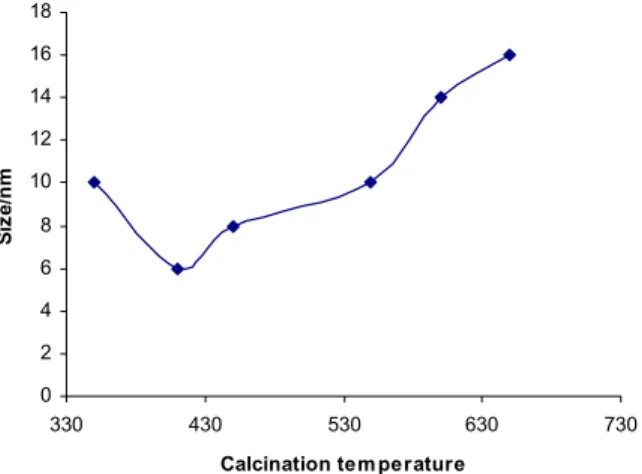
The calcination techniques are crucial to get nano MgO. When the calcination temperature is low, the intermediate might not be decomposed completely. Different size crystals have different surface energies. Crystal size is proportional to its surface energy, so the process of small crystals turning into large crystals happens automatically. The process proceeds very quickly, especially at high temperature. With prolonged calcination time, the process will be achieved completely. The crystal size grows for long times at high calcination temperatures (Shi et al., 2012; Tang and Shi, 2008). Thus, large size nanoparticles can be obtained. For the same reason, although the intermediate can be decomposed completely very quickly at high calcination tempera-ture, large size nanoparticles readily form in order to decrease their surface energy.
For nanoparticles obtained by the sonication method, the smallest size was around 6 nm. When the calcination temperature was above 410 °C, the size began to increase (Fig. 2). With the increase of calcination time (more than 1.5 h), the size of nano-MgO increased too. When the calcination time reached 3.0 h, the size was about 13 nm. Fig. 3 indicates that, when the calcination time was reduced to 1.5 h, the size of 8 nm could be obtained. Thus,
through controlling calcination temperature and calcination time, nano-MgO with different sizes could be obtained. (Shi et al., 2012; Tang and Shi, 2008). In order to get smallest size particles, the calcination temperature was about 410 °C and calcina-tion time was 1.5 h.
The heating rate of calcination can affect the size of nanoparticles. When the heating rate of calcination is very fast, aggregation of nano ZnO happens more easily, due to urgent collapse of the intermediate. When the heating rate of calcination is low, nano MgO exists for a long time at high calcination temperatures, which will decrease production effi-ciency (Shi et al., 2012; Tang and Shi, 2008). So, an optimized heating rate of calcination must exist. The optimized heating rate was 3-5 °C/min.
0 2 4 6 8 10 12 14 16 18
330 430 530 630 730
Calcination tem perature
Si
z
e
/n
m
Figure 2: Effect of calcination temperature on the size Condition: Heating rate 3 °C /min; Calcination time 1.5 h
0 2 4 6 8 10 12 14 16 18
0 5 10 15 20
Calcination tim e
Si
z
e
/
n
m
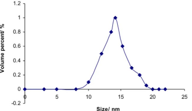
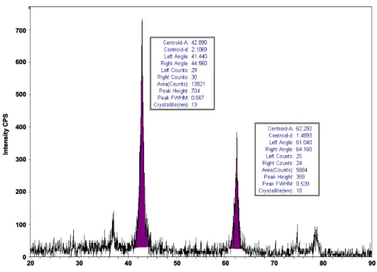
Accordingly, nanoparticles with different sizes could be obtained by controlling the calcination conditions. The size distribution of nano MgO is presented in Figure 4. The size of the nanoparticles was around 15 nm. The distribution was narrow. The resulting size distribution (Figure 4) was a good match for TEM (Figure 5) and the XRD results (Figure 6). From Fig. 5, MgO nanoparticles have an average particle size of about 15 nm. MgO nanoparticles could be dispersed well. Very little aggregation could be found. The structure of nanosize MgO was characterized by XRD (Figure 6). All peaks were consistent with the peaks of standard MgO with high crystallinity. XRD patterns showed broadening of the peaks, indicative of the ultra fine nature of the crystallite material. The crystal sizes calculated using Scherrer’s formula were about 14 nm. No peaks from any other phases of MgO were observed.
The results of liquid N2 adsorption measurements
aid in understanding the formation of nanoparticles. For nanoparticles from the sonication method, the specific surface area is around 44 m2/g (figures not shown). This low surface area is due to the long calcination time.
Ultrasound irradiation has been routinely used in the field of materials science. The chemical effect of ultrasound irradiation arises from acoustic cavitation formation, growth and collapse of microbubbles in liquid. The extremely high temperatures, pressures and cooling rate attained during cavitation lead to unique properties in the irradiated solution (Shi et al., 2012).These extreme conditions have been exploited to prepare nanoscale metal oxides (Huang et al., 2000; Wang et al., 2001), metal sulphides (Jeevanndam et al., 2000), nanocomposites and others (Rassokhin, 1998; Savrun and Toy, 1997; Aymonier et al., 2000; Davydov et al., 2001). We also tried in our lab (results not shown) the thermal decomposition method, but small nanocrystal could only be obtained under a very high stirring rate since mechanical power is used to prepare nanoparticles. For these two methods, the mechanism of preparing the nano-particles is different and the characteristics of the nanoparticles are also different. Acoording to this work, the sonication method for the preparation of nanoparticles is more powerful than the thermal decomposition method.
-0.2 0 0.2 0.4 0.6 0.8 1 1.2
0 5 10 15 20 25
Size/ nm
V
o
lu
m
e
p
e
rc
e
n
t/
%
Figure 4: Distribution of MgO (sonication, C) nanoparticles.
20 30 40 50 60 70 80 90 100
100 200 300 400 500 600 700
In
te
n
s
it
y
C
PS
20 30 40 50 60 70 80 90
20 30 40 50 60 70 80 90
100 100 200 300 400 500 600 700
100 100 200 300 400 500 600 700
In
te
n
s
it
y
C
PS
Figure 6: XRD of nano-MgO (sonication, C)
Antibacterial Activity of Nanosize MgO
The lethal effect of nano-crystalline MgO was evaluated on L. plantarum. The results are shown in Table 1. From Table 1, with the increase of calcination time, the lethal effect of MgO nanoparticles increased after 6 h or 24 h exposure at 100 ppm or 1000 ppm. 2.86 log L. plantarum and 2.89 log L. plantarum
were killed at 1000 ppm after 24 h exposure using the sample MgO, sonication, A, and the sample MgO, sonication, B, respectively. When the sample MgO, sonication, C, was used, the lethal effect on
L. plantarum was increased to a 3.36 log reduction.
Yamamoto et al. (2000) indicated that the higher the concentration, the better the antibacterial activity will be. At 1000 ppm, for the samples MgO, sonication, B, and MgO, sonication, C, respectively, the lethal effect
on L. plantarum was increased to a 0.18 log reduction.
Impurities can conceal active sites of nanoparticles. With increasing calcination time, the antibacterial activity of MgO nanoparticles increased. But, when the calcination time was increased to 10 h, the MgO nanoparticle weight loss from TGA (results not shown) is similar. The antibacterial activities of the sample MgO, sonication, B, and the sample MgO, sonication, C, were similar.
Huang et al. (2000) and Makhluf et al. (2005) proposed that active superoxide ions are generated on the surface of the oxide, which can react with the peptide linkages in the cell wall of bacteria and thus
disrupt them. The bactericidal action of MgO may result from the attack of superoxide ions on the carbonyl group of the peptide linkages, leading to degradation of the proteins. As the surface area of the particles increases, there is an increase in the O2
-concentration in solution, which results in a more effective destruction of the cell wall of the bacteria.
L. plantarum contains mainly mono-unsaturated fatty
acids, which are less susceptible to oxidation. Also, L. plantarum contains glutamic acid-meso-diaminopimelic acid peptide in its peptidoglycan. Nanosize samples MgO exhibited somewhat similar trends in their lethal effects against L. plantarum.
Table 1: Viability of L. plantarum after treatment with nanosize MgO at 100 ppm and 1000 ppm
Log Reduction Samples
6 h 24 h
MgO, sonication , A, 100 ppm 0.07 1.23 MgO, sonication , A, 1000 ppm 0.09 2.86 MgO, sonication , B, 100 ppm 0.04 1.75 MgO, sonication , B, 1000 ppm 0.18 2.89 MgO, sonication , C, 100 ppm 0.00 1.15 MgO, sonication , C, 1000 ppm 0.18 3.36
CONCLUSIONS
time, the lethal effect of MgO nanoparticles was increased after 6 h or 24 h exposure time at 100 ppm or 1000 ppm. 2.86 log L. plantarum and 2.89 log
L. plantarum were killed at 1000 ppm after 24 h
exposure using the sample MgO, sonication, A, and the sample MgO, sonication, B, respectively. When the sample MgO, sonication, C, was used, the lethal effect on L. plantarum was increased to a 3.36 log reduction. The results suggest that nanosize MgO from the sonication method has good antibacterial activity.
REFERENCES
Alvarado, E., Torres-Martinez, L. M., Fuentes, A. F., Quintana, P., Preparation and characterization of MgO powders obtained from different magnesium salts and the mineral dolomite. Polyhedron, 19, 2345-2351 (2000).
Aramendía, M. A., Borau, V., Jiménez, C., Marinas, J. M., Porras, A., Urbano, F. J., Synthesis and characterization of various MgO and related systems. J. Mater. Chem., 6, 1943-1949 (1996). Ardizzone, S., Bianchi, C. L., Vercelli, B., Acid/base
and surface features of pure phase magnesia powders. Colloid. Surfaces A: Physicochem. Eng. Aspects, 144, 9-17 (1998).
Aymonier, C., Bottreau, M., Berdeu, B., Cansell, F., Ultrasound for hydrothermal treatments of aqueous wastes: Solution for overcoming salt precipitation and corrosion. Ind. Eng. Chem. Res., 39, 4734-4740 (2000).
Bhatte, K. D., Fujita, S., Arai, M., Pandit, A. B. and Bhanage, B. M., Ultrasound assisted additive free synthesis of nanocrystalline zinc oxide. Ultrason. Sonochem., 18, 54-58 (2011).
Choi, H., Hwang, S., Sol-gel-derived magnesium oxide precursor for thin-film fabrication. J. Mater. Res., 15, 842-845 (2000).
Davydov, L., Reddy, E. P., France, P., Smirniotis, P. G., Sonophotocatalytic destruction of organic contaminants in aqueous systems on TiO2 powders. Appl. Catal. B. Environ., 32, 95-105 (2001).
Huang, W., Tang, X., Wang, Y., Koltypin, Y., Gedanken, A., Selective synthesis of anatase and rutile via ultrasound irradiation. Chem. Comm., 15, 1415-1416 (2000).
Jeevanandam, P., Koltypin, Y., Gofer, Y., Diamant, Y., Gedanken, A., Sonochemical synthesis of nanocrystallites of ruthenium sulfide. RuS1.7. J. Mater. Chem., 10, 2769-2773 (2000).
López, T., Gómez, R, J., Navarrete, J., López-Salinas, E., Evidence for Lewis and Brønsted acid sites on
MgO obtained by sol-gel. J. Sol-Gel. Sci. Tech., 13, 1043-1047 (1998).
Makhluf, S., Dror, R., Nitzan, Y., Abramovich, Y., Jelinek, R., Gedanken, A., Microwave-assisted synthesis of nanocrystalline MgO and its use as bacteriocide. Adv. Funct. Mater., 15, 1708-1715 (2005).
Kenneth, S. S., Price, G. J., Application of ultrasound to materials chemistry. Annu. Rev. Mater. Sci., 29, 295-326 (1999).
Koper, O. B., Lagadic, I., Volodin, A., Klabunde, K. J., Alkaline-earth oxide nanoparticles obtained by aerogel methods. Characterization and rational for unexpectedly high surface chemical reactivities. Chem. Mater., 9, 2468-2480 (1996).
Koper, O. B., Klabunde, J. S., Marchin, G. L., Klabunde, K. J., Stoimenov, P. L., Nanoscale powders and formulations with biocidal activity toward spores and vegetative cells of bacillus species, viruses, and toxins. Curr. Microbiol., 44, 49-55 (2002).
Rassokhin, D. N., Accumulation of surface-active solutes in the aerosol particles generated by ultrasound. J. Phys. Chem. B., 102, 4337-4341 (1998).
Savrun, E., Toy, C., The effect of sonication for precipitation of hydrated aluminium sulphate in aqueous Al(SO4)3-urea system. J. Mater. Sci. Lett., 16, 1164-1166 (1997).
Sawai, J., Kojima, H., Igarashi, H., Hashimoto, A., Shoji, S., Sawaki, T., Hakoda, A., Kawada, E., Kokugan, T. and Shimizu, M., Antibacterial characteristics of magnesium oxide powder. World J. Microbiol. Biotechnol., 16,187-194 (2000).
Shi, L. E., Fang, X. J., Zhang, Z. L., Zhou, T., Jiang, D., Wu, H. H. and Tang, Z. X., Preparation of nano-ZnO using sonication method and its antibacterial characteristics. Int. J. Food Sci. Tech., 47, 1866-1871(2012).
Shukla, S. K., Parashar, G. K., Mishra, A. P., Misra, P., Yadav, B. C., Shukla, R. K., Bali, L. M., Dubey, G. C., Nano-like magnesium oxide films and its significance in optical fiber humidity. Sensor Actuator B Chem., 98, 5-11 (2004)
Stark, J. V., Park, D. G., Lagadic, I., Klabunde, K. J., Nanoscale metal oxide particles/clusters as chemical reagents. Unique surface chemistry on magnesium oxide as shown by enhanced adsorption of acid gases (sulfur dioxide and carbon dioxide) and pressure dependence. Chem. Mater., 8, 1904-1912 (1996).
bactericidal agents. Langmuir, 18, 6679-6686 (2002).
Suslick, K. S., Hyeon, T., Fang, M., Nanostructured materials generated by high-intensity ultrasound: Sonochemical synthesis and catalytic studies. Chem. Mater., 8, 2172-2179 (1996).
Tang, Z. X., Claveau, D., Corcuff, R., Belkacemi, K., Arul, J., Preparation of nano-CaO using thermal decomposition method. Mater. Lett., 62, 2096-2098 (2008).
Tang, Z. X., Shi, L. E., Preparation of nano-MgO using ultrasonic method and its characteristics. Eclét. Quím., 33, 15-20 (2008).
Wang, Y. Q., Yin, L. X., Palchik, O., Hacohen, Y. R., Koltypin, Y., Gedanken, A., Rapid synthesis of mesoporous yttrium-zirconium oxides with ultrasound irradiation. Langmuir, 17, 4131-4133 (2001).
Wu, S., Zheng, X., Chen, J., Preparation of nano-crystalline magnesia using ethylene glycol as medium. Acta Scientiarum Naturalium Universitatis Nankaiensis, 37, 73-77 (2004).
Xu, B. Q., Wei, J. M., Wang, H. Y., Sun, K. Q., Zhu, Q. M., Nano-MgO: Novel preparation and application as support of Ni catalyst for CO2 reforming of methane. Catal. Today, 68, 217-225 (2001).
Yamamoto, O., Sawai, J., Sasamoto, T., Change in antibacterial characteristics with doping amount of ZnO in MgO–ZnO solid solution. Int. J. Inorg. Mater., 2, 451-454 (2000).
Znaidi, L., Chhor, K., Pommier, C., Batch and semi-continuous synthesis of magnesium oxide powders from hydrolysis and supercritical treat-ment of Mg(OCH3)2. Mater. Res. Bull., 31,