Lectin obtained from the red seaweed
Bryothamnion triquetrum
:
Secondary structure and anti-in
fl
ammatory activity in mice
Thais Pontes Carvalho Fontenelle
a, Glauber Cruz Lima
a,⁎
, Jacilane Ximenes Mesquita
a,
José Luiz de Souza Lopes
b, Tarcísio Vieira de Brito
c, Francisco das Chagas Vieira Júnior
c, Adriano Bezerra Sales
c,
Karoline Saboia Aragão
d, Marcellus Henrique Loiola Ponte Souza
d,
André Luiz dos Reis Barbosa
c, Ana Lúcia Ponte Freitas
aaLaboratory of Proteins and Carbohydrates of Marine Algae, Department of Biochemistry and Molecular Biology, Federal University of Ceará, Fortaleza, CEP, 60440-900, CE, Brazil bDepartment of Applied Physics, Institute of Physics, University of São Paulo, CEP, 05508-090, São Paulo, SP, Brazil
cLAFFEX
–Laboratory of Experimental Physiopharmacology, Biotechnology and Biodiversity Center Research (BIOTEC), Federal University of Piauí, Parnaíba, CEP, 64202-020, PI, Brazil dLEFFAG
–Laboratory of Physiopharmacology Study of Gastrointestinal Tract, Department of Physiology and Pharmacology, Federal University of Ceará, Fortaleza, CEP, 60430-270, CE, Brazil
a b s t r a c t
a r t i c l e
i n f o
Article history:
Received 16 November 2017
Received in revised form 9 February 2018 Accepted 9 February 2018
Available online 13 February 2018
Seaweeds are sources of biomolecules with biological activities and pharmacological potential–for example, lectins, a group of proteins that can bind reversibly to carbohydrates or compounds containing them. The aim of this study was to elucidate the structural properties of a lectin extracted from the red seaweedBryothamnion triquetrum(BtL) and to investigate its anti-inflammatory activity in mice. The lectin was purified by precipitation with ammonium sulfate and ion-exchange chromatography. Its secondary structure and tryptophan (Trp) micro-environment were analyzed by circular dichroism spectroscopy and steady-statefluorescence spectroscopy, re-spectively. The anti-inflammatory effect was evaluated by means of paw edema induced by carrageenan or dextran, myeloperoxidase activity in paw tissue, and by measurement of leukocyte and neutrophil migration and cytokine quantification in a peritonitis model. The secondary structure of BtL is mostly composed ofβ -strands and unordered conformation, and it is quite resistant to extremes of pH and temperature, preserving the exposure of Trp residues under these conditions. In an assessment of biological activities, groups of mice were subjected to pretreatment with BtL before the inflammatory stimulus. BtL had anti-inflammatory effects in the models tested, and hence may be considered a molecule with potential to be used in the pharmaceutical industry.
© 2018 Elsevier B.V. All rights reserved.
Keywords:
Marine algae Structural conformation Anti-inflammatory effect
1. Introduction
Marine macroalgae (seaweeds) are a part of the traditional oriental diet. In Western countries, their major uses are associated with their rheological properties: they serve as gelling agents, thickeners, and sta-bilizers [1,2]. Nonetheless, in the last few decades, there was an increase in the pharmaceutical industry's interest in natural products of marine origin with biological activities, especially seaweeds, owing to the
peculiarity of their chemical structures caused by the special features of the marine environment [3,5]. Many compounds with pharmacolog-ical properties are found in seaweeds, such as polyphenols, peptides, sulfated polysaccharides, and lectins [6,7]. Among these compounds, lectins obtained from seaweeds have shown a substantial therapeutic potential, exhibiting antinociceptive [8,9], antiviral [10], and antitumor effects [11].
Lectins are proteins, or glycoproteins, of non-immune origin which bind reversibly to sugar residues found ubiquitously in nature [12]. The wide structural and functional diversity of lectins in all major taxa of liv-ing organisms has been reported, e.g., in invertebrates, vertebrates, sea-weeds, and microorganisms. These proteins are important glycobiology tools with the ability to recognize and decipher the codes found in oligo-saccharides and glycosyl residues (i.e., glycol-codes) because of their highly specific binding to carbohydrates [13,14].
Inflammation is a complex reaction involving recognition of an in-fectious agent (or injurious stimulus) for its subsequent destruction and reconstruction of the damaged tissue. This recognition initiates an Abbreviations:BSA, bovine serum albumin; BtL,Bryothamnion triquetrumlectin; CD,
circular dichroism; DEAE-Sephacel, diethylaminoethanol covalently linked to Sephacel (a polysaccharide polymer material derived from agarose); ELISA, enzyme-linked immu-nosorbent assay; H2O2, hydrogen peroxide; HCl, hydrochloric acid; HU/mL, hemagglutina-tion units; IL-1β, interleukin 1 beta; MPO, myeloperoxidase; MRW, mean residual weight; NaCl, sodium chloride; NaOH, sodium hydroxide; PBS, phosphate-buffered saline; TNF-α, tumor necrosis factor alpha; UFC, Federal University of Ceará.
⁎ Corresponding author at: Campus do Pici–Bloco 907, CEP 60440-900, Fortaleza, CE,
Brazil.
E-mail address:[email protected](G.C. Lima).
https://doi.org/10.1016/j.ijbiomac.2018.02.058 0141-8130/© 2018 Elsevier B.V. All rights reserved.
Contents lists available atScienceDirect
International Journal of Biological Macromolecules
immune response activating and amplifying the release of infl amma-tory mediators, such as cytokines, chemokines, biogenic amines, and ei-cosanoids by immune-defense cells [15,16]. Nevertheless, when eradication of the pathogen and tissue repair do not proceed in an effi -cient and synchronized manner, the inflammatory response may lead to persistent tissue injury induced by leukocytes and collagenous accumu-lation, in addition to other substances that may be harmful to the organ-ism [17]. Therefore, aberrations in the inflammatory process underlie several health problems, such as sepsis, asthma, arthritis, psoriasis, and autoimmune disorders [18,19] and contribute significantly to the development of cancer, diabetes mellitus, and cardiovascular disorders [20].
Several lectins from plants have been successfully evaluated in
in-flammatory models [21,22]; however, there are only a few studies on anti-inflammatory activities of lectins from seaweeds, in part because of the small amounts of lectins isolated. Nevertheless, this situation is changing, and the interest in seaweed lectins has been increasing nota-bly due to their molecular structure and glycosidic bonding different from those found in plants or other life forms. Indeed, these lectins have unique features, such as low molecular weight, monomeric struc-ture, thermostability (due to the presence of disulfide bridges), and high affinity for glycoproteins instead of monosaccharides [23,24].
A lectin obtained from the green seaweedCaulerpa cupressoides
possesses an anti-inflammatory activity, inhibiting the paw edema induced by carrageenan and the recruitment of polymorphonuclear cells [8]. Among lectins from red seaweeds, those extracted from
Pterocladiella capillacea[25] andHypnea cervicornis[26] also exert anti-inflammatory effects in the paw edema model and in a neutrophil migration model, both based on carrageenan injection. A lectin obtained from the red seaweedSolieriafiliformisyields similar results and can re-duce the inflammation caused by dextran [27].
Lectins from the red seaweedBryothamnion triquetrumwere isolated and initially characterized by Ainouz et al. [28] and Calvete et al. [29]. Thenceforth, several researchers have demonstrated the pharmacologi-cal activities of lectins fromB. triquetrum, for example, antinociceptive [30] and vasorelaxant effects [32]. However, there are no reports re-garding a possible anti-inflammatory effect. Therefore, this study aims to elucidate the secondary structure properties of a lectin extracted from the red seaweedBryothamnion triquetrum(BtL) and investigate its anti-inflammatory activity in mice using several classical models of inflammation.
2. Materials and methods
2.1. Animals
Female Swiss mice (20–25 g) were obtained from the Animal Care
Unit of the Federal University of Ceará in Fortaleza, Brazil. They were housed in a temperature-controlled room and maintained under a 12-h lig12-ht/dark cycle wit12-h free access to water and food. All t12-he procedures and animal treatments were conducted in accordance with the guide-lines set forth by the U.S. Department of Health and Human Services, and were approved by the Ethics Committee on Research at the Federal University of Ceará (protocol No. 12/2012).
2.2. Drugs and reagents
The following drugs and reagents were used: bovine serum albumin (BSA), papain, sulfuric acid, sodium hydroxide (NaOH), hydrochloric acid (HCl), phosphate-buffered saline (PBS), carrageenan, dialysis sacks, indomethacin, dextran 70, o-dianisidine, hexadecyltrimethylammonium bromide, an avidin-peroxidase conjugate, and orthophenylenediamine dihydrochloride from Sigma (St. Louis, MO, USA). ELISA kits for TNF-αand IL-1βquantification were purchased from R&D Systems (Minneapolis, MN, USA), and DEAE-Sephacel from Merck (Darmstadt, Germany). Ammonium sulfate, sodium chloride (NaCl), violet gentian,
and hydrogen peroxide (H2O2) were acquired from Synth (Diadema, SP,
Brazil). All the reagents and lectin (BtL) were dissolved in distilled water or 0.9% sterile NaCl (saline).
2.3. The marine algae
Specimens of the red seaweedB. triquetrumwere collected from the Atlantic coast in northeast Brazil (Fleixeiras Beach, Trairí–Ceará). A
voucher sample ofB. triquetrumwas deposited at Prisco Bezerra Herbar-ium at the Institute of Marine Sciences (Federal University of Ceará, For-taleza, Brazil) under the following identification number (EAC): 31,138.
2.4. BtL extraction and purification
The extraction of BtL was performed, with some modifications, as described by Ainouz et al. [28]. Seaweed samples were washed, dried, and grounded in liquid nitrogen. The powdered seaweed was subjected to protein extraction with 0.02 M phosphate-buffered saline (PBS) con-taining 0.15 M NaCl (pH 7.0) in the ratio 1:6 (w/v). The samples were incubated with the buffer for 4 h, and then, the mixture wasfiltered through a nylon mesh. Thefiltrate was centrifuged (8000 ×gat 4 °C for 30 min), and the pellet was discarded; the supernatant was acidified to pH 1.0 with 2 N HCl and incubated at 10 °C for 16 h. The liquid was centrifuged as mentioned above, and the pellet was discarded. The pH of the supernatant was adjusted to 7.0 with 2 N NaOH. Next, the super-natant was incubated with 60% ammonium sulfate for 16 h at 10 °C followed by centrifugation. The pellet was resuspended in 0.02 M PBS (pH 7.0) and dialyzed using dialysis sacks with an 8 kDa cut off against distilled water and then against the same buffer. The resulting solution was designated as Fraction F (0/60).
Fraction F was subjected to ion-exchange chromatography on a DEAE-Sephacel (19 × 27.5 cm) column equilibrated with 0.02 M PBS (pH 7.0) and was eluted by gravityflow. Solutions with different con-centrations of NaCl were used to release adsorbed proteins from the col-umn. Theflow rate was maintained at 60 mL/h. Chromatographic fractions (3 mL/tube) were monitored for protein content at 280 nm and were analyzed for hemagglutinating activity.
2.5. Erythrocytes
Blood samples for the hemagglutination assays were collected from healthy adult New Zealand albino rabbits. The animals were maintained in the Animal Care Unit of the Federal University of Ceará, Fortaleza, Brazil.
2.6. Hemagglutinating activity
The hemagglutinating activity assay was performed on serial dilu-tions of the chromatographic fracdilu-tions. Into each tube 100 μL of 0.15 M NaCl were pipetted. Into thefirst tube, 100μL of a sample was added and serial twofold dilutions were prepared (1:2, 1:4, 1:8,…
1:64). We added 100μL of 2% rabbit erythrocytes (trypsin-treated cells, at the concentration of 0.1 mg per 10 mL of blood). The reaction mixture was kept at 25 °C for 1 h. The tubes were then centrifuged at 2000 ×gfor 30 s, and the results were expressed as a titer–the reciprocal
of the highest twofold dilution that produced visible agglutination–in
hemagglutination units (HU/mL). The active fractions were pooled, dia-lyzed, freeze-dried, and used for biological assays.
2.7. Circular dichroism (CD) spectroscopy
2.7.1. Structural composition of BtL under native conditions
CD spectroscopy of BtL (0.24 mg/mL) in 0.02 M PBS (pH 7.0) was performed on a Jasco J-815 (Tokyo, Japan) spectropolarimeter with con-stant N2flushing, in a rectangular quartz cuvette (0.1 cm path length),
scans at 25 °C. Final CD spectra were subtracted from the respective baseline and expressed in delta epsilon (Δε) units, using mean residual weight (MRW) of 110. Estimations of BtL secondary structure was done by CD spectrum deconvolution, using CD-Pro Package and ContinLL software.
2.7.2. The effect of pH on the native structure of BtL
BtL (0.24 mg/mL) was incubated for 30 min at extreme pH values by means of 0.02 M sodium acetate and sodium borate buffers at pH 4.0 and 11, respectively. The CD spectrum in each condition was recorded as described above and compared to the spectra at pH 7.0 to evaluate possible structural changes.
2.8. Steady-statefluorescence spectroscopy
2.8.1. The emission spectrum of BtL under native conditions
Thefluorescence emission spectra of the Trp residues in BtL were ac-quired in a quartz cuvette (1-cm path length) at 25 °C in a circulating water bath from Fisher Scientific (Hampton, NH, USA). Analyses were carried out on an ISS K2 multifrequency phasefluorimeter (Champaign, IL, USA) with BtL at 0.1 mg/mL and excitation at 295 nm. The emission spectrum was monitored in the range from 300 to 450 nm. The refer-ence spectra were subtracted in each analysis.
2.8.2. The effect of temperature on the BtL emission spectrum
Thermal stability of BtL was studied by heating the protein at 25 to 75 °C and by registering the successive emission spectra in 10 °C steps, with excitation performed at 295 nm. Thefinal spectra were compared with those obtained under native conditions, and possible conforma-tional changes were evaluated.
2.9. Experimental protocols
2.9.1. The paw edema model
The model proposed by Winter et al. [33] was used for the carrageenan-induced paw edema test. The animals were subdivided into the following groups: saline (negative control), carrageenan, BtL + carrageenan, and indomethacin + carrageenan. Dextran was also used, as a phlogistic agent, and all assays were conducted in accordance with the same protocol design in terms of carrageenan injection. Carrageenan (500μg/paw, 100μL) and dextran (500μg/paw, 100μL) were adminis-tered into the right hind paw by intraplantar injection (i.pl.). BtL was ad-ministered intraperitoneally (i.p.) 30 min before the inflammatory stimulus, at a dose of 1, 5, or 10 mg/kg. Indomethacin (10 mg/kg) was also injected via thei.p.route 30 min before the inflammatory stimulus. Right hind paw edema was measured by hydroplethysmometry (Ugo Basile, Gemonio, Italy) immediately before (time zero) the stimulus and at selected time points: at 1, 2, 3, and 4 h for carrageenan-induced paw edema and at 30 min and 1–4 h for dextran-induced paw edema. The
re-sults are expressed as a change in paw volume (mL) calculated as the dif-ference from the basal volume (time zero).
2.9.2. Determination of myeloperoxidase (MPO) activity
The mice were subdivided into four groups: saline (negative control), carrageenan, BtL + carrageenan, and indomethacin + car-rageenan. Carrageenan (500μg/paw, 100μL) was administeredi.pl., and BtL or indomethacin was injectedi.p.at a dose of 10 mg/kg 1 h before the carrageenan injection. Four hours later, the animals were killed, and 50 mg of skin and subcutaneous cellular tissue were collected from each animal. The samples were resuspended in hexadecyltrimethylammonium buffer (pH 6.0, 50 mg of tissue per milliliter of buffer) and triturated with a tissue homogenizer. After that, the samples were centrifuged at 3000 ×gfor 12 min at 4 °C, and the supernatant was collected. The MPO activity was determined according to the method described by Bradley et al. [34], with 0.0005% hydrogen peroxide (H2O2) as a substrate for MPO. The
MPO unit was defined as the amount of the enzyme able to convert 1μmol of H2O2to water in 1 min at 22 °C. In the assay, the superoxide
anion was produced while H2O2was degraded. This anion reacted
with o-dianisidine, resulting in a brown compound. The MPO activity was assayed by measuring changes in optical density at 450 nm and was expressed in units per milligram of tissue.
2.9.3. The peritonitis model
This experiment was conducted to test whether BtL decreases leukocyte and neutrophil migration (into the peritoneal cavity) induced by carrageenan. The animals were distributed into four groups: sterile saline (negative control), carrageenan, BtL + carrageenan, and indomethacin + carrageenan. Carrageenan (500μg) was dissolved in 1 mL of sterile saline and administeredi.p.; the animals received sterile saline alone (i.p.) or BtL (10 mg/kg) and indomethacin (10 mg/kg) (i.p.) 1 h before the leukocyte migration stimulus. Four hours later, the ani-mals were killed, and the peritoneal cavity was washed with 15 mL of PBS containing 5 IU/mL heparin. Total leukocyte counts were deter-mined in a Neubauer chamber, and differential leukocyte counting was carried out on cytocentrifuge slides stained with hematoxylin and eosin (H&E). The results are expressed in cells per microliter of the peri-toneal wash.
2.9.4. Cytokine quantification
The levels of tumor necrosis factor (TNF)-αand interleukin (IL)-1β
were measured in the carrageenan-induced peritonitis model, as de-scribed above, by a sandwich ELISA. ELISA kits were purchased from the National Institute for Biological Standards and Control (Potters Bar, UK). Briefly, an anti-mouse TNF-αpolyclonal antibody and an anti-mouse IL-1βantibody (2μg/mL) were diluted in 50μL of PBS and used to coat microtiter plates. The blocking of nonspecific binding sites was accomplished by incubation of the plates with PBS containing 2% BSA for 90 min at 37 °C. After incubation and washing of the plates in assay buffer (0.01 M phosphate, 0.05 M NaCl, 0.1% Tween 20, pH 7.2), 50
μL of a standard (TNF-αor IL-1β) or a sample was added into each well and incubated overnight at 4 °C. After washing of the plates, 50μL of the biotinylated rabbit polyclonal anti-mouse TNF-αantibody (1:1000 dilu-tion with assay buffer plus 1% normal sheep serum) or the anti-mouse IL-1βantibody (1:1000) were added into the plate wells and incubated for 1 h at 25 °C. After incubation, the plates were washed again, and 50
μL of an avidin-peroxidase conjugate (1:5000 dilution) was added into all the wells. Next, the wells were incubated for 30 min with 50μL of a substrate (40μg/well, orthophenylenediamine dihydrochloride). After color development, the reaction was stopped by the addition of sulfuric acid (1 M). Absorbance was measured at 490 nm. These ELISA methods consistently detected TNF-αand IL-1βlevels over 4000 pg/mL and did not cross-react with other cytokines. The results are expressed in pico-grams (pg) of each cytokine per milliliter.
2.10. Statistical analysis
The data are expressed as mean ± standard error of the mean (SEM;
n= 6). The statistical analysis was performed by one-way analysis of variance (ANOVA) followed by the Newman–Keuls post hoc test
(Prism 5.0 GraphPad software), when appropriate. Statistical signifi -cance was set topb0.05.
3. Results and discussion
3.1. BtL purification and hemagglutinating activity
with 0.02 M PBS (pH 7.0) containing 1 M NaCl and showed only weak hemagglutinating activity when compared to that of thefirst peak.
The apparent molecular weight was 9 kDa. The lectin isolation pro-cedure was similar to that described by Ainouz et al. [28] for purifying two lectins from the red seaweedsB. triquetrumandBryothamnion seaforthii, with minor modifications in the experimental design; how-ever, they found a lectin inB. triquetrumwith a molecular weight of 3.5 kDa. Therefore, a different molecule was obtained in our study.
TheB. triquetrumlectin used in this study has major (9 kDa) spec-ificity for glycoprotein mucin, as demonstrated by Pinto et al. [35] and Teixeira et al. [36], showing a hemagglutinating activity of 256 HU/mL. Calvete et al. [29], using SDS-PAGE, discovered a lectin inB. triquetrum
with the same molecular weight as in our study, strongly suggesting that they are the same lectin. Gel electrophoresis showed that when treated with a reducing agent, such asβ-mercaptoethanol or dithiothreitol, BtL yields only a single band with the molecular weight of lectin, suggesting that BtL consists of a monomeric unit.
3.2. Secondary structure of BtL and structural analysis under extreme pH conditions
The CD spectra of BtL showed a small-intensity minimum at approx-imately 197 nm, which was assigned to the substantial proportion of a disordered conformation in the structure [37]. Moreover, this band is a feature shared with proteins belonging to theβII-class, which is
charac-terized by a high proportion ofβ-strands and disordered elements [38], but noα-helices (Fig. 1). An additional low-intensity minimum was ob-served in the region of 215 nm and may be assigned to the nπ* transi-tions in theβ-sheet part of the protein structure [39]. Under native conditions (pH 7.0 at 25 °C), the CD spectra of BtL showed 3% ofα -helices, 38% of β-sheets, 21% of turns, and 37% of the disordered conformation.
These data are corroborated by a study by Oliveira et al. [40] on the sameB. triquetrumlectin (9 kDa) as BtL used in our work. They observed a similar CD spectrum, with the minimum at approximately 198 nm and low proportion ofα-helices.
No significant conformational changes were observed in the CD spectra of BtL at pH 4.0 and 11.0. All the three CD spectra (Fig. 1) were nearly identical, albeit a small increase in the intensity at the ~197-nm minimum could be seen at both acidic and basic pH, indicating a small increase in the disordered state of the protein and confirming the struc-tural stability of BtL under these extreme pH conditions. Furthermore, a hemagglutination assay was performed on BtL incubated at the three pH levels. The hemagglutination activity of BtL did not change much
after incubation in the acidic or alkaline medium, thus supporting the results of CD spectroscopy. This high stability may be explained by the presence of intramolecular disulfide bridges [29].
Disulfide bonds are common in lectins from red seaweeds; Nagano et al. [41] found disulfide bridges in lectins extracted from the seaweeds
H. cervicornisandHypnea musciformis. Hori et al. [42] also found
disul-fide bonds in two lectins obtained from another species of the genus
Hypnea,Hypnea japonica. Besides these seaweeds, in the red seaweed
B. seaforthii, Nascimento-Neto et al. [43] detected a lectin with two intrachain disulfide bonds.
3.3. Analysis of intrinsicfluorescence of BtL
Thefluorescence maximum emission of Trp residues in BtL was ob-served at 341 nm (Fig. 2). Maximum emission in the range between 336 and 344 nm is characteristic of partial exposure of Trp residues to an aqueous environment.
The BtL emission spectrum was also analyzed to determine the thermal stability of the protein by increasing the temperature from 25 to 75 °C. The exposure of the Trp residues in BtL to the aqueous environment did not change during the thermal treatment (Fig. 2); thisfinding suggests that the global structure of the protein was sta-ble without drastic changes within this temperature range. The small decrease influorescence intensity observed during the heating pro-cess might be due to the preference for decay in the nonradiative process as the temperature was increased. The thermal stability of BtL in the whole range under study may also be explained by the presence of the disulfide bridges in the protein [29].
The physiological function of lectins occurs by deciphering the gly-cosylation patterns encoded in the structure of sugars attached to cell membrane glycoconjugates [44,45]. Thus, several biological processes, such as host-pathogen interactions, cell-cell communication and many others involving recognizing and binding carbohydrates are attributed to lectins [46]. These properties are common to lectins found in other living organisms; specifically in seaweeds the lectins play an essential role in recognition and adherence of gametes during sexual reproduc-tion [47,48]. Thus, the stability of these molecules is a key-factor to face the hostile marine environment, with high variances of tempera-ture, luminosity and salinity during low tides in coastal areas.
Fig. 1.The effect of pH on the secondary structure of BtL. Circular dichroism measurements of each sample was taken after 30 min incubation in a specific buffer to ensure a respective pH value (4.0, 7.0, and 11.0).
3.4. BtL inhibits paw edema induced by carrageenan and dextran
Paw edema induced by carrageenan is an experimental model widely used for anti-inflammatory studies on compounds with promis-ing anti-inflammatory therapeutic properties [49]. In this model, carra-geenan causes the release of several inflammatory mediators (bioactive amines, prostaglandins, and nitric oxide, among others) and induces edema characterized by a substantial neutrophilic infiltrate [50]. This
in-filtration may be identified by means of MPO enzymatic activity, a well-standardized marker of neutrophilic infiltrates [51].
BtL was administeredi.p.at one of three doses: 1, 5, or 10 mg/kg. In-domethacin (10 mg/kg) served as a positive control. As shown in
Table 1, the three doses of BtL significantly inhibited the paw edema induced by carrageenan. Pretreatment with BtL at a dose of 10 mg/kg was found to be the most effective dose in this experiment (pb0.05)
because it reduced the carrageenan-induced paw edema by 73.52%, 96.51%, 82.41%, and 91.39% at 1, 2, 3, and 4 h after induction, respectively, when compared to the carrageenan group. The anti-inflammatory effect of BtL (at the dose of 10 mg/kg) on carrageenan-induced paw edema is illustrated inFig. 3A.
Similarly, indomethacin significantly inhibited (p b 0.05) the edematogenic response evoked by carrageenan at all the time points evaluated, mainly at 3 h (91.20%) and 4 h (91.39%) after carrageenan induction.
The lectin obtained from the green seaweedC. cupressoides adminis-tered intravenously (i.v.) reduced the inflammation induced by carra-geenan (at the dose of 9 mg/kg), especially in the third hour after induction [8]. The lectin isolated from the red seaweedP. capillacea
yielded a similar result, by significantly reducing the paw edema in-duced by carrageenan; however, it was less effective when compared to the positive control group treated with dexamethasone [25]. Our re-sults obtained with BtL revealed a better activity when compared to
C. cupressoidesandP. capillacealectins.
The edema caused by carrageenan proceeds in three phases. In the
first hour after carrageenan injection, there is an increase in vascular permeability mediated by histamine and serotonin. In the second hour, this phenomenon is mediated by bradykinin. The third and most severe phase (between 3 and 4 h after induction) of edema induced by carrageenan is characterized by the presence of prostaglandins and leukotrienes promoting vascular permeability [52].
The reduction in paw edema at all the doses and time points tested suggests that BtL may interfere with the release of a cascade of infl am-matory mediators. Therefore, the antiedematogenic property of BtL is probably related to the inflammatory events that involve neutrophil migration.
Dextran is a proinflammatory agent that causes a release of vasoac-tive amines, such as histamine and serotonin, thereby increasing vascu-lar permeability, followed by osmotic edema. This edema is associated with low amounts of proteins and low neutrophil counts. Thus, this
model was used here to determine whether BtL can reduce osmotic edema.
As presented inFig. 3B, dextran produced its maximal effect 30 min after administration. BtL (10 mg/kg) reduced the dextran-induced paw edema significantly (pb0.05) during all periods of the experiment. During thefirst 30 min, BtL reduced edema by 90.20%, thus showing a better result than that in the positive control group, which showed an inhibition rate of 46.34%. One hour after induction, BtL yielded 89.28% inhibition. BtL produced an inhibition rate near 100% in subsequent as-says (2, 3, and 4 h after induction with dextran).
Indomethacin (10 mg/kg) also significantly reduced (pb0.05) the
paw edema in all periods of the experiment. By contrast, the edema was reduced by nearly 100% only in the last assay (4 h after induction with dextran).
There are a few studies about the effects of lectins from seaweeds on dextran-induced paw edema. The lectin extracted from the red seaweed
S.filiformissignificantly reduces the paw edema induced by dextran at all tested doses (1, 3, and 9 mg/kg), but without a statistically significant difference among the doses applied [27]. Previously, the lectin from
H. cervicornisalso was evaluated but was unable to reduce dextran-induced paw edema at the doses tested [26].
The model of dextran-induced paw edema allows for evaluation of new anti-inflammatory agents in processes mediated predominantly by histamine. There are three receptors mediating histamine action [53]. H1 receptors are present (among other tissues) in blood vessels, H2 receptors are located predominantly in the gastric mucosa, whereas H3 receptors are expressed predominantly in the central nervous sys-tem. H4 receptors are found in peripheral hematopoietic cells, eosino-phils, neutroeosino-phils, and CD4+T cells [54,55].
Ourfindings indicate that BtL affects vascular events of infl amma-tion. Thus, it is likely that BtL reduces edema by inhibiting the process of histamine release; however, its effects may also be caused by block-age of histaminergic and serotonergic receptors, through a lectin-receptor interaction.
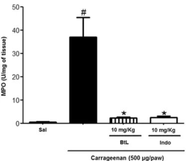
3.5. BtL inhibits MPO activity in paw tissue
MPO is an enzyme present predominantly in azurophilic granules of neutrophils and in other cells of myeloid origin. It is widely known as a quantitative marker of neutrophil infiltration in inflammatory processes [51]. Therefore, the increase in MPO activity is a key indicator of the pro-gression of inflammation. Thus, to confirm the anti-inflammatory effect of BtL, MPO activity was measured in mice with paw edema induced by carrageenan.
As depicted inFig. 4, BtL at 10 mg/kg reduced the MPO activity by 92.93% when compared to the carrageenan group (pb0.05).
Pretreat-ment with indomethacin produced a similar result, reducing MPO activity by 92.65%. Accordingly, this result supports thefindings in the carrageenan-induced paw edema assay at the dose of 10 mg/kg.
Table 1
The effect of BtL on paw edema induced by carrageenan.
Experimental groups Paw edema (mL)
1 h 2 h 3 h 4 h
Saline 0.015 ± 0.005 0.018 ± 0.004 0.013 ± 0.008 0.008 ± 0.004
Carrageenan (Cg) 0.068 ± 0.004 0.086 ± 0.005 0.091 ± 0.004 0.093 ± 0.002
Cg + Indo (10 mg/kg) 0.018 ± 0.004
(73.04)⁎
0.020 ± 0.005 (76.74%)⁎
0.008 ± 0.004 (91.20%)⁎
0.008 ± 0.004 (91.39%)⁎
Cg + BtL (1 mg/kg) 0.036 ± 0.011
(47.05%)⁎
0.020 ± 0.007 (76.74%)⁎
0.017 ± 0.014 (81.31%)⁎
0.024 ± 0.008 (74.19%)⁎
Cg + BtL (5 mg/kg) 0.016 ± 0.007
(76.47%)⁎
0.015 ± 0.006 (82.55%)⁎
0.013 ± 0.007 (85.71%)⁎
0.018 ± 0.008 (80.64%)⁎
Cg + BtL (10 mg/kg) 0.018 ± 0.004
(73.52%)⁎
0.003 ± 0.002 (96.51%)⁎
0.016 ± 0.001 (82.41%)⁎
0.008 ± 0.004 (91.39%)⁎
Values are expressed as mean ± SEM (n = 6). The percentage of inhibition is shown in parentheses.
Similar results were obtained for other lectins isolated from sea-weeds, such as the lectins extracted fromH. cervicornis[24] and from
S.filiformis, where the MPO activity was reduced by 60.00%, 73.00%, and 88.00% at doses of 1, 3, and 9 mg/kg, respectively [27]. These data confirm the anti-inflammatory therapeutic potential of these lectins and demonstrate a possible interaction between lectins from red seaweeds and selectins found on the leukocyte and endothelial cell surface through recognition of specific glycoconjugates. Thus, this anti-inflammatory effect may be explained by the interruption of leukocyte–endothelium interaction through competitive blocking of
selectin binding sites, resulting in inhibition of leukocyte recruitment [56–58].
3.6. BtL decreases cell migration in peritonitis induced by carrageenan
Carrageenan induces neutrophil migration toward the peritoneal cavity through an indirect mechanism, i.e., via macrophage chemotaxis. The inhibition of leukocyte migration is the central mechanism of action of some anti-inflammatory drugs [59].
When administered to mice, BtL (10 mg/kg) significantly decreased the total leukocyte count at an inhibition rate of 91.84% (pb0.05).
Indo-methacin decreased this migration by 86.82% (Fig. 5A). The differential leukocyte counts revealed that BtL (10 mg/kg) significantly decreased the neutrophil migration by 92.02% (pb0.05), whereas indomethacin
decreased neutrophil migration by 82.98% (Fig. 5B).
In another study using the lectin obtained fromB. triquetrum, Neves et al. [60] obtained the opposite result. This lectin provoked neutrophil migration and was thus classified as a proinflammatory protein. None-theless, this phenomenon may be explained by the fact that this lectin is the same as the one found by Ainouz et al. [28], with molecular weight 3.5 kDa and no relation to the lectin used in our study.
In a study on the lectin extracted fromH. cervicornis, Bitencourt et al. [26] observed a 90.00% reduction in neutrophil migration, at the dose of 10 mg/kg (i.p.). This effect was associated with leukocyte–endothelium
interactions and an increase in nitric oxide production. The lectin ob-tained fromP. capillacea, at the dose of 10 mg/kg, can also significantly reduce the neutrophil migration into the peritoneal cavity of rats [25]. Another lectin extracted from a red seaweed (S.filiformis) yielded a
similar result when compared with BtL, by significantly decreasing the neutrophil migration (by 76.20%), when administered at a dose of 9 mg/kg [27].
The mechanisms of action of BtL involved in the reduction of neutro-phil migration are still not completely elucidated; however, it is possible that BtL inhibits rolling and adhesion of neutrophils to endothelial cells and/or affects the release of chemoattractants by resident cells. All these activities are modulated by cytokines; hence, to assess the effects of BtL on the release of proinflammatory cytokines in carrageenan-induced peritonitis, the production of TNF-αand IL-1βwas measured in the peritoneal cavity.
3.7. BtL decreased TNF-αand IL-1βproduction in carrageenan-induced peritonitis
The administration of BtL to mice (at 10 mg/kg) reduced the levels of TNF-αin carrageenan-induced peritonitis by 62.83% (pb0.001), thus
yielding a result similar to that obtained after indomethacin pretreat-ment, in which the TNF-αconcentration decreased by 59.33% when compared to the carrageenan group (pb0.001). The results on IL-1β
are almost identical, the pretreatment of mice with BtL (10 mg/kg) sig-nificantly decreased (pb0.001) the levels of this cytokine in the
perito-neal cavity (by 54.52%). The pretreatment with indomethacin reduced this concentration by 53.17% (pb0.001). All the results are summarized
inFig. 6(A and B).
The peritonitis provoked by carrageenan, in addition to increasing the neutrophil migration into the peritoneal cavity (as demonstrated above), is also associated with an increase in cytokine production [61–63]. TNF-αis thefirst interleukin released by immune cells, mainly monocytes and macrophages, exerting its proinflammatory effects by stimulating transcription of various genes (such as those encoding other cytokines, chemokines, and cell adhesion molecules) via activa-tion of transcripactiva-tion factors, such as nuclear factor (NF)-κB [64,65].
IL-1βis released most commonly by monocytes, macrophages, and mast cells; however, nonimmune cells, such as neuronal and glial cells, can synthesize and release IL-1βduring cell injury or infl amma-tion. Thus, IL-1βis associated with inflammation, pain, and autoim-mune conditions, in addition to upregulating chemokines [66–68].
Lectins extracted from plant seeds possess known effects on cyto-kine levels, decreasing them to the basal level during peritonitis induced
by carrageenan [22,69]. Nevertheless, researches into lectins from red seaweeds that evaluate cytokine production in peritonitis induced by carrageenan are still scarce.
In the present study, BtL inhibited vascular and cellular events of an acute inflammatory response. Therefore, the inhibition of neutrophil migration to inflammation sites via suppression of TNF-αand IL-1β pro-duction, as shown in carrageenan-induced peritonitis, suggests that the modulation of the process of leukocyte recruitment is a major mecha-nism behind the anti-inflammatory action of BtL.
4. Conclusions
According our results, we concluded that the lectin extracted from the red seaweedB. triquetrumis a stable protein, mostly com-posed of disordered secondary structure elements andβ-strands, and possessing Trp residues partially exposed to an aqueous envi-ronment. This lectin is capable to preserve its native secondary struc-ture unchanged even under extreme conditions, such as extreme pH, and high temperatures. The interesting anti-inflammatory activity of BtL in classical models of inflammation holds promise for evaluation in clinical trials.
Fig. 4.Effects of BtL on MPO activity during carrageenan-induced paw edema. Data are expressed as mean ± SEM (n = 6); *pb0.05 when compared with the carrageenan group;#p
b0.05 when compared with the saline group (according to one-way ANOVA followed by the Newman-Keuls post hoc test).
Fig. 5. Effects of BtL on cell migration in the carrageenan-induced peritonitis. (A) Leukocyte migration. (B) Neutrophil migration. Data are expressed as mean ± SEM (n = 6); *pb0.05 when compared with the carrageenan group;#p
Acknowledgments
This study was supported by grants from CNPq (grant 406429/2016-2 to JLSL), CAPES, and Funcap. The authors also acknowledge the group of molecular biophysics“Sérgio Mascarenhas”at IFSC-USP, for conducting
experiments on CD andfluorescence spectroscopy, and the Animal Ethics Committee at UFC, for providing animals for the experiments.
Conflicts of interest
The authors declare that they have no conflicts of interest.
References
[1] S.H.Z. Ariffin, W.W. Yeen, I.Z.Z. Abidin, R.M.A. Abdul Wahab, Z.Z. Ariffin, S. Senafi, Cy-totoxicity effect of degraded and undegradedkappaandiotacarrageenan in human intestine and liver cell lines, BMC Complement. Altern. Med. 14 (2014), 508.https:// doi.org/10.1186/1472-6882-14-508.
[2] A.J. Smit, Medicinal and pharmaceutical uses of seaweed natural products: a review, J. Appl. Phycol. 16 (2004) 245–262,https://doi.org/10.1023/B:JAPH.0000047783. 36600.ef.
[3] E. Shannon, N. Abu-Ghannam, Antibacterial derivatives of marine algae: an over-view of pharmacological mechanisms and applications, Mar. Drugs 14 (2016) 81, https://doi.org/10.3390/md14040081.
[5] Y. Hu, J. Chen, G. Hu, J. Yu, X. Zhu, Y. Lin, S. Chen, J. Yuan, Statistical research on the bio-activity of new marine natural products discovered during the 28 years from 1985 to 2012, Mar. Drugs 13 (2015) 202–221,https://doi.org/10.3390/md13010202.
[6] W.A.J.P. Wijesinghe, Y.J. Jeon, Biological activities and potential industrial applica-tions of fucose rich sulfated polysaccharides and fucoidans isolated from brown sea-weeds: a review, Carbohydr. Polym. 88 (2012) 13–20,https://doi.org/10.1016/j. carbpol.2011.12.029.
[7] I. Wijesekara, R. Pangestuti, S.K. Kim, Biological activities and potential health bene-fits of sulfated polysaccharides derived from marine algae, Carbohydr. Polym. 84 (2011) 14–21,https://doi.org/10.1016/j.carbpol.2010.10.062.
[8] E.S.O. Vanderlei, K.K.N.R. Patoilo, N.A. Lima, A.P.S. Lima, J.A.G. Rodrigues, L.M.C.M. Silva, M.E.P. Lima, V. Lima, N.M.B. Benevides, Antinociceptive and anti-inflammatory activities of lectin from the marine green algaCaulerpa cupressoides, Int. Immunopharmacol. 10 (2010) 1113–1118,https://doi.org/10.1016/j.intimp. 2010.06.014.
[9] S.A. Neves, A.L.P. Freitas, B.W.S. Souza, M.L.A. Rocha, M.V.O. Correia, D.A. Sampaio, G.S.B. Viana, Antinociceptive properties in mice of a lectin isolated from the marine algaAmansia multifidaLamouroux, Braz. J. Med. Biol. Res. 40 (2007) 127–134, https://doi.org/10.1590/S0100-879X2007000100016.
[10] Y. Takebe, C.J. Saucedo, G. Lund, R. Uenishi, S. Hase, T. Tsuchiura, N. Kneteman, K. Ramessar, D.L.J. Tyrrell, M. Shirakura, T. Wakita, J.B. McMahon, B.R. O'Keefe, Antiviral lectins from red and blue-green algae show potentin vitroandin vivoactivity against hepatitis C virus, PLoS One 8 (2013), e64449.https://doi.org/10.1371/ journal.pone.0064449.
[11] Y. Omokawa, T. Miyazaki, P. Walde, K. Akiyama, T. Sugahara, S. Masuda, A. Inada, Y. Ohnishi, T. Saeki, K. Kato,In vitroandin vivoanti-tumor effects of novel span 80 ves-icles containing immobilizedEucheuma serraagglutinin, Int. J. Pharm. 389 (2010) 157–167,https://doi.org/10.1016/j.ijpharm.2010.01.033.
[12] J.C. Manning, A. Romero, F.A. Habermann, G. García-Caballero, H. Kaltner, H.J. Gabius, Lectins: a primer for histochemists and cell biologists, Histochem. Cell Biol. 147 (2017) 199–222,https://doi.org/10.1007/s00418-016-1524-6. [13] G.A. Bezerra, R. Viertlmayr, T.R. Moura, P. Delatorre, B.A.M. Rocha, K.S. Nascimento,
J.G. Figueiredo, I.G. Bezerra, C.S. Teixeira, R.C. Simões, C.S. Nagano, N.M.N. Alencar, K. Gruber, B.S. Cavada, Structural studies of an anti-inflammatory lectin from
Canavalia bolivianaseeds in complex with dimannosides, PLoS One 9 (2014), e97015.https://doi.org/10.1371/journal.pone.0097015.
[14] N. Sharon, Lectins: past, present and future, Biochem. Soc. Trans. 36 (2008) 1457–1460,https://doi.org/10.1042/BST0361457.
[15] C. Nathan, Points of control in inflammation, Nature 420 (2002) 846–852,https:// doi.org/10.1038/nature01320.
[16] R. Medzhitov, Origin and physiological roles of inflammation, Nature 454 (2008) 428–435,https://doi.org/10.1038/nature07201.
[17] M.E. Kotas, R. Medzhitov, Homeostasis, inflammation, and disease susceptibility, Cell 160 (2015) 816–827,https://doi.org/10.1016/j.cell.2015.02.010.
[18] G.R. Burmester, R. Panaccione, K.B. Gordon, M.J. McIlraith, A.P.M. Lacerda, Adalimumab: long-term safety in 23 458 patients from global clinical trials in rheu-matoid arthritis, juvenile idiopathic arthritis, ankylosing spondylitis, psoriatic ar-thritis, psoriasis and Crohn's disease, Ann. Rheum. Dis. 72 (2013) 517–524,
https://doi.org/10.1136/annrheumdis-2011-201244.
[19] Y. Vodovotz, M. Csete, J. Bartels, S. Chang, G. An, Translational systems biology of in-flammation, PLoS Comput. Biol. 4 (2008), e1000014.https://doi.org/10.1371/ journal.pcbi.1000014.
[20] S.M. Lucas, N.J. Rothwell, R.M. Gibson, The role of inflammation in CNS injury and disease, Br. J. Pharmacol. 147 (2006) S232–S240,https://doi.org/10.1038/sj.bjp. 0706400.
[21] N.M.N. Alencar, R.S.B. Oliveira, J.G. Figueiredo, I.J.M. Cavalcante, M.P.V. Matos, F.Q. Cunha, J.V.S. Nunes, L.R. Bomfim, M.V. Ramos, An anti-inflammatory lectin from
Luetzelburgia auriculataseeds inhibits adhesion and rolling of leukocytes and mod-ulates histamine and PGE2action in acute inflammation models, Inflamm. Res. 59 (2010) 245–254,https://doi.org/10.1007/s00011-009-0092-9.
[22] A.F. Pires, N.V.F.C. Rodrigues, P.M.G. Soares, R. de A. Ribeiro, K.S. Aragão, M.M. Marinho, M.T.L. da Silva, B.S. Cavada, A.M.S. Assreuy, A novelN-acetyl-glucosamine lectin ofLonchocarpus araripensisattenuates acute cellular inflammation in mice, Inflamm. Res. 65 (2016) 43–52,https://doi.org/10.1007/s00011-015-0889-7. [23] D. Praseptiangga, Algal lectins and their potential uses, Squalen Bull. of Mar. & Fish.
Postharvest & Biotech. 10 (2015) 89–98,https://doi.org/10.15578/squalen.v10i2. 125.
[24] J.G. Figueiredo, F.S. Bitencourt, T.M. Cunha, P.B. Luz, K.S. Nascimento, M.R.L. Mota, A.H. Sampaio, B.S. Cavada, F.Q. Cunha, N.N.M. Alencar, Agglutinin isolated from the red ma-rine algaHypnea cervicornisJ. Agardh reduces inflammatory hypernociception: in-volvement of nitric oxide, Pharmacol. Biochem. Behav. 96 (2010) 371–377,https://
doi.org/10.1016/j.pbb.2010.06.008.
[25] L.M.C.M. Silva, V. Lima, M.L. Holanda, P.G. Pinheiro, J.A.G. Rodrigues, M.E.P. Lima, N.M.B. Benevides, Antinociceptive and anti-inflammatory activities of lectin from marine red algaPterocladiella capillacea, Biol. Pharm. Bull. 33 (2010) 830–835, https://doi.org/10.1248/bpb.33.830.
[26] F.S. Bitencourt, J.G. Figueiredo, M.R.L. Mota, C.C.R. Bezerra, P.P. Silvestre, M.R. Vale, K.S. Nascimento, A.H. Sampaio, C.S. Nagano, S. Saker-Sampaio, W.R.L. Farias, B.S. Cavada, A.M.S. Assreuy, N.M.N. Alencar, Antinociceptive and anti-inflammatory ef-fects of a mucin-binding agglutinin isolated from the red marine algaHypnea
Fig. 6.Effects of BtL on TNF-αand IL-1βproduction during the carrageenan-induced peritonitis. (A) TNF-αlevels. (B) IL-1βlevels. Data are expressed as mean ± SEM (n = 6); *pb0.001 when compared with the carrageenan group;#p
cervicornis, Naunyn Schmiedeberg's Arch. Pharmacol. 377 (2008) 139–148,https:// doi.org/10.1007/s00210-008-0262-2.
[27] T.M. Abreu, N.A. Ribeiro, H.V. Chaves, R.J.B. Jorge, M.M. Bezerra, H.S.A. Monteiro, I.M. Vasconcelos, E.F. Mota, N.M.B. Benevides, Antinociceptive and anti-inflammatory ac-tivities of the lectin from marine red algaSolieriafiliformis, Planta Med. 82 (2016) 596–605,https://doi.org/10.1055/s-0042-101762.
[28] I.L. Ainouz, A.H. Sampaio, A.L.P. Freitas, N.M.B. Benevides, S. Mapurunga, Compara-tive study on hemagglutinins from the red algaeBryothamnion seaforthiiand
Bryothamnion triquetrum, Braz. J. Plant Physiol. 7 (1995) 15–19http://agris.fao.org/
agris-search/search.do?recordID=BR9510062.
[29] J.J. Calvete, F.H.F. Costa, S. Saker-Sampaio, M.P.M. Murciano, C.S. Nagano, B.S. Cavada, T.B. Grangeiro, M.V. Ramos, C. Bloch Jr, S.B. Silveira, B.T. Freitas, A.H. Sampaio, The amino acid sequence of the agglutinin isolated from the red marine alga
Bryothamnion triquetrumdefines a novel lectin structure, Cell. Mol. Life Sci. 57 (2000) 343–350,https://doi.org/10.1007/PL00000696.
[30] G.S.B. Viana, A.L.P. Freitas, M.M.L. Lima, L.A.P. Vieira, M.C.H. Andrade, N.M.B. Benevides, Antinociceptive activity of sulfated carbohydrates from the red algae
Bryothamnion seaforthii(Turner) Kütz. andB. triquetrum(S.G. Gmel.) M. Howe, Braz. J. Med. Biol. Res. 35 (2002) 713–722, https://doi.org/10.1590/S0100-879X2002000600012.
[32] R.F. Lima, D.N. Criddle, E.P. Souza, A.H. Sampaio, K.S. Nascimento, B.S. Cavada, A.M.S. Assreuy, Red marine algaBryothamnion triquetrumlectin induces endothelium-dependent relaxation of the rat aorta via release of nitric oxide, J. Pharm. Pharmacol. 56 (2004) 1415–1421,https://doi.org/10.1211/0022357044616.
[33] C.A. Winter, E.A. Risley, G.W. Nuss, Carrageenin-induced edema in hind paw of the rat as an assay for antiiflammatory drugs, Proc. Soc. Exp. Biol. Med. 111 (1962) 544–547,https://doi.org/10.3181/00379727-111-27849.
[34] P.P. Bradley, D.A. Priebat, R.D. Christensen, G. Rothstein, Measurement of cutaneous inflammation: estimation of neutrophil content with an enzyme marker, J. Invest. Dermatol. 78 (1982) 206–209,https://doi.org/10.1111/1523-1747.ep12506462.
[35] V.P.T. Pinto, H. Debray, D. Dus, E.H. Teixeira, T.M. de Oliveira, V.A. Carneiro, A.H. Teixeira, G.C. Filho, C.S. Nagano, K.S. Nascimento, A.H. Sampaio, B.S. Cavada, Lectins from the red marine algal speciesBryothamnion seaforthiiandBryothamnion triquetrumas tools to differentiate human colon carcinoma cells, Adv. Pharmacol. Sci. 2009 (2009), 862162.https://doi.org/10.1155/2009/862162.
[36] E.H. Teixeira, M.H. Napimoga, V.A. Carneiro, T.M. de Oliveira, K.S. Nascimento, C.S. Nagano, J.B. Souza, A. Havt, V.P.T. Pinto, R.B. Gonçalves, W.R.L. Farias, S. Saker-Sampaio, A.H. Saker-Sampaio, B.S. Cavada,In vitroinhibition of oral streptococci binding to the acquired pellicle by algal lectins, J. Appl. Microbiol. 103 (2007) 1001–1006, https://doi.org/10.1111/j.1365-2672.2007.03326.x.
[37] S.M. Kelly, T.J. Jess, N.C. Price, How to study proteins by circular dichroism, Biochim. Biophys. Acta 1751 (2005) 119–139,https://doi.org/10.1016/j.bbapap.2005.06.005. [38] N. Sreerama, R.W. Woody, Structural composition ofβI- andβII-proteins, Protein
Sci. 12 (2003) 384–388,https://doi.org/10.1110/ps.0235003.
[39] N. Sreerama, R.W. Woody, Computation and analysis of protein circular dichroism spectra, Methods Enzymol. 383 (2004) 318–351, https://doi.org/10.1016/S0076-6879(04)83013-1.
[40] S.C.B. Oliveira, F.V. Fonseca, E. Antunes, E.A. Camargo, R.P. Morganti, R. Aparício, D.O. Toyama, L.O.S. Beriam, E.V. Nunes, B.S. Cavada, C.S. Nagano, A.H. Sampaio, K.S. Nascimento, M.H. Toyama, Modulation of the pharmacological effects of enzymatically-active PLA2by BTL-2, an isolectin isolated from theBryothamnion triquetrumred alga, BMC Biochem. 9 (2008) 16, https://doi.org/10.1186/1471-2091-9-16.
[41] C.S. Nagano, H. Debray, K.S. Nascimento, V.P.T. Pinto, B.S. Cavada, S. Saker-Sampaio, W.R.L. Farias, A.H. Sampaio, J.J. Calvete, HCA and HML isolated from the red marine algaeHypnea cervicornisandHypnea musciformisdefine a novel lectin family, Pro-tein Sci. 14 (2005) 2167–2176,https://doi.org/10.1110/ps.051498505.
[42] K. Hori, K. Matsubara, K. Miyazawa, Primary structures of two hemagglutinins from the marine red alga,Hypnea japonica, Biochim. Biophys. Acta 1474 (2000) 226–236, https://doi.org/10.1016/S0304-4165(00)00008-8.
[43] L.G. Nascimento-Neto, R.F. Carneiro, S.R. Silva, B.R. Silva, F.V.S. Arruda, V.A. Carneiro, K.S. Nascimento, S. Saker-Sampaio, V.A. Silva Jr, A.L.F. Porto, B.S. Cavada, A.H. Sampaio, E.H. Teixeira, C.S. Nagano, Characterization of isoforms of the lectin iso-lated from the red algaeBryothamnion seaforthiiand its pro-healing effect, Mar. Drugs 10 (2012) 1936–1954,https://doi.org/10.3390/md10091936.
[44]H.J. Gabius, S. Gabius, Glycosciences: Status and Perspectives,first ed. Weinheim, Chapman & Hall GmbH, 1997.
[45] G.H. Kim, T.A. Klochkova, K.S. Yoon, Y.S. Song, K.P. Lee, Purification and characteriza-tion of a lectin, bryohealin, involved in the protoplast formacharacteriza-tion of a marine green algaBryopsis plumosa(Chlorophyta), J. Phycol. 42 (2005) 86–95,https://doi.org/ 10.1111/j.1529-8817.2006.00162.x.
[46] N.E. Ziółkowska, A. Wlodawer, Structural studies of algal lectins with anti-HIV activ-ity, Acta Biochim. Pol. 53 (2006) 617–62617128290.
[47] W.R. Liao, J.Y. Lin, W.Y. Shieh, W.L. Jeng, R. Huang, Antibiotic activity of lectins from marine algae against marine vibrios, J. Ind. Microbiol. Biotechnol. 30 (2003) 433–439,https://doi.org/10.1007/s10295-003-0068-7.
[48] S.H. Kim, G.H. Kim, Cell-cell recognition during fertilization in the red alga,
Aglaothamnion oosumiense(Ceramiaceae, Rhodophyta), Hydrobiologia 398 (1999) 81–89,https://doi.org/10.1007/978-94-011-4449-0_10.
[49] M.D. García, M.A. Fernández, A. Alvarez, M.T. Saenz, Antinociceptive and anti-inflammatory effect of the aqueous extract from leaves ofPimenta racemosavar. ozua (Mirtaceae), J. Ethnopharmacol. 91 (2004) 69–73,https://doi.org/10.1016/j.
jep.2003.11.018.
[50] M.R. Morris, S. Dewitt, I. Laffafian, M.B. Hallett, Phagocytosis by inflammatory phagocytes. Experimental strategies for stimulation and quantification, in: P.G. Winyard, D.A. Willoughby (Eds.),Inflammation Protocols, Humana Press Inc., Totowa 2003, pp. 35–46,https://doi.org/10.1385/1-59259-374-7:35.
[51] I. Posadas, M. Bucci, F. Roviezzo, A. Rossi, L. Parente, L. Sautebin, G. Cirino, Carrageenan-induced mouse paw oedema is biphasic, age-weight dependent and displays differential nitric oxide cyclooxygenase-2 expression, Br. J. Pharmacol. 142 (2004) 331–338,https://doi.org/10.1038/sj.bjp.0705650.
[52] M. Di Rosa, J.P. Giroud, D.A. Willoughby, Studies on the mediators of the acute in-flammatory response induced in rats in different sites by carrageenan and turpen-tine, J. Pathol. 104 (1971) 15–29,https://doi.org/10.1002/path.1711040103. [53] T.N. Lo, A.P. Almeida, M.A. Beaven, Dextran and carrageenan evoke different
inflam-matory responses in rat with respect to composition of infiltrates and effect of indo-methacin, J. Pharmacol. Exp. Ther. 221 (1982) 261–267 (6174730).
[54] L. Maintz, N. Novak, Histamine and histamine intolerance, Am. J. Clin. Nutr. 85 (2007) 1185–1196 (17490952).
[55] J. Raber, Histamine receptor-mediated signaling during development and brain function in adulthood, Cell. Mol. Life Sci. 64 (2007) 735–741,https://doi.org/10. 1007/s00018-007-6442-2.
[56] B.A.M. Rocha, P. Delatorre, T.M. Oliveira, R.G. Benevides, A.F. Pires, A.A.S. Sousa, L.A.G. Souza, A.M.S. Assreuy, H. Debray, W.F. de Azevedo Jr, A.H. Sampaio, B.S. Cavada, Structural basis for both pro- and anti-inflammatory response induced by mannose-specific legume lectin fromCymbosema roseum, Biochimie 93 (2011) 806–816,https://doi.org/10.1016/j.biochi.2011.01.006.
[57] D. Impellizzeri, S. Cuzzocrea, Targeting selectins for the treatment of inflammatory diseases, Expert Opin. Ther. Targets 18 (2014) 55–67,https://doi.org/10.1517/
14728222.2013.841140.
[58] K. Ley, The role of selectins in inflammation and disease, Trends Mol. Med. 9 (2003) 263–268,https://doi.org/10.1016/S1471-4914(03)00071-6.
[59] G.E.P. Souza, F.Q. Cunha, R. Mello, S.H. Ferreira, Neutrophil migration induced by in-flammatory stimuli is reduced by macrophage depletion, Agents Actions 24 (1988) 377–380,https://doi.org/10.1007/BF02028296.
[60] S.A. Neves, M. Dias-Baruffi, A.L.P. Freitas, M.C. Roque-Barreira, Neutrophil migration inducedin vivoandin vitroby marine algal lectins, Inflamm. Res. 50 (2001) 486–490,https://doi.org/10.1007/PL00000222.
[61] R. Herwig, B. Glodny, C. Kühle, B. Schlüter, O.A. Brinkmann, H. Strasser, N. Senninger, G. Winde, Early identification of peritonitis by peritoneal cytokine measurement, Dis. Colon Rectum 45 (2002) 514–521,https://doi.org/10.1007/s10350-004-6231-z. [62] R.O. Silva, S.R.B. Damasceno, T.V. Brito, J.M. Dias, A.M. Fontenele, I.S. Braúna, J.S.C. Júnior, J.S. Maciel, R.C. de Paula, R.A. Ribeiro, M.H.L.P. Souza, A.L.P. Freitas, J.V.R. Medeiros, D.C. Silva, A.L.R. Barbosa, Polysaccharide fraction isolated fromPassiflora edulisinhibits the inflammatory response and the oxidative stress in mice, J. Pharm. Pharmacol. 67 (2015) 1017–1027,https://doi.org/10.1111/jphp.12399.
[63] Q. Pan, J. Cai, Y. Peng, H. Xiao, L. Zhang, J. Chen, H. Liu, Protective effect of a novel an-tibody against TLR2 on zymosan-induced acute peritonitis in NF-κB transgenic mice, Am. J. Transl. Res. 9 (2017) 692–699 PMCID: PMC5340704.
[64] J. Jiao, R. Mao, D. Teng, X. Wang, Y. Hao, N. Yang, X. Wang, X. Feng, J. Wang,In vitro
andin vivoantibacterial effect of NZ2114 againstStreptococcus suistype 2 infection in mice peritonitis models, AMB Express 7 (2017), 44.https://doi.org/10.1186/ s13568-017-0347-8.
[65] G. Malleo, E. Mazzon, A.K. Siriwardena, S. Cuzzocrea, Role of tumor necrosis factor-alpha in acute pancreatitis: from biological basis to clinical evidence, Shock 28 (2007) 130–140,https://doi.org/10.1097/shk.0b013e3180487ba1.
[66] A.A.C. Almeida, R.O. Silva, L.A.D. Nicolau, T.V. Brito, D.P. Sousa, A.L.R. Barbosa, R.M. Freitas, L.S. Lopes, J.V.R. Medeiros, P.M.P. Ferreira, Physio-pharmacological investiga-tions about the anti-inflammatory and antinociceptive efficacy of (+)-limonene ep-oxide, Inflammation 40 (2017) 511–522, https://doi.org/10.1007/s10753-016-0496-y.
[67] K. Ren, R. Torres, Role of interleukin-1beta during pain and inflammation, Brain Res. Rev. 60 (2009) 57–64,https://doi.org/10.1016/j.brainresrev.2008.12.020. [68] R. Natoli, N. Fernando, M. Madigan, J.A. Chu-Tan, K. Valter, J. Provis, M. Rutar,
Microglia-derived IL-1βpromotes chemokine expression by Müller cells and RPE in focal retinal degeneration, Mol. Neurodegener. 12 (2017), 31.https://doi.org/ 10.1186/s13024-017-0175-y.