Cop
yright
© ABE&M t
odos os dir
eit
os r
eser
vados
.
Prevalence of goiter and
thyroid nodular disease in
patients with class III obesity
Prevalência de bócio e doença nodular tireoidiana em pacientes com obesidade grau III
Priscila Alves Medeiros de Sousa¹, Mario Vaisman¹, João Regis Ivar Carneiro2,
Lorena Guimarães¹, Heloisa Freitas¹, Maria Fernanda Castellar Pinheiro3, Sally Liechocki4, Clarissa Menezes Maya
Monteiro4, Patrícia de Fátima dos Santos Teixeira¹
ABSTRACT
Objectives: To evaluate the prevalence of goiter and nodular disease in patients with class III obesity, and to correlate results with serum leptin levels and insulin resistance (IR) param-eters. Subjects and methods: A cross-sectional study was performed to assess thyroid ultra-sound (US) patterns, HOMA-IR, serum leptin, and TSH levels in obese patients and controls.
Results: Thyroid volume was positively correlated with body mass index (BMI) (r = 0.240, p = 0.039) and with HOMA-IR (r = 0.329; p < 0.01). Thyroid US patterns were similar between groups. However, when data from the male group was considered, greater thyroid volume was detected in the obese group compared with controls (10.8 vs. 8.5 cm3; p = 0.04). Also,
nodules were more frequently detected (67% vs. 18%), as were nodules requiring FNAB (33.3% vs. 0%, p ≥ 0.05-0.09), in this group. Conclusion: Although IR did not correlate directly with the presence of nodules, the results support the hypothesis of a direct association be-tween insulin resistance and thyroid volume. Arq Bras Endocrinol Metab. 2013;57(2):120-5
Keywords
Obesity; insulin resistance; goiter; thyroid nodule
RESUMO
Objetivos: Avaliar a prevalência de bócio e doença nodular tireoidiana em pacientes com obesidade grau III e correlacionar os resultados com os níveis de leptina e parâmetros de resistência à ação da insulina (RI). Sujeitos e métodos: Estudo seccional foi desenvolvi-do realizandesenvolvi-do ultrassonograia (US) tireoidiana e níveis séricos de HOMA-IR e TSH nos pacien tes obesos e nos controles. Resultados: Volume tireoidiano foi positivamente cor-relacionado com índice de massa corporal (IMC) (r = 0,240, p = 0,039) e com HOMA (r = 0,329; p < 0,01). Volume tireoidiano e prevalência de doença nodular tireoidiana foram similares entre os grupos. Quando avaliado o subgrupo masculino, maiores volumes tir-eoidianos foram detectados no grupo dos obesos comparados aos controles (10,8 vs. 8,5 cm3; p = 0,04), nódulos foram mais frequentes (67% vs. 18%), assim como nódulos com indi-cação de punção (33,3% vs. 0%, p ≥ 0,05-0,09). Conclusão: Embora RI não se correlacione diretamente com a presença de nódulos, os resultados suportam a hipótese da direta associação entre resistência à ação da insulina e volume tireoidiano. Arq Bras Endocrinol Metab. 2013;57(2):120-5
Descritores
Obesidade; resistência à insulina; bócio; nódulo da glândula tireoide
1 Endocrinology Service, Medicine
School, Universidade Federal do Rio de Janeiro, Hospital Universitário Clementino Fraga Filho (UFRJ/ HUCFF), Rio de Janeiro, RJ, Brazil
2 Nutrology Service and
Radiology Department, Medicine School, UFRJ/HUCFF, Rio de Janeiro, RJ, Brazil
3 Sergio Franco Medicina
Diagnóstica, Duque de Caxias, RJ, Brazil
4 Fundação Oswaldo Cruz (Fiocruz),
Laboratório de Imunofarmacologia, Rio de Janeiro, RJ, Brazil
Correspondence to:
Priscila Alves Medeiros de Sousa [email protected]
Cop
yright
© ABE&M t
odos os dir
eit
os r
eser
vados
.
INTRODUCTION
T
he signiicant increase in the worldwide prevalence of overweight and obese individuals is an impor-tant public health issue at the start of this century, both in developed and developing countries. Additionally, the prevalence of more severe forms of obesity, deined as class III obesity, has been increasing worldwide (1). Over recent years, various publications have focused on the association between insulin resistance (IR) and thy-roid abnormalities (2-8). Causes of thythy-roid dysfunction are positively associated with IR, not only in subjects diagnosed with diabetes mellitus (DM), but also insub-jects without this diagnosis (9).
Thyroid nodular disease and differentiated thyroid carcinoma have also been regarded as a possible new spectrum of IR (2). It has been suggested that subjects with hyperinsulinemia may present greater thyroid size and greater prevalence of nodules (2). This inding is in accordance with reported studies from patients with ac-romegaly, in which the prevalence of goiter and nodu-les was found to be greater, probably as a result of the increased activity of IGF-1 (10). Elevated prevalence of IR in patients with differentiated thyroid carcinoma has also been demonstrated (11).
The aim of this study was to describe thyroid ul-trasound (US) indings of subjects with class III obe-sity. Another aim was to correlate thyroid volume and prevalence of thyroid nodules with BMI, IR evaluated by homeostatic model assessment (HOMA-IR), serum leptin and thyroid stimulating hormone (TSH) levels.
SUBJECTS AND METHODS
A cross-sectional study was conducted. Both the expe-rimental group and the control group subjects were submitted to thyroid US and laboratory analyses to de-termine levels of serum TSH, free T4, anti-TPO, leptin, glucose, and insulin (for HOMA-IR calculation). The experimental group was composed of patients who were followed up in the bariatric surgery program at Clementino Fraga Filho Hospital (HUCFF), Rio de Ja-neiro, Brazil, before they underwent any bariatric sur-gical procedure. At the moment, the main indication for bariatric surgery at this hospital is body mass index (BMI) ≥ 40 kg/m². The control group was selected from healthy volunteers with similar demographic characteristics regarding age and sex. Male and female patients were selected and matched between groups. In the experimental group, all individuals had class III
obesity (BMI ≥ 40 kg/m²), and all individuals in the control group had normal BMI levels (< 25 kg/m²). The exclusion criteria were diagnosis of DM type 2, use of medication that could interfere with insulin sensiti-vity, and known thyroid disease, and were the same for both groups. All research subjects signed an informed consent form, and the study was approved by the local ethics committee.
Participants in the study were submitted to spe-ciic anamnesis and a physical examination. They were weighed wearing light clothing and footwear in Filizo-la® scales. BMI was calculated by means of the
follow-ing formula: BMI = weight/height².
All ultrasound exams were performed by the same radiologist, using a high-frequency SIEMENS- ACUSON X300 multi-frequency transducer (12 MHz). Thyroid volume was calculated by the formula: length x width x thickness x 0.52 of each lobe and the isthmus (11). The characteristics of the thyroid paren-chyma were described according to their echogenicity and homogeneity. Focal lesions were only considered present when their diameter exceeded 3.0 mm. These lesions were classiied as cysts and nodules, which could be mists or solid. Solid lesions with a diameter of 10 mm or greater, and smaller solid lesions with suspicious US pattern (hypoechogenicity with microcalciications) were described as nodules, and required ine needle as-piration biopsy (FNAB).
Serum samples for laboratorial assays were obtained by venipuncture, after an 8-hour fast.
Serum levels of TSH, FT4, and TPO-Ab were determined by immunochemiluminesce (Immulite, Diagnostic Products, Los Angeles, CA). Reference ranges for TSH and FT4 were 0.4-4.0 µUI/mL and 0.9-1.8 ng/dL, respectively. Serum levels of TPO-Ab > 35 UI/mL were considered positive. The in-tra-assay coeficients of variation were 3.8%-12.5%, 4.4%-7.5% and 4.3%-5.6% for TSH, FT4 and TPO-Ab, respectively, while inter-assay coeficients of vari-ation were 4.6%-12.5%, 4.8%-9.0% and 7.8%-10.5%, respectively.
Serum leptin was measured by a sandwich ELISA method (Human Leptin Duo Set Kit). Glucose was as-sayed by the colorimetric enzymatic method. The refe-rence range for glucose was based on the guidelines of the American Diabetes Association (ADA) and the Bra-zilian Diabetes Society (SBD) (12,13).
Cop
yright
© ABE&M t
odos os dir
eit
os r
eser
vados
.
Analytics E 170, Roche) with a reference range of 2.0-23.0 mcU/mL, an intra-assay range of 0.7%-1.5% and an inter-assay range of 2.6%-4.9%. HOMA-IR was calculated with the following formula: [Fasting blood glucose (mmol/L) x Fasting insulin (mU/L)]/22.5. Fasting blood glucose was then converted to mmol/L by multiplying results in mg/dL by 0.05551.
Statistical analysis was performed with the SPSS software, version 13.0 for Windows. Continuous vari-ables were expressed as means and standard devia-tions, and categorical variables were expressed as per-centages. The Student’s t-test or the Mann-Whitney
test were used to compare two continuous variables according to the distribution of the variable in the stu-di ed sample, as assessed by the Kolmogorov-Smirnov test. Comparisons of the proportions between two groups were performed by the Chi-square test or Fisher’s exact test. Correlation between two variables was assessed by Pearson’s correlation coeficient (r) af-ter data were log-transformed. Partial correlation was used to detect the inluence of confounding variables in speciic correlations.
RESULTS
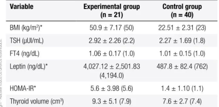
Between February 2010 and October 2011, thirty pa-tients were admitted to the bariatric surgery program at Clementino Fraga Filho Hospital (HUCFF), Rio de Janeiro, Brazil. Nine patients were excluded (four were > 50 years old, and ive had DM), and 21 patients were included in the study. A control group of 40 healthy volunteers that was well-matched with the experimen-tal group regarding gender and age, but with normal weight (IMC < 25 kg/m²) was recruited. The demo-graphic characteristics of the sample are presented in Table 1.
To assess the impact of gender in the results, a strati-ied analysis was performed after an exploratory analysis of baseline demographic data according to gender. These data are shown in table 2. Serum leptin and HOMA-IR were greater among obese subjects, independent of gen-der. Furthermore, thyroid volume was greater in male obese patients compared with control subjects.
Table 1. Demographic characteristics of obese subjects and the control group
Variable Experimental group (n = 21)
Control group (n = 40)
BMI (kg/m2)* 50.9 ± 7.17 (50) 22.51 ± 2.31 (23)
TSH (µUI/mL) 2.92 ± 2.26 (2.2) 2.27 ± 1.69 (1.8) FT4 (ng/dL) 1.06 ± 0.17 (1.0) 1.01 ± 0.15 (1.0) Leptin (ng/dL)* 4,027.12 ± 2,501.83
(4,194.0)
487.8 ± 82.4 (762)
HOMA-IR* 5.6 ± 3.98 (5.6) 1.4 ± 1.10 (1.1)
Thyroid volume (cm3) 9.3 ± 5.1 (7.9) 7.6 ± 2.7 (7.4)
* p < 0.001 in the comparison between the experimental and control group.
Table 2. Demographic characteristics of obese and control subjects, according to gender
Control group Experimental group p-value
Men
TSH (uUI/mL) 2.6 ± 2.5 (2.0) 2.5 ± 2.4 (0.9) NS HOMA* 1.2 ± 0.8 (0.7) 8.7 ± 5.7 (4.0) < 0.01
Thyroid volume* (cm3)
9.2 ± 1.7 (8.5) 15.7 ± 9.6 (10.8) 0.04
Leptin* (ng/dL) 373.0 ± 2.0 (343.0) 5261.0 ± 2.0 (4700.0) < 0.01
Women
TSH (uUI/mL) 2.1 ± 1.3 (1.3) 3.0 ± 2.7 (2.1) NS HOMA* 1.1 ± 1.1 (0.7) 4.3 ± 2.2 (2.7) < 0.01
Thyroid volume (cm3)
7.0 ± 2.8 (6.8) 7.6 ± 2.2 (7.5) NS
Leptin* (ng/dL) 919.0 ± 4.0 (859.0) 2796.0 ± 2.0 (2548.0) < 0.01
Thyroid nodular disease was found in 35% of the patients of the experimental group and in 20% of the control group (p = 0.171). There was a non-signiicant trend towards larger nodules, in average, in the experi-mental group (1.4 ± 1.4 cm3vs. 0.9 ± 0.5 cm3).
The stratiied analysis of gender revealed that 67% of obese men had abnormal US indings, in compari-son with 28% in non-obese men (Table 3). Comparing males in the experimental group with those in control group, we found a relative risk of 3.67 for any kind of US abnormality (CI95%:0.93-14.5, p = 0.05). In obese men, we found a prevalence of 33.3% of nodules that required FNAB in comparison with 0% in the control group. These comparisons showed borderline signii-cant p-values. Greater prevalence of US abnormalities associated with obesity was not found in the female subgroup (Table 3).
HOMA-IR did not differ between subjects who required FNAB or not (3.13 ± 3.3 vs. 2.6 ± 3.0; p =
0.696) and between those patients with normal vs.
ab-normal US indings (2.8 ± 2.4 vs. 2.5 ± 3.5; p = 0.765).
However, HOMA-IR levels were greater, without sta-tistical signiicance, in men who required FNAB (6.6 ± 2.2 vs. 3.5 ± 5.1; p = 0.136). For women, these
Cop
yright
© ABE&M t
odos os dir
eit
os r
eser
vados
.
BMI and serum leptin were positively correlated (r = 0.331, p = 0.016) and BMI correlated quite accurate-ly with HOMA-IR (r = 0.708, p < 0.001), as expected. BMI was also positively correlated with TSH serum levels (r = 0.438, p = 0.023) in obese patients. Serum TSH did not correlate with HOMA-IR or serum leptin levels (r = 0.06, p = 0.325; r = 0.132, p = 0.190, respectively).
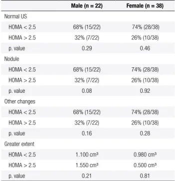
Thyroid volumes did not correlate with serum TSH, but were positively correlated with BMI (r = 0.240, p = 0.039) and HOMA-IR (r = 0.329, p = 0.007) in the en-tire group (Table 4). This latter association was stronger in the obese group (r = 0.571, p = 0.01), but was still present among the controls (r = 0.251, p = 0.05). This correlation was independent from confounding param-eters as detected in the partial correlation controlled for BMI (r = 0.230, p = 0.04), leptin (r = 0.340, p = 0.01) and TSH (r = 0.330, p = 0.01). However, this was not an independent association, as it was absent when leptin and HOMA-IR were added as confounding variables. When the obese patient group was analyzed separately, leptin levels were positively correlated with thyroid vo-lume (r = 0.771, p = 0. 036). Finally, we evaluated the prevalence of US indings in obese and control subjects according to gender stratiied by HOMA-IR. We con-sidered a predictor of an IR value of HOMA-IR > 2.5, and the results are shown in table 4.
DISCUSSION
The results of this study demonstrate a positive cor-relation between thyroid volume and IR, as evaluated by HOMA-IR. Partial correlations, which considered TSH, BMI, and leptin as confounding factors, demons-trated that this correlation between thyroid volume and IR persisted, suggesting an independent correlation.
Table 3. Prevalence of US abnormalities in obese and control subjects, according to gender
Male Experimental group (n = 6)
Control group (n = 11)
Abnormal indings in US 67% (4:6)* 18% (2:11)*
Nodules requiring FNAB** 33,3% (2:6)* 0% (0:11)* Nodular thyroid disease 66.7% (4:6) 18.2% (2:11)
Female group (n = 15)Experimental Control group (n = 29)
Abnormal indings in US 47.5% (7:15) 28% (8:29) Nodules requiring FNAB 6.7% (1:15) 10.3% (3:29) Nodular thyroid disease 21.4% (3-15)* 30.7% (6:29)*
* p-values ≥ 0.05-0.09; ** Solid lesions with a diameter of 10 mm or greater, and smaller solid lesions with a suspicious US pattern (hypoechogenicity with microcalciications).
Table 4. Prevalence of US indings in obese and control subjects, according to gender, stratiied by HOMA-IR
Male (n = 22) Female (n = 38)
Normal US
HOMA < 2.5 68% (15/22) 74% (28/38)
HOMA > 2.5 32% (7/22) 26% (10/38)
p. value 0.29 0.46
Nodule
HOMA < 2.5 68% (15/22) 74% (28/38)
HOMA > 2.5 32% (7/22) 26% (10/38)
p. value 0.08 0.92
Other changes
HOMA < 2.5 68% (15/22) 74% (28/38)
HOMA > 2.5 32% (7/22) 26% (10/38)
p. value 0.16 0.28
Greater extent
HOMA < 2.5 1.100 cm³ 0.980 cm³
HOMA > 2.5 1.550 cm³ 0.500 cm³
p. value 0.21 0.81
Increased insulin levels, which are consequence of IR, reduce the levels of IGF-1 binding proteins, thus raising free IGF-1 levels; IGF-1 can then bind to IGF-1 receptors, which are overexpressed in most cancer cells (14). IRS-1 is essential to the activation of cellular mitosis and its overexpression is linked to neoplastic transformation (15). Additionally, studies in acromegaly show that 64%-92% have thyroid changes in US (10,16,17).
Various authors have previously suggested a link between IR and increased thyroid volume. Rezzónico and cols. were the irst to describe the link between thy-roid nodular disease and IR, and have reported that pa-tients with differentiated thyroid carcinoma present an increase in IR parameters (2). However, IR could act by increasing thyroid proliferation regardless of the pa-tient’s BMI (3). The present study showed that IR was associated with thyroid volume, independent of BMI.
Cop
yright
© ABE&M t
odos os dir
eit
os r
eser
vados
.
characterization of this population lacked a description of serum TSH levels and nodule size, as well as any characterization of the goiter. Last, as this study was performed in a tertiary care reference center, thyroid-ectomy may have been performed as a result of other prognostic factors than just FNAB diagnosis. Obesity might play a role in the pre-operative evaluation (5).
Other studies have demonstrated an association between thyroid function and IR. Chiovatto and cols. concluded that metformin treatment decreases signii-cantly serum TSH concentrations in patients with poly-cystic ovary syndrome (PCOS) and hypothyroi dism, regardless of the concomitant thyroid replacement with levothyroxine therapy (6). The precise mechanisms of metformin action remain debated. The present study showed a positive and signiicant logarithmic correla-tion between thyroid volume and HOMA-IR, with TSH as a confounding factor (p = 0.01). Recently, another study demonstrated the impact of metformin, either alone or combined with levothyroxine, in reduc-ing the volume of hyperplasic thyroid nodules (7). Chi-ovatto and cols. also evaluated the accuracy of US in detecting autoimmune thyroid diseases in patients with class III obesity, and detected that class III obesity may affect thyroid morphology (8).
As there are differences in the prevalence of thyroid nodular disease between sexes and leptin levels may also differ, we performed a stratiied evaluation according to gender. We detected that thyroid volume was signii-cantly greater among obese men than in normal weight controls. Despite the absence of a higher prevalence of abnormal US indings in obese women, these results were more frequent among obese men in comparison with male controls, with borderline signiicance. The same occurred for the prevalence of nodules that re-quired FNAB. Cappelli and cols. showed low preva-lence of thyroid nodular disease in women with class III obesity (18). Therefore, among men, IR appears to play an important role in thyroid volume and in the development of thyroid nodular disease.
We did not ind any independent correlation bet-ween TSH and thyroid volume. This was in accordance with previous reports that TSH alone is not a mito-genic factor (14).
Leptin, produced by the adipose tissue, is respon-sible for informing the central nervous system (CNS) of the energy reserves in adipose tissue. Various stu-dies have been performed to understand the action of leptin on the thyroid axis. The main hypothesis is that
leptin positively regulates TRH expression (19). In the experimental group, we found a strong positive cor-relation between thyroid volume and leptin, but such a correlation was lost in the control group. With a larger sample, it may be possible to document greater inlu-ence of leptin in thyroid nodular disease. Nevertheless, it is important to determine whether such association is independent of or related to IR.
These results strengthen the hypothesis of direct association between obesity and increased thyroid volumes, by means of IR. Further studies are neces-sary to conirm the association between leptin action and thyroid nodular disease. One of the limitations of the study is related to the limited number of pa-tients, and to the sectional design, which interferes in the evidence of direct cause-consequence relationship between obesity, and/or insulin resistance, and goiter. In conclusion, despite the similar prevalence of thyroid nodular disease between the obese and con-trol groups of patients, there was a trend towards a higher frequency of the disease in obese male sub-jects. Although IR did not correlate directly with the presence of thyroid nodules, results also support the hypothesis of a direct association between insulin re-sistance and thyroid volume.
Acknowledgments: we are thankful for Patricia T. Bozza contri-butions and to Coordenação de Aperfeiçoamento de Pessoal de Nível Superior, Sistema Único de Saúde (Capes/SUS), Conselho Nacional de Desenvolvimento Cientíico e Tecnológico (CNPq), Fundação de Amparo à Pesquisa do Estado do Rio de Janeiro (Faperj), and Fundação Oswaldo Cruz (Fiocruz) support.
Disclosure: no potential conlict of interest relevant to this article was reported.
REFERENCES
1. Porto MCV, Brito IC, Calfa ADF, Amoras M, Villela NB, Araújo MB. Peril do obeso classe III do ambulatório de um hospital universitário em Salvador, Bahia. Arq Bras Endocrinol Metabol. 2002;46:668-73.
2. Rezzónico JN, Rezzónico M, Pusiol E, Pitoia F, Niepomniszcze H. Increased prevalence of insulin resistance in patients with dif-ferentiated thyroid carcinoma. Metab Syndr Relat Disord. 2009; 7(4):375-80.
3. Rezzónico J, Rezzónico M, Pusiol E, Pitoia F, Niepomniszcze H. Introducing the thyroid gland as another victim of the insulin re-sistance syndrome. Thyroid. 2008;18(4):461-4.
4. Ayturk S, Gursoy A, Kut A, Anil C, Nar A, Tutuncu NB. Metabolic syndrome and its components are associated with increased thy-roid volume and nodule prevalence in a mild-to-moderate iodine-deicient area. Eur J Endocrinol. 2009;161(4):599-605.
Cop
yright
© ABE&M t
odos os dir
eit
os r
eser
vados
.
6. Rotondi M, Cappelli C, Magri F, Botta R, Dionisio R, Iacobello C, et al. Thyroidal effect of metformin treatment in patients with poly-cystic ovary syndrome. Clin Endocrinol (Oxf). 2011;75(3):378-81. 7. Rezzónico J, Rezzónico M, Pusiol E, Pitoia F, Niepomniszcze H.
Metformin treatment for small benign thyroid nodules in patients with insulin resistance. Metab Syndr Relat Disord. 2011;9:69-75. 8. Rotondi M, Cappelli C, Leporati P, Chytiris S, Zerbini F, Fonte R,
et al. A hypoechoic pattern of the thyroid at ultrasound does not indicate autoimmune thyroid disease in patients with morbid obesity. Eur J Endocrinol. 2010;163(1):105-9.
9. Fernández-Real JM, López-Bermejo A, Castro A, Casamitjana R, Ricart W. Thyroid function is intrinsically linked to insulin sensitivi ty and endothelium dependent vasodilation in healthy euthyroid subject. J Clin Endocrinol Metab. 2006;91:3337-43. 10. Miyakawa M, Saji M, Tsushima T, Wakai K, Shizume K. Thyroid
vo-lume and serum thyroglobulin levels in patients with acromega-ly: correlation with plasma insulin like growth factor 1 levels. J Clin Endocrinol Metab. 1988;67:973-8.
11. Michalaki MA, Vagenakis AG, Leonardou AS, Argentou MN, Habeos IG, Makri MG, et al. Thyroid functions in humans with morbid obesity. Thyroid. 2006;16:73-8.
12. Position Statement: Standards of Medical Care in Diabetes – 2012. Diabetes Care. 2012;5(Suppl. 1):S11-63.
13. Milech A, Angelucci AP, Golbert A, et al. Metodos e critérios para o diagnóstico de diabtes mellitus. In: Gomes MB, Lerario AC, edi-tors. Diretrizes da Sociedade Brasileira de Diabetes 2009/Socie-dade Brasileira de Diabetes. 3th ed. São Paulo: A. Araújo Silva Farmacêutica; 2009. p. 18-20.
14. Vella V, Sciacca L, Pandini G, Mineo R, Squatrito S, Vigneri R, et al. The IGF system in thyroid cancer: new concepts. Mol Path. 2001;54:121-4. 15. Brunn J, Block U, Ruf G, Bos I, Kunze WP, Scriba PC. Volumet-ric analysis of thyroid lobes by real time ultrasound. Dtsch Med Wochenschr. 1981;41:1338-40.
16. Cheung NW, Boyages SC. Increased incidence of neoplasias in females with acromegaly. Clin Endocrinol. 1997;47:323-7. 17. Ruchala M, Skiba A, Gurgul E, Uruski P, Wasko R, Sowinski J.
The occurrence of thyroid focal lesions and a need for ine needle aspiration biopsy in patients with acromegaly due to an increased risk of thyroid cancer. Neuro Endocrinol Lett. 2009;30:382-6.
18. Cappelli C, Pirola I, Mittempergher F, De Martino E, Casella C, Agosti B, et al. Morbid obesity in women is associated to a lower prevalence of thyroid nodules. Obes Surg. 2012;22(3):460-4. 19. Zimmermann-Blesing T, Brabant G, Holst JJ, Feldt-Rasmussen