Association between asthma and female sex hormones
Associação entre asma e hormônios sexuais femininos
Raquel Prudente de Carvalho Baldaçara
I, Ivaldo Silva
IIUniversidade Federal do Tocantins (UFT), Palmas (TO), and Universidade Federal de São Paulo (Unifesp), São Paulo, Brazil
ABSTRACT
CONTEXT AND OBJECTIVE: The relationship between sex hormones and asthma has been evaluated in several studies. The aim of this review article was to investigate the association between asthma and female sex hormones, under diferent conditions (premenstrual asthma, use of oral contraceptives, meno-pause, hormone replacement therapy and pregnancy).
DESIGN AND SETTING: Narrative review of the medical literature, Universidade Federal do Tocantins (UFT) and Universidade Federal de São Paulo (Unifesp).
METHODS: We searched the CAPES journal portal, a Brazilian platform that provides access to articles in the MEDLINE, PubMed, SciELO, and LILACS databases. The following keywords were used based on Medi-cal Subject Headings: asthma, sex hormones, women and use of oral contraceptives.
RESULTS: The associations between sex hormones and asthma remain obscure. In adults, asthma is more common in women than in men. In addition, mortality due to asthma is signiicantly higher among fe-males. The immune system is inluenced by sex hormones: either because progesterone stimulates pro-gesterone-induced blocking factor and Th2 cytokines or because contraceptives derived from progester-one and estrogen stimulate the transcription factor GATA-3.
CONCLUSIONS: The associations between asthma and female sex hormones remain obscure. We specu-late that estrogen luctuations are responsible for asthma exacerbations that occur in women. Because of the anti-inlammatory action of estrogen, it decreases TNF-α production, interferon-γ expression and NK cell activity. We suggest that further studies that highlight the underlying physiopathological mechanisms contributing towards these interactions should be conducted.
RESUMO
CONTEXTO E OBJETIVO: A relação entre os hormônios sexuais e a asma tem sido investigada em diversos estudos. Esta revisão tem como objetivo descrever a relação entre hormônios sexuais (endógenos e exó-genos) e a inlamação nas vias aéreas, especialmente na asma, em eventos diferentes (na asma pré-mens-trual, durante o uso de anticoncepcionais, na menopausa, no uso de terapia hormonal e na gestação).
TIPO DE ESTUDO E LOCAL: Revisão narrativa da literatura médica, Universidade Federal do Tocantins (UFT) e Universidade Federal de São Paulo (Unifesp).
MÉTODO: Pesquisamos o Portal de Periódicos Capes, uma plataforma brasileira que fornece acesso a artigos nas bases de dados MEDLINE, PubMed, SciELO e LILACS. Os descritores utilizados foram asma, hor-mônios sexuais, mulheres e uso de anticoncepcionais, com base no “Medical Subject Headings”.
RESULTADOS: As associações entre hormônios sexuais e asma ainda permanecem obscuras. Em adultos, a asma é mais frequente em mulheres do que em homens. Além disso, a mortalidade por asma é sig-niicativamente maior no sexo feminino, destacando-se que o sistema imunológico sofre inluência de hormônios sexuais, seja porque a progesterona estimula o fator bloqueador induzido pela progesterona e citocinas Th2 ou porque contraceptivos derivados de progesterona e estrógeno estimulam o fator de transcrição GATA-3.
CONCLUSÕES: A associação entre asma e hormônios sexuais femininos permanece obscura. Nós espe-culamos que as lutuações do estrogênio são responsáveis pelas exacerbações da asma que ocorrem nas mulheres. Devido à ação anti-inlamatória do estrogênio há redução da produção de TNF-α, da expressão do interferon-γ e da atividade das células NK. Sugerimos que sejam realizados novos estudos para esclare-cer os mecanismos isiopatológicos dessas interações.
IMD. Assistant Professor, Medicine, Universidade
Federal do Tocantins (UFT), Palmas (TO), Brazil.
IIMD, PhD. Adjunct Professor, Gynecology,
Universidade Federal do São Paulo (SP), Brazil.
KEY WORDS:
Asthma.
Gonadal steroid hormones. Women.
Contraceptives, oral. Cytokines.
PALAVRAS-CHAVE:
Asma.
Hormônios sexuais. Mulheres.
INTRODUCTION
Asthma is a heterogeneous process that displays considerable phenotypic variability and afects 300 million people globally.1,2 It is characterized by the presence of inlammation, hyperrespon-siveness and reversible airway obstruction. It is considered to be a public health problem that afects 21% of the Brazilian popula-tion.3,4 In Brazil, the mortality rate due to asthma among women is 0.241 per 100,000 inhabitants, whereas among men, it is 0.193 per 100,000 inhabitants.5 Among adults, epidemiological stud-ies have demonstrated that the prevalence of asthma is higher among females than among males.6-8
he relationship between sex hormones and asthma has been eval-uated in several studies.9,10 Sex-related diferences in the risk, incidence and pathogenesis of a variety of lung diseases exist in humans.11 Among children, the prevalence is higher in boys than in girls.12 Interestingly, ater puberty, the frequency and severity of asthma increase among girls, such that it becomes more common among women by the age of 20 years.13,14 Ater the menopause, the diference in asthma preva-lence between men and women decreases.14 hus, in the United States, 65% of all deaths due to asthma occur among women.11
he current paradigm for the pathogenesis of asthma is directly related to gene-environment interaction. Production of h2 cells (T helper 2) involves the 5q32 region, which is located on the long arm of chromosome 5, in a cluster of genes encoding IL-4 (inter-leukin 4), IL-5 (inter(inter-leukin 5), IL-13 (inter(inter-leukin 13) and IgE (immunoglobulin E) levels.15 he transcription factors that relate to increased h2 cytokine levels include STAT-5 (signal transducer and activator transducing-5) and GATA-3 (a transcription factor that promotes diferentiation of h2 cells from naïve T lymphocytes). GATA-3 stimulates growth of h2 cells and inhibits diferentia-tion to h1 (T helper 1).16,17 T lymphocytes are important efector cells in relation to asthma, and activation of h2 cells is considered to be important, especially in cases of asthma relating to atopy. However, immune responses to h1 lymphocyte activation may be responsible for epithelial changes and activation of airway smooth muscle. In addition, as the disease becomes chronic, it may cause activation of h1 lymphocytes with increased TNF-α expression (tumor necrosis factor) and IFN-γ (interferon gamma). In non-atopic asthma, a neutrophil inlammatory process may occur.18
Tregs (regulatory T cells) reduce proliferation and decrease h2 levels and hence the inlammatory process in asthma cases.19 Tregs are essential for induction and maintenance of tolerance against antigens.20 In asthmatic patients, Tregs become reduced in number and function.20 Recently, other T helper cells were dis-covered (h9 and h17), and these cells are related to the phys-iopathological process and worsening asthma.21 he role of IL-17 in asthma is oten investigated in patients with non-IgE-mediated non-atopic asthma with a predominance of neutrophils, because h17 cell levels correlate with disease severity.22
Sex hormones play an important role in respiratory health, and hormone luctuations may be responsible for exacerbations of asthma in women. Hormone luctuations occur cyclically in reproductive-age women. For four days ater menstruation, fol-licle-stimulating hormone (FSH), luteinizing hormne (LH) and 17-β-estradiol levels are low. During the follicular phase of the menstrual cycle (days 12-16), progesterone levels remain low, while FSH, LH and 17-β-estradiol levels reach a peak. Finally, during the luteal phase (days 24-28 of the cycle), FSH and LH levels are low, whereas progesterone and 17-β-estradiol levels are moderately high.23 If pregnancy occurs, luteolysis is prevented and the pro-gesterone and 17-β-estradiol levels remain high. Ater many years, as follicles are depleted and women reaches menopausal status, their sex hormone concentrations decrease to very low levels. In women using oral contraceptives, the progestin component sup-presses secretion of LH, and the estrogenic component supsup-presses secretion of FSH, thus preventing ovulation.12
Asthmatic women need to be monitored for hormonal changes.24 In a study conducted by Scichilone that included eight healthy women, the progesterone levels during the menstrual cycle inluenced the concentration of nitric oxide in exhaled air (FeNO) and alveolar exhaled nitric oxide (CANO).25 here is evidence sug-gesting that both endogenous and exogenous sex steroids modu-late inlammatory processes in the lungs and in smooth muscle tissue during diferent phases of the hormonal cycle in women.26,27
A relationship between sex hormones and inlammatory responses in the lower airways, especially with regard to asthma, has been observed in several studies.9-14 However, the mechanism for this interaction remains obscure. hus, it is very important to review the main indings regarding interactions between sex hor-mones and to understand the pathophysiological mechanisms of this association.
OBJECTIVE
To investigate the association between asthma and female sex hormones, at diferent conditions (premenstrual asthma, use of oral contraceptives, menopause, hormone replacement therapy and pregnancy).
METHODS
from various locations around the world, including the ing: MEDLINE, PubMed, SciELO, and LILACS. he follow-ing keywords were used (based on Medical Subject Headfollow-ings: https://www.nlm.nih.gov/mesh/): asthma and sex hormones (for the initial search); and women and oral contraceptives (included to reine the analysis). he inclusion criteria were the following: complete articles, published over the last 20 years and written in English or French. he exclusion criteria were the following: items for which the full content was not available, letters to the editor, editorials and articles published in non-scientiic journals.
he search was performed in four steps: 1. Keywords search.
2. Preliminary search to include and exclude articles by using their abstracts.
3. Complete articles were read and additional exclusions were made. 4. Synthesis.
RESULTS
Results from search
In the initial search, we identiied 447 references. However, through the preliminary analysis, only 68 references were selected. Only 16 were original articles. he process of study selection is presented in a low diagram (Figure 1).
Results from studies included
Menstrual cycle and asthma
here is little data about airway physiology and hormonal luc-tuations.28 Exacerbation of asthma in the form of premenstrual asthma (PMA) afects 30% to 40% of women with asthma.29,30 PMA was described for the irst time by Frank in 1931, who reported on a woman who experienced severe attacks of asthma that occurred before her menstrual period.31 Some studies have reported a decrease in pulmonary function during the premen-strual portion of the cycle, with a decreased peak expiratory low rate.24 here is also evidence for increased airway inlam-mation in patients with PMA, as demonstrated by increased lev-els of eosinophils in sputum and increased levlev-els of fractionated exhaled nitric oxide.32
Tan et al. reported on abnormal regulation of beta2-adreno-receptors, which was proposed as a possible mechanism for PMA during the period of the cycle when progesterone levels are high.33 he peak incidence of PMA complaints is two to three days before the onset of menstruation, but this phenomenon can also occur during both the menstrual and premenstrual intervals.31 In a pro-spective study on 182 female patients with asthma, 46% of all admis-sions to emergency departments due to acute asthma occurred during the perimenstrual period.29,34 Murphy reported that use of
338 records assessed using full text
437 records after duplicates removed, screening by title and abstract 447 records identiied
through database search
Additional records identiied through other sources
270 records excluded, with reasons 99 records eliminated
0 -10
Iden
tiica
tion
Scr
eening
Elig
ibilit
y
Included
68 records included in qualitative synthesis
oral contraceptives was not protective, and further investigation was required to determine the mechanisms involved in PMA.35
A few studies have described treatments for PMA, with con-licting results. Several small series have described use of leukotri-ene receptor antagonists, exogenous intramuscular progesterone, xanthines,14,24 increased doses of inhaled corticosteroids, addition of long-acting beta2 agonists during the second half of the men-strual cycle, oral contraceptives, a single dose of estradiol (2 mg) during the luteal phase and gonadotropin-releasing-hormone (GnRH) analogues.29 However, more studies are needed in order to determine the appropriate treatment for PMA.
Use of hormone contraceptives and asthma
Contraceptives have frequently been used over the last 50 years for indications including hirsutism, irregular menstruation, dysmenorrhea, polycystic ovarian disease and contraception. Recently, clinical evidence has suggested that use of contracep-tives is associated with impaired lung function.7,36 Some stud-ies have suggested that use of contraceptives is a risk factor for development or exacerbation of asthma crises.7,36 he associa-tion between asthma and use of combined contraceptives (estro-gen and progesterone) is unclear. he indings in the literature are divergent, given that some studies have reported that estro-gen and progesterone improve total lung capacity and reduce the exacerbation of asthma symptoms, such as coughing, wheez-ing and dyspnea.37-40 In a study by Carlson et al., use of oral con-traceptives (combined concon-traceptives) and unopposed forms of estrogens reduced hormone luctuations and decreased premen-strual asthma.41 In a study by Lange, no relationship was found between use of oral contraceptives and asthma.42
Erkoçoğlu et al.45 found in a survey on 487 women by means of a questionnaire that 196 (40.2%) reported using oral contracep-tives. his use was associated with higher risk of current wheezing among adolescents and young adults, but only among those who had taken the oral contraceptives recently during the previous year. In a study by Macsali et al.,7 women taking oral contracep-tives had more asthma and allergies, but this association was not present in lean women, and there was an additional association with body mass index (BMI).
he association between asthma, obesity and sex hormones has been discussed in the medical literature. Obesity has been correlated with higher estrogen levels and with the enzyme aro-matase, which in adipose tissue can convert androgens into estro-gens.43,44 he Tucson Children’s Respiratory Study showed a sig-niicant positive association between obesity and wheezing among women who reached puberty when they were under 11 years of age, while obesity was not associated with wheezing among women in whom puberty occurred ater they were 11 years old. In the study by Erkoçoğlu et al., there was no evidence of a relationship
between BMI and current wheezing.45 In a study by Nwaru and Sheikh, hormonal contraceptives reduced exacerbation of asthma and the number of episodes requiring care. hat study also showed that overweight and obese women who do not use contraceptives may be at higher risk of asthma.38 In a study by Dratva et al., oral contraceptives also appeared to have a protective efect, through decreasing bronchial hyperreactivity.39
The cohort study by Jenkins et al. was the first to report an association between parity, use of oral contraceptives and the onset of asthma among women. In this study, women without asthma or wheezing by the age of seven years showed a lower risk of developing asthma, and the risk decreased by 7% per year of oral contraceptive pill use, independent of parity his-tory. In this group (women without previous asthma or wheez-ing), the risk of current asthma increased for each birth (odds ratio, OR: 1.50; CI: 1.03-2.23; P = 0.04). Moreover, in the same group, the risk of current asthma was greater among women who were parous, according to the number of births. Women with one birth were at lower risk than nulliparous women. Among women who did have asthma or wheezy breathing by the age of seven years, neither reproductive history nor oral contracep-tive pill use predicted current asthma.46
Some authors have suggested mechanisms to explain the complex interaction between hormonal contraceptives and asthma. Velez-Ortega reported on the impact of oral contra-ceptives on generation of induced regulatory T cells (iTregs).37 Dysregulation of iTregs plays a major role in the pathophysiol-ogy of asthma. In this study, patients taking oral contraceptives showed reduced serum sex hormone levels, and this was associ-ated with higher rates of iTreg induction, better asthma control test scores and a tendency towards lower exhaled nitric oxide (eNO) levels.37 On the other hand, Guthikonda et al.47 reported that oral contraceptives and early menarche (via exogenous or endogenous hormones) were associated with the DNA meth-ylation level of the Th2 transcription factor gene and GATA-3 and that they increased the risk of asthma among girls, pos-sibly through interaction with genetic variants. This factor may explain how endogenous and exogenous hormones can, in women, increase the prevalence of asthma after puberty.47
Another mechanism was reported by Tan et al., who proposed that exogenous progesterone but not estradiol induces paradoxi-cal downregulation and desensitization of β2-adrenoceptors in asthmatic women, compared with non-asthmatic subjects.48,49 Moreover, in another study on eleven women with stable mild to moderate asthma, Tan et al. reported that oral contraceptives did not alter β2-adrenoceptor regulation and function in stable female asthmatic patients.33
outcomes from their study demonstrated that among women without asthma, oral contraceptive use was associated with higher risk of current wheezing. In contrast, in the same study, oral contraceptive use was associated with reduced prevalence of current wheezing among women with asthma. This para-dox between hormonal contraceptives and immunologically unclear characteristics of sex hormones emphasizes the need for further research and the importance of knowing a patient’s medical history, including the gynecological and hormonal characteristics of asthmatic women.26
In Table 1, we have summarized the diferences between the results from diferent studies on asthma and hormone contracep-tives. In Table 2, we have reported the main outcomes from animal model studies on sex hormones and asthma.
Postmenopausal hormone replacement therapy (HRT) and asthma
Among women over 50 years of age, the menopause can either coincide with the onset of asthma or be associated with deterioration of a pre-existing asthma condition.50 The definition of menopause is the cessation of menstruation for 12 months.51 The overall incidence of asthma decreases after the menopause,14 although in the Nurses’ Health Study, use of hormone replacement therapy (HRT) approximately doubled the risk of asthma, compared with postmenopausal women without HRT. In that study, a 35% decrease in the incidence of asthma was observed among postmenopausal women without HRT.10 In a cohort study, Romieu et al. reported that the increase in the risk of asthma onset at the
Authors Method Results and conclusions
Hellings et al.63
BALB/c male mice of 6 weeks of age were sensitized to ovalbumin (Ova) using intraperitoneal injections. Medroxyprogesterone or placebo was instilled daily into the esophagus before and during the inhalatory Ova challenge phase.
Progesterone worsened allergic airway inlammation in Ova-challenged mice. Progesterone increased IL-5 levels and elevated airway eosinophilia. Progesterone did not inluence allergen-speciic IgE production. Progesterone aggravates the phenotype of eosinophilic airway inlammation in mice by enhancing systemic IL-5 production.
Degano et al.64
Ovariectomized seven-week-old female
Wistar rats received either placebo or 17β-estradiol (E2) (10 to 100 mcg/kg/day) for 21 days. They were administered an aerosol of saline and increasing concentrations of acetylcholine (Ach) until lung resistance was observed.
Rats treated with low-dose E2 were less responsive to Ach than rats given either placebo or high-dose E2 were.
Treatment with E2 had a diferential, dose-dependent efect on airway responsiveness to Ach.
de Oliveira et al.65
The authors evaluated the roles of estradiol and
progesterone in allergic lung inlammation. Female Wistar rats were ovariectomized (Ovx) and then sensitized with ovalbumin (OA). They received estradiol and progesterone.
In Ovx-allergic rats, treatment with estradiol decreased the amount of IL-10 and increased the amount of IL-4 produced by bone marrow (BM) cells. Estradiol increased IL1β and TNFα levels in BAL (bronchoalveolar lavage) cells. Progesterone increased the release of IL-10, IL-1β and TNFα by BAL cells and increased the production of IL-4 by BM cells.
The existence of such dual hormonal efects suggests that hormone therapy in asthmatic postmenopausal women and women who sufer from premenstrual asthma should take into account the possibility that these treatments may worsen pulmonary conditions.
Mitchell et al.66
Adult female BALB/c mice were ovariectomized and implanted with time-release progesterone pallets. They were housed in iltered air or ETS (environmental tobacco smoke) for 6 weeks and exposed to HDMA (house dust mite allergen) by inhalation.
Progesterone alone did not increase mucous cell mass or abundance of eosinophils, but ETS coupled with progesterone exposure resulted in a signiicant increase in mucous cell metaplasia and increased accumulation of eosinophils in the asthma model.
Progesterone, in the absence of estrogen, exacerbated the airway inlammation and airway remodeling that was induced by the toxicant ETS.
Matsubara et al.67
The authors compared sex diferences in the development of airway hyperresponsiveness (AHR) following allergen exposure exclusively via the airways. Ovalbumin was administered via nebulization on 10 consecutive days in 8 to 10-week male and female BALB/c mice. After methacholine challenge, significant AHR developed in male mice but not in female mice. Ovariectomized female mice showed significant AHR after 10 days of Ova inhalation. ICI182,780, an estrogen antagonist, similarly enhanced airway responsiveness even when administered 1 hour before the assay.
The results showed that 17 beta-estradiol dose-dependently suppressed AHR in male mice. In all cases, airway responsiveness was inhibited by administration of a neurokinin 1 receptor antagonist. The neurokinin 1 receptor antagonist attenuated the efect that the estrogen receptor antagonist had in enhancing AHR in female mice in vivo. Endogenous estrogen may regulate the neurokinin 1–dependent prejunctional activation of airway smooth muscles in allergen-exposed mice.
Authors and
type of study Method Results and conclusions
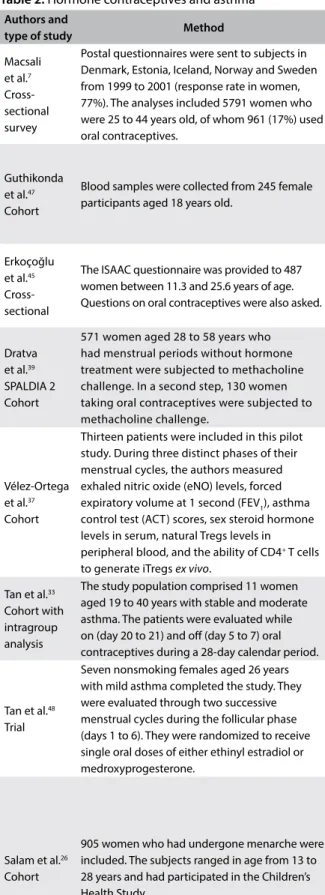
Macsali et al.7 Cross-sectional survey
Postal questionnaires were sent to subjects in Denmark, Estonia, Iceland, Norway and Sweden from 1999 to 2001 (response rate in women, 77%). The analyses included 5791 women who were 25 to 44 years old, of whom 961 (17%) used oral contraceptives.
Oral contraceptive pills were associated with an increased risk of asthma, asthma with hay fever, wheezing and shortness of breath, hay fever and ≥ 3 asthma symptoms. Associations were present. Women using oral contraceptive pills had more asthma. This was found only in the normal weight and overweight women and not in lean women, thus indicating an interplay between sex hormones and metabolic status in their efects on airways.
Guthikonda et al.47 Cohort
Blood samples were collected from 245 female participants aged 18 years old.
Subjects with genotype AG showed an increase in the average risk ratio (RR) from 0.31 (95% CI: 0.10 to 0.8) to 11.65 (95% CI: 1.71 to 79.5) when the methylation level increased from 0.02 to 0.12 relative to the risk in genotype AA. A two-stage model that takes into account genetic variants of the GATA-3 gene, oral contraceptive use, age at menarche and DNA-methylation may explain how sex hormones can increase the prevalence of asthma after puberty.
Erkoçoğlu et al.45 Cross-sectional
The ISAAC questionnaire was provided to 487 women between 11.3 and 25.6 years of age. Questions on oral contraceptives were also asked.
In this study, n = 487 (ages ranged from 11.3 to 25.6 years old), 196 (40%) reported using an oral contraceptive, 7.4% had a diagnosis of asthma from a physician and 10.3% of them were active smokers. Young women taking oral contraceptives had a higher rate of current wheezing, thus suggesting that sex steroids may be important for respiratory health.
Dratva et al.39 SPALDIA 2 Cohort
571 women aged 28 to 58 years who had menstrual periods without hormone treatment were subjected to methacholine challenge. In a second step, 130 women taking oral contraceptives were subjected to methacholine challenge.
An efect of modiication according to asthma status and oral contraceptive use was found, with a lower odds ratio (OR) among subjects without asthma. An OR < 1 was found among woman taking oral contraceptives. Oral contraceptives appeared to have a protective efect through which they decreased bronchial hyperreactivity.
Vélez-Ortega et al.37 Cohort
Thirteen patients were included in this pilot study. During three distinct phases of their menstrual cycles, the authors measured exhaled nitric oxide (eNO) levels, forced expiratory volume at 1 second (FEV1), asthma control test (ACT) scores, sex steroid hormone levels in serum, natural Tregs levels in peripheral blood, and the ability of CD4+ T cells to generate iTregs ex vivo.
Patients taking oral contraceptives showed reduced serum sex hormone levels in association with higher levels of iTreg induction, better ACT scores and a tendency to have lower eNO levels. The impact of sex hormones on the capacity of T cells to polarize towards a regulatory phenotype suggests that regulation of peripheral T cell lineage plasticity is a potential mechanism that may underlie the beneicial efects of oral contraceptives among women with asthma.
Tan et al.33 Cohort with intragroup analysis
The study population comprised 11 women aged 19 to 40 years with stable and moderate asthma. The patients were evaluated while on (day 20 to 21) and of (day 5 to 7) oral contraceptives during a 28-day calendar period.
Baseline FEV1did not difer between patients who were on and of oral
contraceptives. These did not alter beta2-adrenoreceptor regulation or function in stable female asthmatic patients.
Tan et al.48 Trial
Seven nonsmoking females aged 26 years with mild asthma completed the study. They were evaluated through two successive menstrual cycles during the follicular phase (days 1 to 6). They were randomized to receive single oral doses of either ethinyl estradiol or medroxyprogesterone.
The results showed that exogenous progesterone, but not estrogen, when given during the follicular phase, decreased beta2- adrenoreceptor density and cyclic-adenosine monophosphate (AMP) responses in female asthmatics. The beta2-adrenoreceptor was abnormally regulated in female asthmatics, and this might be a potential mechanism through which premenstrual asthma could be triggered when progesterone levels are high.
Salam et al.26 Cohort
905 women who had undergone menarche were included. The subjects ranged in age from 13 to 28 years and had participated in the Children’s Health Study.
In women without asthma, oral contraceptive use was associated with higher risk of current wheezing. In contrast, oral contraceptive use was associated with reduced prevalence of current wheezing in women with asthma. These associations showed signiicant trends with duration of oral contraceptive use.
Age at menarche was associated with new-onset asthma after puberty. Compared with women who had their menarche after they were 12 years old, women who reached their menarche before they were 12 years old were at higher risk of asthma after puberty. Because women have a higher risk of asthma after puberty, and because oral contraceptive use is common among young women, clinicians should inform women with asthma about the potential efects of oral contraceptives on asthma-related respiratory symptoms.
Table 2. Hormone contraceptives and asthma
Authors and
type of study Method Results and conclusions
Jenkins et al.46 Cohort
681 women aged 29-32 years were randomly sampled from participants who were irst surveyed at the age of 7 years in the 1968 Tasmanian Asthma Survey, which was a study of all children born in 1961 who attended school. Current asthma was deined as reporting asthma or wheezy breathing during the past 12 months.
The risk of current asthma in individuals who were parous increased with the number of births, while women with one birth were at lower risk than nulliparous women. Independent of parity, the risk decreased by 7% per year of oral
contraceptive pill use. In women who had asthma or wheezy breathing by the age of 7 years old, neither reproductive history nor oral contraceptive pill use predicted current asthma.
Parity and decreased oral contraceptive use predicted asthma in women, and these results are consistent with the hypothesis that the asthma that develops after childhood is in part a response to endogenous and exogenous female hormones. Nwaru and
Sheikh38 Cross-sectional survey
A population-based analysis using serial data from the Scottish general population. A total of 3257 non- pregnant, 16-45-year-old women were included.
The use of any hormonal contraceptive was associated with a reduced risk of current physician-diagnosed asthma.
The use of a hormonal contraceptive may reduce asthma exacerbations. Overweight and obese non-contraceptive-using women may be at increasing risk of asthma.
Lange et al.42 Cross-sectional
Data from a study on women who were selected from the general population were used to correlate the efect of treatment with oral contraceptives and hormonal replacement therapy (HRT) with asthma indications. 377 women were on oral contraceptives (24.5% of the premenopausal women) and 458 were on HRT (15.2% of the postmenopausal women). The age span of the premenopausal women was 21-49 years and of the postmenopausal women, 27-90 years.
A weak association was observed between HRT and self-reported asthma. No relationship was found between the use of oral contraceptives and asthma, although an association was observed between asthma and HRT.
Table 2. Continues...
time of the menopause was only significant among women who reported using estrogen alone, especially among those who had never been smokers and those who had had an allergic disease before the onset of asthma. A small increase in the risk of asthma among women who used estrogen/pro-gestogen was found in these subgroups.52 In a systematic review and meta-analysis, Zemp et al. found that there was no significant association between menopause with asthma prevalence or incidence except for women who reported using HRT.53
In a study by Carlson et al., HRT was associated with bet-ter lung function and an increase in forced expiratory volume at one second (FEV1).41 he mechanisms that link asthma and the menopause are unclear. Ater the menopause, FSH and LH levels are elevated, and estrogen levels decrease to the levels observed in patients with surgical oophorectomy, who also show extremely low progesterone levels. he incidence of asthma may be associ-ated with decreased estrogen levels and a protective efect against the relative androgen excess that occurs during the menopausal transition.53,54 Clinical studies have indicated that the menopause is associated with exacerbation of pre-existing asthma. hus, the onset of asthma is characterized by absence of atopy, absence of a family history and associations with urticaria and/or recurrent sinusitis of high severity.23 Balzano et al.55 showed that eosinophil
levels were higher in the induced sputum of menopausal asthmat-ics, but Foschino Barbaro et al. reported that there were high spu-tum levels of neutrophils and exhaled interleukin (IL)-6 in women with menopausal asthma.50
Few studies have explored the link between the menopause and asthma. Hormonal processes and other factors, including genetics and inlammatory and metabolic characteristics, need to be taken into consideration. Studies have indicated that obe-sity has an efect on the severity of asthma and that this relation-ship is modiied by gender. Estrogen and leptin levels (which have been correlated with increased airway inlammation in animal models)56 are higher in obese women than in non-obese women.54 Moreover, obesity has been shown to increase the risk of developing asthma. Interestingly, Gómez Real reported that lean women presented a higher risk of postmenopausal asthma than did obese women using HRT.57 his phenomenon can be explained by the notion that in lean women without insulin resis-tance, the pro-inlammatory efect of estrogens may predominate; while in obese women, the pro-inlammatory efects of estrogens are decreased through insulin resistance.53
Pregnancy and asthma
maternal and fetal adverse perinatal outcomes,58 such that 20%-30% of women with asthma experience exacerbations that require medi-cal intervention during pregnancy.43 here is also evidence of an increased risk of maternal mortality among some asthmatic women.59
A number of the physiological changes that occur during preg-nancy can afect asthma status, including mechanical, immuno-logical and hormonal alterations. Estradiol and progesterone lev-els are highest during pregnancy.60 Moreover, one third of women experience improved asthma, while another third of women retain the same asthma status and the remaining third experience worse asthma. Pregnancy is also marked by a state of h2 dominance, and asthma is generally characterized by h2 inlammation. Progesterone receptors are present in large quantities on the surface of lympho-cytes, and binding of progesterone to its receptor induces stimula-tion and release of progesterone-induced blocking factor (PIBF) in a h2 cytokine expression pattern (IL-4, IL-5, IL-6, IL-9, IL-10 and IL-13). he efects of these proteins are reduced in natural killer (NK) cells, in which expression of IFN-γ is decreased. NK cells are mainly observed in the endometrium of pregnant women.12,61 During the irst trimester of pregnancy, the numbers of circulating and decidual regulatory T cells (Tregs) increase to promote toler-ance at the maternal-fetal interface.62
Interestingly, fetal sex may inluence asthma. Kwon et al. exam-ined pregnant asthmatic women and found that carrying a female fetus was associated with worse maternal asthma than carrying a male fetus was.60 he mechanism that contributes towards this result is unclear, but there is evidence showing that testosterone potentiates the β-adrenergic-mediated relaxation of bronchial tis-sues and inhibits responses to histamine. Female sex is associated with higher maternal circulation of monocytes and upregulation of maternal inlammatory pathways.58
he mechanisms through which sex hormones inluence asthma and the immunological characteristics of pregnancy at the mater-nal-fetal interface remain obscure, and new studies are needed in order to increase our understanding of and ability to manage asthmatic women.
DISCUSSION
Studies examining the role of hormonal factors in asthma among women have been conducted on human subjects and animal models, and the results have been described in reviews. In an attempt to understand the inluence of sex hormones on pulmo-nary inlammatory responses, we discuss the main immunologi-cal aspects of sex hormones here.
Studies using animal models have demonstrated that both pro-gesterone and estrogen can directly afect the lungs.63-67 Sex steroid hormones inluence the immune system by acting on the struc-ture and function of the thymus, thereby modulating the activ-ity of B and T cells, mast cells and natural killer cells (NK cells),
and afecting phagocytic cells and cytokine production. hese hormones act via a variety of receptors (including the estrogen receptors ERα and ERβ; and the progesterone receptors PR-A and PR-B), and these steroid receptors have been described as nuclear receptors that act as transcription factors to regulate gene expres-sion.23 However, it has been shown that some steroid receptors are located at the plasma membrane (e.g. membrane-bound G-protein-coupled receptors).68,69 hese receptors are also expressed in the human lungs, such that sex hormones play a role in development of the lungs and androgen receptors are expressed in the mesen-chymal and epithelial cells of the lungs.
Gender diferences have been observed in relation to develop-ment of the lungs. For example, production of surfactants appears earlier in female than in male neonatal lungs, and male preterm infants are at higher risk of experiencing developmental distress syndrome. In addition, before puberty, the prevalence of asthma is higher among boys.43 Both male and female fetuses express androgen receptors (AR-A, AR-B) in non-reproductive tissues, with signiicantly higher numbers of AR-B than AR-A recep-tors expressed in the lungs. However, few studies have examined expression of androgens in inlammatory airways, and testosterone has been shown to cause relaxation of airway smooth muscles.70 Testosterone may increase apoptosis in T cells, thus resulting in a lower percentage of T lymphocytes in the total pool of lympho-cytes in males than in females.12
In allergic asthma, airway inlammation is mainly character-ized by h2-mediated processes, including secretion of the cyto-kines IL-4, IL-5, IL-6, IL-9 and IL-13, secretion of chemocyto-kines, regulation of the activation of normal T cells (RANTES), and production of granulocyte macrophage colony-stimulating factor (GM-CSF). In patients with asthma and in allergic animal models (e.g. allergen-challenged mice), bronchoalveolar lavage contains large numbers of eosinophils, M2-polarized macrophages and acti-vated mast cells. In several cases, the numbers of neutrophils in the bronchoalveolar lavage have been found to be higher as a result of h17-mediated responses and production of IL-8.68,69 he airway epithelium in asthmatic patients recruits innate and adaptive cells via cytokines, including IL-25 and IL-33, and chemokines such as CCL2, CCL17 and CCL20, and it secretes transforming growth factor beta (TGFβ), which is responsible for airway remodelling.69
Few studies have examined the effects of sex hormones on the bronchial epithelium. The human bronchial epithelium expresses both ERα and ERβ. In patients with asthma, estro-gens facilitate dissociation of endothelial nitric oxide synthetase, which results in activation of the NO pathway, vasodilatation and increased inflammation.72 In another study, treatment of bronchial epithelial cells with 10 nM estrogen induced expres-sion of NOS and production of nitric oxide, thus resulting in bronchodilation.69,73 In a study by Mandhane et al., among women who were not using oral contraceptives, an increase in progesterone level was associated with an increase in exhaled nitric oxide levels, thus indicating that an inflammatory pro-cess was associated with progesterone.74
Stimulation of h2-mediated inlammatory responses and asthma by progesterone has been considered by many studies to represent a typical h2 disorder.69,75 In a study by Loza et al., increased accumulation of IL-13+T cells (h2) was observed in female but not in male asthmatics, and this association was main-tained when the analysis was restricted to atopic subjects.75 In an animal model, ovariectomized or estradiol antagonist-treated mice developed reduced IL-5 dependent eosinophilia during aller-gic inlammation.76 However, depending on the concentration of estrogen, it may play dual pro and anti-inlammatory roles.64,77
CONCLUSIONS
We have attempted to discuss the characteristics that are afected by sexual hormones during pulmonary inlammatory responses. However, the associations between these factors remain obscure. We speculate that estrogen luctuations are responsible for asthma exacerbations that occur in women. Because of the anti-inlam-matory action of estrogen, as this hormone decreases TNF-α pro-duction, it reduces IFN-γ expression, and NK cell activity. We suggest that further studies that highlight the underlying phys-iopathological mechanisms contributing towards these interac-tions should be conducted.
REFERENCES
1. Global Strategy for Asthma Management and Prevention (GINA).
Available from: http://ginasthma.org/wp-content/uploads/2016/04/
GINA-2016-main-report_tracked.pdf. Accessed in 2016 (Aug 30).
2. National Institute of Allergy and Infectious Diseases. Asthma. Available
from: http://www.niaid.nih.gov/topics/asthma/Pages/default.aspx.
Accessed in 2016 (Aug 30).
3. IV Diretrizes Brasileiras para o Manejo da Asma [IV Brazilian Guidelines for
the management of asthma]. J Bras Pneumol.2006;32(supl 7):s447-s474.
4. Solé D, Yamada E, Vana AT, et al. International Study of Asthma and
Allergies in Childhood (ISAAC): prevalence of asthma and
asthma-related symptoms among Brazilian schoolchildren. J Investig Allergol
Clin Immunol.2001;11(2):123-8.
5. Lotufo PA, Bensenor IM. Temporal trends of asthma mortality rates in
Brazil from 1980 to 2010. J Asthma.2012;49(8):779-84.
6. Siroux V, Oryszczyn MP, Paty E, et al. Relationships of allergic sensitization,
total immunoglobulin E and blood eosinophils to asthma severity in
children of the EGEA Study. Clin Exp Allergy.2003;33(6):746-51.
7. Macsali F, Real FG, Omenaas ER, et al. Oral contraception, body mass
index, and asthma: a cross-sectional Nordic-Baltic population survey.
J Allergy Clin Immunol.2009;123(2):391-7.
8. Melgert BN, Ray A, Hylkema MN, Timens W, Postma DS. Are there
reasons why adult asthma is more common in females? Curr Allergy
Asthma Rep.2007;7(2):143-50.
9. Barr RG, Wentowski CC, Grodstein F, et al. Prospective study of
postmenopausal hormone use and newly diagnosed asthma
and chronic obstructive pulmonary disease. Arch Intern Med.
2004;164(4):379-86.
10. Troisi RJ, Speizer FE, Willett WC, Trichopoulos D, Rosner B. Menopause,
postmenopausal estrogen preparations, and the risk of adult-onset
asthma. A prospective cohort study. Am J Respir Crit Care Med.
1995;152(4 Pt 1):1183-8.
11. Matteis M, Polverino F, Spaziano G, et al. Efects of sex hormones
on bronchial reactivity during the menstrual cycle. BMC Pulm Med.
2014;14:108.
12. Bouman A, Heineman MJ, Faas MM. Sex hormones and the immune
response in humans. Hum Reprod Update.2005;11(4):411-23.
13. Varraso R, Siroux V, Maccario J, et al. Asthma severity is associated with
body mass index and early menarche in women. Am J Respir Crit Care
Med.2005;171(4):334-9.
14. Kynyk JA, Mastronarde JG, McCallister JW. Asthma, the sex diference.
Curr Opin Pulm Med.2011;17(1):6-11.
15. Wills-Karp M, Ewart SL. Time to draw breath: asthma-susceptibility
genes are identiied. Nat Rev Genet.2004;5(5):376-87.
16. Zhu J, Yamane H, Cote-Sierra J, Guo L, Paul WE. GATA-3 promotes Th2
responses through three diferent mechanisms: induction of Th2
cytokine production, selective growth of Th2 cells and inhibition of
Th1 cell-speciic factors. Cell Res.2006;16(1):3-10.
17. Höfer T, Nathansen H, Löhning M, Radbruch A, Heinrich R. GATA-3
transcriptional imprinting in Th2 lymphocytes: a mathematical model.
Proc Natl Acad Sci U S A.2002;99(14):9364-8.
18. Magnan AO, Mély LG, Camilla CA, et al. Assessment of the Th1/
Th2 paradigm in whole blood in atopy and asthma. Increased
IFN-gamma-producing CD8(+) T cells in asthma. Am J Respir Crit Care
Med.2000;161(6):1790-6.
19. Lowder TW, Kunz HE. Regulatory T Cells in Asthma and Airway
Hyperresponsiveness. Journal of Allergy & Therapy.2011;S1-002.
Available from:
http://www.omicsonline.org/regulatory-t-cells-in-asthma-and-airway-hyperresponsiveness-2155-6121.S1-002.pdf.
Accessed in 2016 (Aug 30).
20. Langier S, Sade K, Kivity S. Regulatory T cells in allergic asthma. Isr Med
21. Vock C, Hauber HP, Wegmann M. The other T helper cells in asthma
pathogenesis. J Allergy (Cairo).2010;2010:519298.
22. Cosmi L, Liotta F, Maggi E, Romagnani S, Annunziato F. Th17 cells: new
players in asthma pathogenesis. Allergy.2011;66(8):989-98.
23. Balzano G, Fuschillo S, Melillo G, Bonini S. Asthma and sex hormones.
Allergy.2001;56(1):13-20.
24. Karpel JP, Wait JL. Asthma in women, Part 3: Perimenstrual asthma,
efects of hormone therapy. Journal of Critical Illness.
2000;15(5):265-72. Available from: http://go.galegroup.com/ps/anonymous?id=GAL
E%7CA76609703&sid=googleScholar&v=2.1&it=r&linkaccess=fulltex
t&issn=10400257&p=AONE&sw=w&authCount=1&isAnonymousEn
try=true. Accessed in 2016 (Aug 30).
25. Scichilone N, Battaglia S, Braido F, et al. Exhaled nitric oxide is associated
with cyclic changes in sexual hormones. Pulm Pharmacol Ther.
2013;26(6):644-8.
26. Salam MT, Wenten M, Gilliland FD. Endogenous and exogenous sex
steroid hormones and asthma and wheeze in young women. J Allergy
Clin Immunol.2006;117(5):1001-7.
27. Haggerty CL, Ness RB, Kelsey S, Waterer GW. The impact of estrogen and
progesterone on asthma. Ann Allergy Asthma Immunol.
2003;90(3):284-91; quiz 291-3, 347.
28. Macsali F, Svanes C, Sothern RB, et al. Menstrual cycle and respiratory
symptoms in a general Nordic-Baltic population. Am J Respir Crit Care
Med.2013;187(4):366-73.
29. Tan KS. Premenstrual asthma: epidemiology, pathogenesis and
treatment. Drugs.2001;61(14):2079-86.
30. Pereira-Vega A, Sánchez Ramos JL, Vázquez Oliva R, et al. Premenstrual
asthma and female sex hormones. J Investig Allergol Clin Immunol.
2012;22(6):437-9.
31. Vrieze A, Postma DS, Kerstjens HA. Perimenstrual asthma: a syndrome
without known cause or cure. J Allergy Clin Immunol.2003;112(2):271-82.
32. Redmond AM, James AW, Nolan SH, Self TH. Premenstrual asthma:
emphasis on drug therapy options. J Asthma.2004;41(7):687-93.
33. Tan KS, McFarlane LC, Lipworth BJ. Beta2-adrenoceptor regulation and
function in female asthmatic patients receiving the oral combined
contraceptive pill. Chest.1998;113(2):278-82.
34. Skobelof EM, Spivey WH, Silverman R, et al. The efect of the menstrual
cycle on asthma presentations in the emergency department. Arch
Intern Med.1996;156(16):1837-40.
35. Murphy VE, Gibson PG. Premenstrual asthma: prevalence, cycle-to-cycle
variability and relationship to oral contraceptive use and menstrual
symptoms. J Asthma.2008;45(8):696-704.
36. Real FG, Svanes C, Macsali F, Omenaas ER. Hormonal factors and respiratory
health in women--a review. Clin Respir J.2008;2 Suppl 1:111-9.
37. Vélez-Ortega AC, Temprano J, Reneer MC, et al. Enhanced generation
of suppressor T cells in patients with asthma taking oral contraceptives.
J Asthma.2013;50(3):223-30.
38. Nwaru BI, Sheikh A. Hormonal contraceptives and asthma in women of
reproductive age: analysis of data from serial national Scottish Health
Surveys. J R Soc Med.2015;108(9):358-71.
39. Dratva J, Schindler C, Curjuric I, et al. Perimenstrual increase in bronchial
hyperreactivity in premenopausal women: results from the
population-based SAPALDIA 2 cohort. J Allergy Clin Immunol.2010;125(4):823-9.
40. Caracta CF. Gender diferences in pulmonary disease. Mt Sinai J Med.
2003;70(4):215-24.
41. Carlson CL, Cushman M, Enright PL, et al. Hormone replacement therapy
is associated with higher FEV1 in elderly women. Am J Respir Crit Care
Med.2001;163(2):423-8.
42. Lange P, Parner J, Prescott E, Ulrik CS, Vestbo J. Exogenous female sex
steroid hormones and risk of asthma and asthma-like symptoms: a cross
sectional study of the general population. Thorax.2001;56(8):613-6.
43. Carey MA, Card JW, Voltz JW, et al. It’s all about sex: gender,
lung development and lung disease. Trends Endocrinol Metab.
2007;18(8):308-13.
44. Weiss ST. Obesity: insight into the origins of asthma. Nat Immunol.
2005;6(6):537-9.
45. Erkoçoğlu M, Kaya A, Azkur D, et al. The efect of oral contraceptives on
current wheezing in young women. Allergol Immunopathol (Madr).
2013;41(3):169-75.
46. Jenkins MA, Dharmage SC, Flander LB, et al. Parity and decreased use
of oral contraceptives as predictors of asthma in young women. Clin
Exp Allergy.2006;36(5):609-13.
47. Guthikonda K, Zhang H, Nolan VG, et al. Oral contraceptives
modify the effect of GATA3 polymorphisms on the risk of asthma
at the age of 18 years via DNA methylation. Clin Epigenetics.
2014;6(1):17.
48. Tan KS, McFarlane LC, Lipworth BJ. Paradoxical down-regulation and
desensitization of beta2-adrenoceptors by exogenous progesterone
in female asthmatics. Chest.1997;111(4):847-51.
49. Tan KS, McFarlane LC, Lipworth BJ. Loss of normal cyclical beta 2
adrenoceptor regulation and increased premenstrual responsiveness
to adenosine monophosphate in stable female asthmatic patients.
Thorax.1997;52(7):608-11.
50. Foschino Barbaro MP, Costa VR, Resta O, et al. Menopausal asthma: a
new biological phenotype? Allergy.2010;65(10):1306-12.
51. Macsali F, Svanes C, Bjørge L, Omenaas ER, Gómez Real F. Respiratory
health in women: from menarche to menopause. Expert Rev Respir
Med.2012;6(2):187-200; quiz 201-2.
52. Romieu I, Fabre A, Fournier A, et al. Postmenopausal hormone therapy
and asthma onset in the E3N cohort. Thorax. 2010;65(4):292-7.
53. Zemp E, Schikowski T, Dratva J, Schindler C, Probst-Hensch N. Asthma
and the menopause: a systematic review and meta-analysis. Maturitas.
2012;73(3):212-7.
54. Zein JG, Erzurum SC. Asthma is Diferent in Women. Curr Allergy Asthma
Rep.2015;15(6):28.
55. Balzano G, Fuschillo S, De Angelis E, et al. Persistent airway inlammation
and high exacerbation rate in asthma that starts at menopause. Monaldi
Arch Chest Dis.2007;67(3):135-41.
56. Shore SA, Fredberg JJ. Obesity, smooth muscle, and airway
57. Gómez Real F, Svanes C, Björnsson EH, et al. Hormone replacement
therapy, body mass index and asthma in perimenopausal women: a
cross sectional survey. Thorax.2006;61(1):34-40.
58. Bakhireva LN, Schatz M, Jones KL, et al. Fetal sex and maternal asthma
control in pregnancy. J Asthma.2008;45(5):403-7.
59. Tan KS, Thomson NC. Asthma in pregnancy. Am J Med.
2000;109(9):727-33.
60. Kwon HL, Belanger K, Holford TR, Bracken MB. Efect of fetal sex on
airway lability in pregnant women with asthma. Am J Epidemiol.
2006;163(3):217-21.
61. Broide DH. Molecular and cellular mechanisms of allergic disease. J
Allergy Clin Immunol.2001;108(2 Suppl):S65-71.
62. Oertelt-Prigione S. Immunology and the menstrual cycle. Autoimmun
Rev.2012;11(6-7):A486-92.
63. Hellings PW, Vandekerckhove P, Claeys R, et al. Progesterone increases
airway eosinophilia and hyper-responsiveness in a murine model of
allergic asthma. Clin Exp Allergy.2003;33(10):1457-63.
64. Degano B, Mourlanette P, Valmary S, et al. Diferential efects of low
and high-dose estradiol on airway reactivity in ovariectomized rats.
Respir Physiol Neurobiol.2003;138(2-3):265-74.
65. de Oliveira AP, Domingos HV, Cavriani G, et al. Cellular recruitment and
cytokine generation in a rat model of allergic lung inlammation are
diferentially modulated by progesterone and estradiol. Am J Physiol
Cell Physiol.2007;293(3):C1120-8.
66. Mitchell VL, Van Winkle LS, Gershwin LJ. Environmental tobacco smoke
and progesterone alter lung inlammation and mucous metaplasia in
a mouse model of allergic airway disease. Clin Rev Allergy Immunol.
2012;43(1-2):57-68.
67. Matsubara S, Swasey CH, Loader JE, et al. Estrogen determines sex
diferences in airway responsiveness after allergen exposure. Am J
Respir Cell Mol Biol.2008;38(5):501-8.
68. Zierau O, Zenclussen AC, Jensen F. Role of female sex hormones, estradiol
and progesterone, in mast cell behavior. Front Immunol.2012;3:169.
69. Keselman A, Heller N. Estrogen Signaling Modulates Allergic
Inlammation and Contributes to Sex Diferences in Asthma. Front
Immunol.2015;6:568.
70. Chang HY, Mitzner W. Sex diferences in mouse models of asthma. Can
J Physiol Pharmacol.2007;85(12):1226-35.
71. Ito A, Bebo BF, Jr., Matejuk A, et al. Estrogen treatment down-regulates
TNF-alpha production and reduces the severity of experimental
autoimmune encephalomyelitis in cytokine knockout mice. J Immunol.
2001;167(1):542-52.
72. Sathish V, Martin YN, Prakash YS. Sex steroid signaling: implications for
lung diseases. Pharmacol Ther.2015;150:94-108.
73. Townsend EA, Meuchel LW, Thompson MA, Pabelick CM, Prakash
YS. Estrogen increases nitric-oxide production in human bronchial
epithelium. J Pharmacol Exp Ther.2011;339(3):815-24.
74. Mandhane PJ, Hanna SE, Inman MD, et al. Changes in exhaled nitric
oxide related to estrogen and progesterone during the menstrual
cycle. Chest.2009;136(5):1301-7.
75. Loza MJ, Foster S, Bleecker ER, Peters SP, Penn RB. Asthma and gender
impact accumulation of T cell subtypes. Respir Res.2010;11:103.
76. Rifo-Vasquez Y, Ligeiro de Oliveira AP, Page CP, Spina D,
Tavares-de-Lima W. Role of sex hormones in allergic inlammation in mice. Clin
Exp Allergy.2007;37(3):459-70.
77. Straub RH. The complex role of estrogens in inlammation. Endocr Rev.
2007;28(5):521-74.
Sources of funding: None
Conlict of interest: None
Date of irst submission: May 6, 2016
Last received: June 19, 2016
Accepted: June 27, 2016
Address for correspondence:
Raquel Prudente de Carvalho Baldaçara
Universidade Federal do Tocantins
Quadra 401 Sul, Avenida LO 11, Conjunto 2, Bloco 2
Edifício Palmas Medical Center, sala 504
Plano Diretor Sul — Palmas (TO) — Brasil
Tel. (+55 63) 3217-7288