Mammographic density among indigenous women in forested
areas in the state of Amapá, Brazil: a cross-sectional study
Densidade mamográica em mulheres indígenas de áreas da loresta do estado do
Amapá, Brasil: estudo transversal
José Mauro Secco
I, Simone Elias
II, Cristina Valletta de Carvalho
III, Ismael Dale Cotrim Guerreiro da Silva
IV,
Kátia Jung de Campos
V, Gil Facina
VI, Afonso Celso Pinto Nazário
VIIUniversidade Federal de São Paulo (Unifesp), São Paulo (SP), Brazil
ABSTRACT
CONTEXT AND OBJECTIVE: There is no register of breast cancer cases among indigenous populations in Brazil. The objective here was to evaluate the association of clinical and demographic characteristics with mammographic density among indigenous women.
DESIGN AND SETTING: Cross-sectional analytical study conducted in indigenous territories in the state of Amapá, Brazil.
METHODS: Women were recruited from three indigenous territories and underwent bilateral mammogra-phy and blood collection for hormonal analysis. They were interviewed with the aid of an interpreter. Mam-mographic density was calculated using computer assistance, and was expressed as dense or non-dense.
RESULTS: A total of 137 indigenous women were included in this study, with an average age of 50.4 years, and an average age at the menarche of 12.8 years. Half (50.3%) of the 137 participants had not reached the menopause at the time of this study. The women had had an average of 8.7 children, and only two had never breastfed. The average body mass index of the population as a whole was 25.1 kg/m2. The
mam-mographic evaluation showed that 82% of women had non-dense breasts. The clinical characteristics associated with mammographic density were age (P = 0.0001), follicle-stimulating hormone (FSH) (P < 0.001) and estrogen levels (P < 0.01).
CONCLUSIONS: The majority of the indigenous women had non-dense breasts. Age, menopausal status and FSH and estrogen levels were associated with mammographic density.
RESUMO
CONTEXTO E OBJETIVO: Não há registro de casos de câncer de mama em populações indígenas no
Brasil. O objetivo foi avaliar a associação de características clínicas e demográicas com a densidade ma-mográica em mulheres indígenas.
TIPO DE ESTUDO E LOCAL: Estudo transversal, analítico, realizado em territórios indígenas no estado do Amapá, Brasil.
MÉTODOS: Mulheres foram recrutadas de três territórios indígenas e submetidas a mamograia bilateral e a coleta de sangue para análise hormonal. As participantes foram entrevistadas com a ajuda de um in-térprete. A densidade mamográica foi calculada com assistência de computador, e expressa como densa ou não densa.
RESULTADOS: 137 mulheres foram incluídas no estudo, com média de 50,4 anos e média de idade à me-narca de 12,8 anos. Metade (50,3%) das 137 participantes não havia entrado na menopausa no momento do estudo. As mulheres tinham em média 8,7 ilhos, e duas nunca haviam amamentado. O índice de massa corpórea médio da população como um todo foi de 25,1 kg/m2. A análise mamográica mostrou
que 82% das mulheres tinham mamas não densas. As características clínicas associadas com a densidade mamográica foram idade (P = 0.0001), hormônio folículo-estimulante (FSH, P < 0,001) e níveis de estro-gênio (P < 0,01).
CONCLUSÃO: A maioria das indígenas tinha mamas não densas. Idade, status menopausal e níveis de estrógeno e FSH foram associados com a densidade mamográica.
IMD, PhD. Researcher, Universidade Federal de São Paulo (Unifesp), São Paulo (SP), and Adjunct Professor, Universidade Federal do Amapá (Unifap), Amapá (AP), Brazil.
IIMD, PhD. Researcher, Universidade Federal de São Paulo (Unifesp), São Paulo (SP), Brazil. IIIBSc, PhD. Researcher, Universidade Federal de São Paulo (Unifesp), and Adjunct Professor, Department of Biological Sciences, Centro Universitário Fundação Santo André, and Department of Genetics, Fundação ABC, São Paulo (SP), Brazil.
IVMD, PhD. Researcher, Universidade Federal de São Paulo (Unifesp); Adjunct Professor and Coordinator of Molecular Gynecology Laboratory, Department of Gynecology; and Coordinator of Research and Technological Innovation within Biology, Universidade Federal de São Paulo (Unifesp), São Paulo (SP), Brazil. VMD, PhD. Researcher, Universidade Federal de São Paulo (Unifesp), São Paulo (SP), and Attending Physician and Residency Coordinator,
Department of Gynecology, Universidade Federal do Amapá, Amapá (AP), Brazil.
VIMD, PhD. Full Professor, Department of Gynecology and Head of Department of Mastology, Universidade Federal de São Paulo (Unifesp), São Paulo (SP), Brazil.
VIIMD, PhD. Researcher and Full Professor, Universidade Federal de São Paulo (Unifesp), São Paulo (SP), Brazil.
KEY WORDS: Polymorphism, genetic. Receptors, estrogen. Receptors, progesterone. Health Services, indigenous. Population groups. Breast neoplasms.
INTRODUCTION
It is known that breast cancer is less prevalent among African-American women than among white women. However, the dis-ease onset is earlier among African-Americans, and these women
have more aggressive tumors.1,2 Women of indigenous origin
clearly present lower incidences of breast cancer than women
with no indigenous descent.3-6
he incidence of breast cancer in the indigenous population in Brazil is very low, and absolutely no cases have been found in some ethnic groups, such as the Xavantes group, living in the state of
Mato Grosso.7 his phenomenon can be explained by lifestyle
hab-its: some peculiarities of indigenous populations are indeed protec-tive factors, e.g. irst pregnancy early in life, multiparity, prolonged breastfeeding and absence of hormone therapy. he hypotheses for explaining the lower incidence of breast cancer among indigenous
women may include lower life expectancy and underreporting.7
However, indigenous populations also present some risk factors for breast cancer: sedentary lifestyle, overweight and obesity are
observed in more than half of these women.3
Breast density, as evaluated by mammography (mammographic density), is an independent risk factor for breast cancer, which is
independent of age, menopausal status or exogenous steroid use.8-12
To our knowledge, there is no other study in the literature address-ing the relationship between demographic and clinical factors and mammographic density among indigenous women in Brazil.
OBJECTIVE
his study aimed to evaluate clinical and demographic character-istics and their association with mammographic density among indigenous women.
METHODS
Study design, participants and ethics
In this cross-sectional analytical study, indigenous women in Oiapoque, in the Brazilian state of Amapá, were evaluated. hese women were recruited in three indigenous territories, Uaça, Galiby and Juminã, which are home to four indigenous ethnici-ties: Karipuna, Galibi Marworno, Palikúr and Galibi Kalina, dis-tributed in 38 villages. he total population of these indigenous territories in 2010 was 7,021, according to igures from the local technical coordination oice of the National Indian Foundation, in Oiapoque.
To evaluate these women, it was necessary to obtain autho-rizations from the indigenous leaders of Oiapoque, from the National Indian Foundation (FUNAI) and from the National Health Foundation (FUNASA). Permission from the indigenous leaders was obtained for this study ater we participated in the annual meeting of the Association of Indigenous Peoples of Oiapoque
(APIO), at which the research project was presented to the com-munity. An informed consent form was signed by all the women to be examined, ater the study objectives had been explained to them by a local interpreter, in the local Indian Community Center during the interview. Ethical approvals were obtained from the National Committee for Research Ethics (CONEP) and the local Ethics Committee of the university hospital.
he inclusion criteria were that the women needed to be 40 years of age or older, living in indigenous villages (and not in cities) and not using hormonal medications (including plant hormones) to treat menopausal symptoms during the 12 months preceding the interview. Women sufering from endocrine, liver or kidney disorders, as ascertained from the local medical records kept by FUNAI and FUNASA, were also excluded. Mammograms that were classiied as Breast Imaging Reporting and Data System (BI-RADs) category 3 or above also constituted an exclusion
cri-terion a priori.
From January to December 2009, it was possible to obtain authorizations and arrange transportation to examine 150 indig-enous women. All of them, except for nine with age disparities and two with BI-RADs category 3, underwent blood collection for follicle-stimulating hormone (FSH) and estradiol tests and underwent mammography.
he subjects were transported by bus to the nearest big city to undergo mammography exams. Because they live in villages far from each other, the women were transported by boat or car from their villages to the Indian House, in the municipality of Oiapoque, and from this place they were transported by bus, for about 450 km, to another Indian House, in the city of Macapá, where they were interviewed by the principal investigator and a nurse technician. Ater the women had signed the informed consent form, blood samples were collected from them to assay for FSH and estradiol (E2). About 5 ml of peripheral blood were obtained from each subject, using a vacuum extraction tube con-taining the anticoagulant EDTA (ethylenediaminetetraacetic acid). For mammography, the women were then transported to Hospital São Camilo in Macapá.
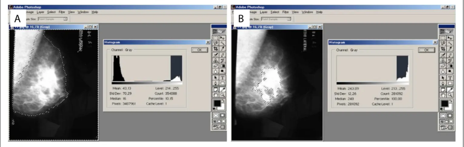
Figure 1. (A) Use of the “Lasso Tool” in Adobe Photoshop software, on a mammogram scan, for selection of the area to be calculated; and (B) “Magic Tool” for selection of dense tissue.
Mammographic density evaluation
Firstly, two independent examiners evaluated the scans to deter-mine the mammographic density. hey worked subjectively, based on the mammographic patterns described in the ACR’s
BI-RADS 2003 manual.1 he ratings D1, D2, D3 and D4 were
grouped two-by-two to enable statistical analysis: D1 + D2 were considered to represent non-dense breasts and D3 + D4, dense breasts.
he mediolateral oblique incidence scans were then digitized (scanner CX312.T, Radiographic Digital Imaging, Compton, CA, USA). Following this, a third evaluator assessed mammographic den-sity by using computer sotware for image analysis. Mammographic density was calculated using the gray-scale histogram tool of the Adobe Photoshop CS3 version 10.0 sotware as follows. he kappa coeicient was used to calculate the concordance between the three observers, two-by-two. Because the concordance was high (r > 0.78; P < 0.001), the third observer (called “computer-assisted evaluation” henceforth) was used as reference from then on.
Mammographic density was objectively determined using imaging analysis computer sotware. By convention, the mid-lateral let oblique incidence was scanned and captured using a CX312.T scanner (Radiographic Digital Imaging, Compton, CA, USA).
Initially, using the “Lasso Tool” (Figure 1), the image of the
entire breast was selected, taking care to exclude the pectoral mus-cle. hrough this procedure, the sotware generated a numerical value corresponding to the number of pixels in the overall breast area. hen, using the “Magic Tool”, the densest area of the breast, corresponding to ibroglandular tissue, was selected to obtain the
number of pixels of this area. Finally, the Equation 1 was applied,
to determine the percentage of ibroglandular tissue:
Mammographic density (MD) = dense area (DA)× 100
total area (1)
he values obtained for the dense area were compared with the subjective values registered by the other two evaluators, and the con-cordance between them was calculated using the kappa coeicient.
Statistical analysis
he concordance between the three observers was calculated by means of the kappa coeicient, two-by-two. Because the con-cordance was high (kappa > 0.75), the evaluations of the third observer (henceforth referred to as “computer-assisted evalua-tion”) were used as a reference. To decrease the number of classes within the computer-assisted evaluation variable, mammogram patterns were added as follows: D1 + D2 with the group of non-dense breasts; and D3 + D4 with the group of non-dense breasts.
he chi-square test was used for comparing qualitative vari-ables (frequency and proportions). To compare quantitative data (mammographic density), the Mann-Whitney U test was used where necessary. When the expected frequency was less than 5, we used Fisher’s exact test. To calculate odds ratios and conidence intervals, binary logistic regression was performed. Statistical sig-niicance was set at 5% or P < 0.05. he statistical sotware SPSS (Statistical Package for the Social Sciences), version 14.0, was used.
RESULTS
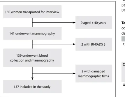
At the beginning of the study period, 150 women were recruited in the indigenous villages and were transported to undergo mam-mography. However, it was found that 9 of them were actually younger than 40 years, which prevented their inclusion in the study. Mammography was performed on the remaining 141 women, but 2 of them were classiied as BI-RADS 3, which probably indicated benign lesions, for which follow-ups six months aterwards were suggested. Nevertheless, because this inding also constituted an exclusion criteria of the study, these 2 women were excluded. hus, a total of 139 indigenous women whose mammograms
showed no signs of breast lesions were initially included in the study. However, in two cases, the mammogram ilm was damaged by moisture before it could be digitized for the analysis on
mam-mographic density (Figure 2). he average age of the remaining
137 women initially included was 50.4 years, with the menarche at an average of 12.8 years (the earliest was at 10 years of age). About half (50.3%) of the women were premenopausal.
Only 10 of the women (0.7%) had a history of using hor-monal contraceptives. Only one of them did not have any children. he total number of ofspring of the 138 women who had chil-dren was 1,209, an average of 8.7 per mother; but more than half of them (57.2%) had 9 or more children, and one of them had 19. he average age at which they had their irst child was 15.4 years, and this ranged from 12 years of age (three women) to 22 years of age. All of the women except for ive had had children before reaching 18 years of age. Half of them (50%) had had children before the age of 15.
Only two women (1.4%) had never breastfed: one without children and the other with six children. Ninety-eight women (70.5%) had breastfed for a minimum of 10 years. In relation to body mass index (BMI), 66 (47.4%) had a BMI indicating that they
were overweight (≥ 25 kg/m2). he average BMI of the population
as a whole was 25.1 kg/m2. None of the women interviewed had
any history of alcoholism (deined as the equivalent of two doses of distilled spirits per day); 44 of them reported occasional alco-hol use (31.6%), which was only at annual community festivals.
Most (68%) of the indigenous women were from two villages: Kumarumã (the Galibi Marworno people) and Espírito Santo (the Karipuna people). However, neither the subjects’ tribe nor their village showed any signiicant association with any of the
other variables of this study. Alcoholism (characterized as the equivalent of two servings of distilled liquor per day) was non-existent in the sample (only occasional consumption of alcohol could be veriied), which prevented calculation of any association with mammographic density. Nor did the distribution of origin (town or village) show any association with the main variables of the study. he Shapiro-Wilk test revealed that the sample data did not have normal distribution regarding the sociodemographic vari-ables, so the Mann-Whitney test was used for the mammographic density association tests (as described below).
Mammographic density
Among the 137 women for whom evaluation of mammographic density was possible, the distribution of the evaluations was classiied as D1, D2, D3 and D4 (D = mammographic density).
Table 1 shows the number of evaluations made by each observer
and the inal average. As shown in Table 2, the three observations
150 women transported for interview
141 underwent mammography
137 included in the study
9 aged < 40 years
2 with BI-RADS 3
2 with damaged mammographic ilms 139 underwent blood
collection and mammography
Figure 2. Flowchart of patients’ recruitment and inclusion in the study.
Mammographic density
Classiication Observer 1 Observer 2
Computer-assisted Average
D1 44 (32.1%) 43 (31.3%) 51 (37.2%) 46 (33.5%)
D2 63 (45.9%) 65 (47.44) 60 (43.7%) 63 (45.9%)
D3 28 (20.4%) 27 (19.7%) 24 (17.5%) 26 (18.9%)
D4 2 (1.45%) 2 (1.45%) 1 (0.72%) 2 (1.45%)
Total 137 137 137 137 Breast density patterns
Density Observer 1 Observer 2 Computer-assisted
Non-dense 107 (78.1%) 108 (78.8%) 112 (81.7%)
Dense 30 (21.8%) 29 (21.16%) 25 (18.2%)
Total 137 137 137
Table 1. Distribution of mammographic density according to the observers
D1 to D4 refer to breast density classiication; the least dense breast is D1 and most dense is D4.
Coeicient P-value Computer-assisted versus observer 1
Pearson’s R 0.892 < 0.001
Spearman’s correlation 0.892 < 0.001
Kappa 0.886 < 0.001
Computer-assisted versus observer 2
Pearson’s R 0.912 < 0.001
Spearman’s correlation 0.912 < 0.001
Kappa 0.908 < 0.001
Observer 1 versus observer 2
Pearson’s R 0.979 < 0.001
Spearman’s correlation 0.979 < 0.001
Kappa 0.978 < 0.001
(observers 1, 2 and the computer-assisted) were highly correlated (coeicient r > 0.8, with P < 0.001).
he D1, D2, D3 and D4 classiications were gathered into two groups to enable statistical analysis: thus, D1 + D2 were considered to be non-dense breasts and D3 + D4 to be dense
breasts. Hence, as shown in Table 1, the majority of the cases
were of non-dense breasts. he inter-observer concordance was also high when the classiications were grouped (D1 + D2) and
(D3 + D4) (Table 3).
Mammographic density and association with other variables
Considering the high agreement (kappa) between the observers, we started to investigate associations with other variables, using the computer-assisted evaluation as a standard. Mammographic density was not signiicantly associated with any history of hor-monal contraceptive use (P = 0.08) or with alcohol consumption (P = 1.00). Several other variables were also not associated with
mammographic density (Table 4).
Two variables were signiicantly associated with mammographic density: serum levels of follicle-stimulating hormone (FSH) and estradiol (E2). Higher levels of FSH were associated with non-dense breasts (P = 0.001) and higher levels of E2, to non-dense breasts (P = 0.01). he participants’ average age was signiicantly
associ-ated with mammographic density (Table 4).
here were 69 postmenopausal women and 68 were premeno-pausal. Most of the postmenopausal women had non-dense breasts (67; or 97%). Dense breasts were seen in two women ater the menopause (3%) and 23 before (34%). he mammographic density
was associated with the menopausal status (Table 5; P < 0.0001).
DISCUSSION
here is a high incidence of certain types of malignant tumors, such as colon, uterine and gastric cancers, in the Amazon region. However, absence of breast cancer among the indig-enous women of several Brazilian states, such as Mato Grosso,
Mato Grosso do Sul and Paraná, has been reported in the
lit-erature.3,6,7 he National Health Foundation (FUNASA) keeps
up-to-date records of the diseases that occur in the indigenous population, which undermines the hypothesis that underreport-ing is responsible for non-registration of cases of breast cancer
among indigenous women in Brazil.13 In the international
lit-erature, lower incidence of breast cancer has been reported in indigenous than in non-indigenous populations in the United
States14,15 and extremely low incidence was reported among
indigenous women in Ecuador.5 Breast cancer is more prevalent
among women living in large cities.2 Because dense breasts are
an independent risk factor for breast cancer (and this risk
per-sists for 10 years or more16), lower mammographic density might
be a protective factor against breast cancer in indigenous
pop-ulations.17,18 hat hypothesis inspired the present investigation,
which was undertaken among women living in indigenous vil-lages in the Amazon forest, far from urban areas. Because of this, these women were probably not subject to the lifestyle and dietary modiications seen in urban regions.
Computer-assisted (3)
Hormonal contraceptive
Total P no yes
D1 + D2 N (%) 105 (94.6%) 6 (5.4%) 111 (100%)
0.08
D3 + D4 N (%) 20 (83.3%) 4 (16.7%) 24 (100.0%)
Total N (%) 125 (92.6%) 10 (7.4%) 135 (100.0%)
Computer-assisted (3)
Alcohol
Total P no yes
D1 + D2 N (%) 75 (67.6%) 36 (32.4%) 111 (100.0%)
1.00
D3 + D4 N (%) 16 (66.7%) 8 (33.3%) 24 (100.0%)
Total N (%) 91 (67.4%) 44 (32.6%) 135 (100.0%)
Table 3. Descriptive statistics on the frequency of hormonal contraceptive and alcohol use, and comparison with mammographic density using Fisher’s exact test
SD = standard deviation; BMI = body mass index; FSH = follicle-stimulating hormone; E2 = estradiol. *Mann-Whitney (z) test for non-parametrical variables.
N Mean SD P*
Age (years) ND 112 51.95 10.19 < 0.001
D 25 44.36 5.19
BMI (kg/m2) ND 112 24.83 4.61 0.98
D 25 26.21 9.98
Age at menarche (years) ND 112 12.9 1.07 0.52
D 25 12.72 1.34
Parity (N) ND 112 8.82 3.15 0.27
D 25 7.72 4.13
FSH (mIU/ml) ND 112 34.83 26.94 0.001
D 25 14.77 21.14
E2 (pg/ml) ND 112 74.48 112.23 0.01
D 25 83.12 83.06
Age at irst term pregnancy (years) ND 110 15.38 1.38 0.43
D 25 15.84 1.68
Duration of breastfeeding (months)
ND 112 13.75 7.26
0.34
D 25 11.86 7.28
Table 4. Clinical and hormonal characteristics of the indigenous women with non-dense (ND) and dense (D) breasts
Menopause Dense Non-dense Total P
Yes 2 67 69
3% 97% 100%
No 23 45 69
34% 66% 100%
Total 25 112 137 < 0.0001 18% 82% 100%
Several studies have shown lower mammographic density in women of indigenous ethnicity. his may have been due not only to their reproductive pattern of bearing many children, with pro-longed periods of breastfeeding, but also perhaps to their indig-enous ethnicity itself. he indigindig-enous women of New Mexico, United States, have early liposubstitution of the breasts, compared
with Hispanic and non-Hispanic white women.19,20 Roubidoux
et al. also observed lower mammographic density among
indig-enous women in the southwestern United States.15 hese authors
also made comparisons between diferent ethnic groups in Alaska and observed that indigenous and Aleut women had less dense
breasts than Eskimos.21 In all of these studies, lower breast
den-sity was associated with lower incidence of breast cancer. In the present study, we conirmed this inding: the majority of the cases were indeed of non-dense breasts: the mammographic density in our study was well below what has been reported in the literature
for non-indigenous populations.22-24
he importance of accurate determination of mammographic
density was emphasized by Boyd et al.,25 who observed a 2% increase
in the relative risk of breast cancer for each 1% increase in the per-centage of mammographic density. We sought to conduct an objec-tive evaluation in the present study by scanning and capturing one of the mammographic views and determining the percentage area of ibroglandular tissue, and consequently, the mammographic density. he lower subjectivity was due to the calculation method, but the deinition of the area to be calculated remained subjective, since it was demarcated using the computer mouse. In our study, when we grouped D1 + D2 as non-dense breasts and D3 + D4 as dense breasts, we had a high degree of inter-observer concordance, of 0.97 (< 0.0001). his level of concordance also resulted from the care with which the images were obtained, using the same techni-cal process in all cases.
In this study, the association between age and mammographic
density was statistically signiicant (P ≤ 0.001). It has been
dem-onstrated in the literature that breast tissue becomes replaced by fat with advancing age. Nonetheless, the mammographic density in our study was much lower than that of non-indigenous
popu-lations.15,25-27 In our series, the frequency of non-dense breasts was
82%; and among the 69 postmenopausal women, only two (3%) had dense breasts, with an average age of 50.4 years. In non-indigenous populations in Brazil, three studies have shown that approximately 30% of women aged 50 years and over who were not using hormonal
contraceptives had dense breasts.22-24 In another Brazilian study,
in which the participants had a mean age of 54 years, the frequency
of observation of dense breasts was 45%.18 In Sweden, Bergkvist et
al.28 observed that 65% of the women aged 35 years had high
mam-mographic density and 15-20% of those aged 55 years.
Mammographic density is inluenced by several factors, such as
reproductive history, BMI, hormonal patterns and genetic factors.29
In the present study, only age, menopausal status and FSH and estra-diol levels were associated with mammographic density. Indigenous women with dense breasts had lower FSH levels and higher estra-diol levels than those with non-dense breasts. he estraestra-diol levels in postmenopausal women were equivalent to those observed in
other studies,29,30 which concluded that in postmenopausal women,
mammographic density was inversely related to estradiol levels. First childbirth before the age of 24 years, having more than two children and breastfeeding for more than two years are con-sidered by many authors to be protective factors against breast
cancer.3,21,27 Reproductive behavior is quite diferent in
indige-nous populations, and this probably contributes towards creating a protective efect against breast cancer. his, together with other factors, might explain the low incidence of this disease among these women. Both in our study and in others, the number of children per indigenous woman was high, and the irst delivery happened at an early age, thus resulting in many years of
breast-feeding.3,21,27,30 Parity is inversely associated with mammographic
density,31 and thus represents a protective factor against breast
cancer.32 It seems that the state of involution depends, in part, on
parity: ater successive pregnancies, stem cells and/or progenitor cells would accumulate in the mammary glands, and this has been observed in multiparous female mice. his is a valid hypothesis that
would also explain the relationship between density and parity.33
Lactation has consistently been inversely correlated with the risk of breast cancer: the risk decreases by 4.3% for every 12 months
of breastfeeding.34 Currently, the relationship between
breastfeed-ing and mammographic density is a matter of controversy, given
that both positive and inverse associations have been found.32,35-37
he women in our study nursed for a long period (mean of 12.9 years), and their breasts were predominantly non-dense, but despite this, breastfeeding was not signiicantly associated with mammo-graphic density.
information was considered to be quite reliable. Hence, it is unlikely that these women were sufering from diseases that could interfere with the results from the present study.
CONCLUSIONS
In this population of indigenous women in the municipality of Oiapoque, in the state of Amapá, Brazil, there were no cases of breast cancer and mammographic density was predominantly low. Age, menopausal status and FSH and estrogen levels were associated with mammographic density.
REFERENCES
1. American College of Radiology. Breast imaging reporting and data system (BI-RADS®). 4th ed. Reston: American College of Radiology; 2003.
2. Parkin DM, Sitas F, Chirenje M, et al. Part I: Cancer in Indigenous Africans--burden, distribution, and trends. Lancet Oncol. 2008;9(7):683-92. 3. Lima MG, Koifman S, Scapulatempo IL, et al. Fatores de risco para câncer de mama em mulheres indígenas Teréna de área rural, Estado do Mato Grosso do Sul, Brasil [Risk factors for breast cancer among rural Teréna Indian women in the State of Mato Grosso do Sul, Brazil]. Cad Saúde Pública. 2001;17(6):1537-44.
4. Condon JR, Armstrong BK, Barnes A, Cunningham J. Cancer in Indigenous Australians: a review. Cancer Causes Control. 2003;14(2):109-21. 5. San Sebastián M, Hurtig AK. Cancer among indigenous people in
the Amazon Basin of Ecuador, 1985-2000. Rev Panam Salúd Pública. 2004;16(5):328-33.
6. Silva EP, Pelloso SM, Carvalho MDB, Toledo MJO. Exploração de fatores de risco para câncer de mama em mulheres de etnia Kaingáng, Terra Indígena Faxinal, Paraná, Brasil, 2008 [Exploring breast cancer risk factors in Kaingáng women in the Faxinal Indigenous Territory, Parana State, Brazil, 2008]. Cad Saúde Pública. 2009;25(7):1493-500. 7. Castro GB, Fontes CJF, De-Lamonica-Freire EM, Hamada GS. The absence of
death by breast cancer among indigenous women living in Mato Grosso, Brazil 2000: protection or under notiication. Molecular Cancer Research Center. 2000;109-12. Available from http://www.academia.edu/5641651/ Letter_to_the_Editor_The_Absence_of_Death_by_Breast_Cancer_ Among_Indigenous_Women_Living_in_Mato_Grosso_Brazil_2000_ Protection_or_Under_Notiication. Accessed in 2017 (Apr 12). 8. Boyd NF, Lockwood GA, Martin LJ, et al. Mammographic densities and
breast cancer risk. Breast Dis. 1998;10(3-4):113-26.
9. Mandelson MT, Oestreicher N, Porter PL, et al. Breast density as a predictor of mammographic detection: comparison of interval- and screen-detected cancers. J Natl Cancer Inst. 2000;92(13):1081-7. 10. Wolfe JN. Breast patterns as an index of risk for developing breast
cancer. AJR Am J Roentgenol. 1976;126(6):1130-7.
11. Saftlas AF, Szklo M. Mammographic parenchymal patterns and breast cancer risk. Epidemiol Rev. 1987;9:146-74.
12. Saftlas AF, Hoover RN, Brinton LA, et al. Mammographic densities and risk of breast cancer. Cancer. 1991;67(11):2833-8.
13. FUNASA. Sistema de Informação da Saúde Indígena. Brasília: Ministério da Saúde; 2010. Available from: http:sis.funasa.gov.br/siasi. Accessed in 2012 (Sep 21).
14. Partin MR, Korn JE, Slater JS. Questionable data and preconceptions: reconsidering the value of mammography for American Indian Women. Am J Public Health. 1997;87(7):1100-2.
15. Roubidoux MA, Kaur JS, Griith KA, et al. Correlates of mammogram density in southwestern Native-American women. Cancer Epidemiol Biomarkers Prev. 2003;12(6):552-8.
16. Byrne C, Schairer C, Wolfe J, et al. Mammographic features and breast cancer risk: efects with time, age, and menopause status. J Natl Cancer Inst. 1995;87(21):1622-9.
17. Boyd NF, Byng JW, Jong RA, et al. Quantitative classification of mammographic densities and breast cancer risk: results from the Canadian National Breast Screening Study. J Natl Cancer Inst. 1995;87(9):670-5.
18. Byrne C, Schairer C, Wolfe J, et al. Mammographic features and breast cancer risk: efects with time, age, and menopause status. J Natl Cancer Inst. 1995;87(21):1622-9.
19. Hart BL, Steinbock RT, Mettler FA Jr., Pathak DR, Bartow SA. Age and race related changes in mammographic parenchymal patterns. Cancer. 1989;63(12):2537-9.
20. Adams-Cameron M, Gilliland FD, Hunt WC, Key CR. Trends in incidence and treatment for ductal carcinoma in situ in Hispanic, American Indian, and non-Hispanic white women in New Mexico, 1973-1994. Cancer. 1999;85(5):1084-90.
21. Roubidoux MA, Kaur JS, Griith KA, et al. Relationship of mammographic parenchymal patterns to breast cancer risk factors and smoking in Alaska Native women. Cancer Epidemiol Biomarkers Prev. 2003;12(10):1081-6. 22. Chambô D, Kemp C, Costa AM, Souza NC, Guerreiro da Silva ID.
Polymorphism in CYP17, GSTM1 and the progesterone receptor genes and its relationship with mammographic density. Braz J Med Biol Res. 2009;42(4):323-9.
23. Ramos EHM, Elias S, Silva IDCG, et al. Association between estrogen receptor gene polymorphisms and breast density in postmenopausal women. Climateric. 2009;12:490-501.
24. Baldisserotto FD, Elias S, Silva ID, Nazario AC. The relationship between estrogen receptor gene polymorphism and mammographic density in postmenopausal women. Climateric. 2013;16(3):369-80.
25. Boyd NF, Greenberg C, Lockwood G, et al. Efects at two years of a low-fat, high-carbohydrate diet on radiologic features of the breast: results from a randomized trial. Canadian Diet and Breast Cancer Prevention Study Group. J Natl Cancer Inst. 1997;89(7):488-96.
26. Siqueira RFCB, Sá DSB, Pinto Neto AM, et al. Fatores associados à densidade mamográica de mulheres na pós-menopausa [Factors associated with mammographic density in postmenopausal women]. Rev Bras Ginecol Obstetr. 2004;26(1):45-52.
28. Bergkvist L, Tabàr L, Bergström R, Adami HO. Epidemiologic determinants of the mammographic parenchymal pattern. A population-based study within a mammographic screening program. Am J Epidemiol. 1987;126(6):1075-81.
29. Boyd NF, Stone J, Martin LJ, et al. The association of breast mitogens with mammographic densities. Br J Cancer. 2002;87(8):876-82. 30. Aiello EJ, Tworoger SS, Yasui Y, et al. Associations among circulating
sex hormones, insulin-like growth factor, lipids, and mammographic density in postmenopausal women. Cancer Epidemiol Biomarkers Prev. 2005;14(6):1411-7.
31. Lope V, Pérez-Gómez B, Sánchez-Contador C, et al. Obstetric history and mammographic density: a population-based cross-sectional study in Spain (DDM-Spain). Breast Cancer Res Treat. 2012;132(3):1137-46. 32. Woolcott CG, Koga K, Conroy SM, et al. Mammographic density, parity
and age at irst birth, and risk of breast cancer: an analysis of four case-control studies. Breast Cancer Res Treat. 2012;132(3):1163-71. 33. Ferretti G, Felici A, Cognetti F. Re: Age-related lobular involution
and risk of breast cancer. J Natl Cancer Inst. 2007;99(7):571-2; author reply 572.
34. Butler LM, Gold EB, Greendale GA, et al. Menstrual and reproductive factors in relation to mammographic density: the Study of Women’s Health Across the Nation (SWAN). Breast Cancer Res Treat. 2008;112(1):165-74.
35. McCormack VA, Perry N, Vinnicombe SJ, Silva Idos S. Ethnic variations in mammographic density: a British multiethnic longitudinal study. Am J Epidemiol. 2008;168(4):412-21.
36. Riza E, dos Santos Silva I, De Stavola B, et al. Correlates of high-density mammographic parenchymal patterns by menopausal status in a rural population in Northern Greece. Eur J Cancer. 2005;41(4):590-600. 37. Masala G, Ambrogetti D, Assedi M, et al. Dietary and lifestyle
determinants of mammographic breast density. A longitudinal study in a Mediterranean population. Int J Cancer. 2006;118(7):1782-9.
Sources of funding: None
Conlict of interests: None
Date of irst submission: June 10, 2016
Last received: March 2, 2017
Accepted: March 15, 2017
Address for correspondence:
Simone Elias
Departamento de Ginecologia da Universidade Federal de São Paulo (Unifesp)
Rua Botucatu, 740
Vila Clementino — São Paulo (SP) — Brasil CEP 04023-062