63
REV. HOSP. CLÍN. FAC. MED. S. PAULO 59(2):63-66, 2004
From the General Pathology Division of the Triângulo Mineiro Medical School -Uberaba/MG, Brazil.
Received for publication on April 02, 2003.
ORIGINAL RESEARCH
RELATIONSHIP BETWEEN THE MORPHOLOGIC
ALTERATIONS OF VOCAL CORDS FROM ADULT
AUTOPSIES AND THE CAUSE OF DEATH
Ana Karina Marques Salge, Eumenia Costa da Cunha Castro, Mara Lúcia Fonseca Ferraz, Marlene Antônia dos Reis and Vicente de Paula Antunes Teixeira
SALGE AKM et al. - Relationship between the morphologic alterations of vocal cords from adult autopsies and the cause of death. Rev. Hosp. Clín. Fac. Med. S. Paulo 59(2):63-66, 2004.
PURPOSE: The purpose of this study was to identify the possible alteration in the thickness of the epithelium basal membrane of the vocal cords and correlate it with the cause of death.
METHOD: Larynxes collected from adult autopsies during the period of 1993 to 2001 were utilized. We used the hematoxylin-eosin and periodic acid-Schiff staining methods for the morphological and morphometric analysis.
RESULTS: Sixty-six vocal cords were analysed; increased thickness was identified in 14 cases (21.2%), with equal proportions between the genders. Increased vocal-cord thickness was more frequent in patients of the white ethnicity (12 cases, 85.7%). Respiratory alterations were found in 10 (71.4%) of the cases with increased vocal-cord thickness. Of the patients that were maintained with mechanical ventilation before death, 7 (18.4%) had thickening of the basal membrane. Among the smokers, 9 (19.63%) had basal membrane thickening.
CONCLUSION: No statistically significant differences were found between the cases in which the cause of death was related to respiratory diseases as compared to non-respiratory diseases and the thickening of the basal membrane of the vocal cords. However, new studies are needed in order to verify the etiopathogenesis of this thickening.
KEY WORDS: Adults. Autopsy. Basal membrane. Thickness. Vocal cords.
Vocal cords are pearl-white colored structures found in the glottis cavity 1-3. The ventricular folds or false vocal
cords are transversal folds that stand out in the mucous membrane of the larynx. The true vocal cordshave a membrane-like part called an elastic cone or thyroarytenoid ligament4. The
true vocal cords are covered by a non-keratinized stratified epithelium that rests atop the basal membrane.
Study of the vocal cords has revealed the existence of lesions related to altera-tions in the basal membrane. Aside from its role among the normal biological phenomena, the role of the basal mem-brane has been described by several
au-thors in innumerous diseases, such as the blistering disease of the skin5, renal
dis-eases such as the Alport syndrome6, and
the diabetic glomerulopathy1-3. Lesions
of the vocal cords are common in mate-rial from pediatric autopsy7. In earlier
studies the role of the basal membrane of the vocal cords was thought to be re-lated to sudden infant death syndrome. However, more recent studies have shown that this lesion is not related to sudden infant death syndrome8 and that
it is the most commonly found lesion in autopsies of children below 1 year of age who have died for a variety of rea-sons7. In adults, the thickening of the
basal membrane of the vocal cords has been found to be related to benign le-sions of the vocal cords, most often as a response to vibrating effects1,2, infectious
diseases such as tuberculosis9, and
acci-dents or surgeries10.
64
REV. HOSP. CLÍN. FAC. MED. S. PAULO 59(2):63-66, 2004 Relationship between the morphologic alterations of vocal cords
Salge AKM et al.
METHOD
Larynxes from 66 adults autopsied during 1993 to 2001 in our school hospital were used. All subjects under-went a complete autopsy that included the collection of the larynxes. Informa-tion about gender, ethnicity, age, his-tory of smoking, and intubation was gathered from the subjects’ medical records, and the final cause of death was obtained from the autopsy report. Subjects that presented cause of death as diseases related to the respiratory tract such as pneumonia, tuberculosis, pulmonary edema, and others were classified in the Respiratory Alterations group. All other subjects with the cause of death not related to respiratory dis-eases were classified in the Non-respi-ratory Alterations group.
The larynx was transversely sec-tioned at points above and below the glottis cavity, with a 3 cm distance be-tween the 2 points. The fragment was placed in a 10% formalin solution. Af-ter fixation, sections were cut with a 2 mm thickness and were embedded in paraffin11. The fragment was then
histologically processed.
Sections were processed using hematoxylin and eosin, and periodic acid-Schiff stains. Afterwards the basal membrane of the epithelium of the vo-cal cords was measured. Measure-ments were taken by using an interac-tive image analyzer system (MOP-VIDEOPLAN®; Kontron Eletronik;
Germany). The thickness of the basal membrane of the epithelium of the vo-cal cords from each adult was esti-mated by making 5 uniformly distrib-uted measurements of the basal mem-brane in the visible area of the screen. The measurements were performed un-der 40X light microscopy.
An electronic databank was pre-pared for the statistical analysis. After-wards, the normal and homoscedastic distribution were analyzed using the parametric tests, Student’s t-test and
analysis of variance. Otherwise, non-parametric tests were used, such as the Mann-Whitney test and the Kruskal-Wallis test. The proportions were com-pared using the chi-square test along with Fisher’s exact test. In the correla-tion between the variables with a nor-mal distribution, Pearson’s correlation coefficient was used; otherwise, Spearman’s coefficient was applied. The differences were considered to be statistically significant whenever P
was smaller than 5% (P <0.05).
RESULTS
Sixty-six vocal cords were ana-lyzed, and information concerning gender, smoking habits, age, and eth-nicity of the patients is listed on table 1. The thickening of the basal mem-brane of the vocal cords was identified in 14 cases (21.2%) in which the in-flammatory reaction was the most
fre-quently found lesion (Table 2). Among the cases studied, 31 (46.96%) vocal cords were considered normal (Table 2; Fig. 1) and 14 (21.21%) vocal cords considered thickened (Table 2; Fig. 2). The thick-ening of the basal membrane of the vocal cords was more frequent in white patients, 12 cases (85.7%). The median age in the cases without or with thick-ening of the basal membrane of the vocal cords was 48 and 55 years, re-spectively. Of the 35 (53%) cases with respiratory alterations, thickening of the basal membrane of the vocal cords was identified in 10 (28.6%) cases (Ta-ble 3). Among the 38 (57.5%) intu-bated patients, 7 (18.4%) presented vocal cord basal membrane thicken-ing. There was no statistically signifi-cant relationship of gender, ethnicity, age group, smoking, or intubation to the thickening of the basal membrane of the vocal cords. Concerning the cause of death, there were no statisti-cally significant differences between the cases in which the cause of death was related to either respiratory dis-eases or non-respiratory disdis-eases and the diagnosis of thickened basal mem-brane of the vocal cords.
DISCUSSION
This study demonstrated that the thickening of the basal membrane of the vocal cords is found in adults
with-Table 1 - Demographic analysis of autopsied subjects from which vocal cords were collected.
Sample (N= 66)
Age Ethnicity Gender Intubation Smoking
Median MAX MIN White Not White Female Male Yes No Yes No
5 2 9 1 2 3 5 5 1 1 2 4 4 2 3 8 2 8 4 6 2 0
Basal membrane vocal cords thickening sample. (N= 14)
Age Ethnicity Gender Intubation Smoking
Median MAX MIN White Not White Female Male Yes No Yes No
5 5 9 1 2 3 1 2 2 7 7 7 7 9 5
65
REV. HOSP. CLÍN. FAC. MED. S. PAULO 59(2):63-66, 2004 Relationship between the morphologic alterations of vocal cords Salge AKM et al.
out any influence of gender, ethnicity, or age group. Some authors report dif-ficulty in analyzing information con-cerning data contained in autopsy re-ports, which may lead to difficulty in quantifying the incidence of certain diseases in a specific ethnic group12.
Many lesions have been described as a consequence of intubation, for exam-ple the relationship between the
ul-Table 2 - Description of lesions of vocal cords from autopsied subjects.
Lesion N (%)
Normal 31 (46.96%)
Inflammatory infiltration 17 (25.75%)
Vocal cord basement membrane thickening 14 (21.21%)
Hemorrhage 4 (6.06%)
Table 3 - Type of vocal-cord lesion found in the subjects at autopsy and its relationship with respiratory and non-respiratory diseases as a cause of death.
Cause of Death Normal Inflammation Thickness Hemorrhage N (%)
Respiratory Diseases 1 6 8 1 0 1 35 (53)
NRDG Heart Diseases 5 3 0 1 9 (13.6)
Metastatic neoplasm 1 3 1 1 6 (9.0)
CNSH 2 0 1 0 3 (4.5)
Sepsis 2 1 0 0 3 (4.5)
Cirrhosis 3 0 0 0 3 (4.5)
Brain Edema 2 0 0 0 2 (3.0)
Neurotoxoplasmosis 0 1 0 0 1 (1.5)
Gastric Hemorrhage 0 1 0 0 1(1.5)
Pancreatitis 0 0 1 0 1(1.5)
Total 3 1 1 7 13* 3 *
*In these groups, we had a subject for which we could not find the medical record; CNSH = Central Nervous System Hemorrhage; NRDG = Non-Respiratory Diseases Group.
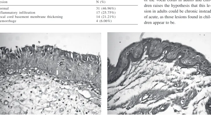
Figure 2 - Vocal-cord thickening basal membrane (arrow). The basal membrane thickening is not related with the cause of death and was diagnosed in 21.2% of the examined subjects (PASX40).
Figure 1 - Vocal cord with normal basal membrane thickening (arrow). The morphometric analysis was performed in 5 points (PASX40).
ceration of the epithelium of the vo-cal cords and intubation13.
Neverthe-less, upon analyzing the relationship between the thickening of the basal membrane of the vocal cords, smok-ing, and intubation, no significant as-sociation was found.
In spite of the fact that lesions to the vocal cords are common in pediatric autopsy14,7, there are few
studies published on this subject. Our study found the same incidence of the thickening of the basal membrane of the vocal cords in adults as those pub-lished about children younger than 1 year of age7. The ultrastructure of the
vocal cords has been well studied, demonstrating its intrinsic constitu-tion1. Nevertheless, the mechanism for
66
REV. HOSP. CLÍN. FAC. MED. S. PAULO 59(2):63-66, 2004 Relationship between the morphologic alterations of vocal cords
Salge AKM et al.
The basal membrane of the vocal cords is directly involved in various biological processes2. In adults, the
thickening of the basal membrane of the vocal cords was found to be re-lated to benign lesions of the vocal cords, most often in response to vibra-tory effects1,2, infectious diseases such
as tuberculosis9, accidents or
surger-ies10, and traumas from a variety of
causes9. In our study, the thickening of
the basal membrane of the vocal cords was found most frequently in subjects whose cause of death was of respira-tory alteration, among which were in-fectious causes such as pneumonia and
tuberculosis.
In conclusion, this study shows that the thickening of the basal membrane of the vocal cords is frequent in adult subjects at autopsy. However, new stud-ies are needed in order to verify the as-sociation of this thickening with dis-eases of the respiratory system.
RESUMO
SALGE AKM e col. - Avaliação Mor-fológica da membrana basal das cordas vocais de adultos autopsia-dos e sua correlação com as causas de óbito. Rev. Hosp. Clín. Fac. Med. S. Paulo 59(2):63-66, 2004.
OBJETIVO: O objetivo deste tra-balho foi identificar as possíveis alte-rações na espessura da membrana basal das cordas vocais e relacionar estas com a causa de morte.
MÉTODO: Foram utilizadas larin-ges coletadas de adultos autopsiados, no período de 1993 até 2001. Reali-zamos as colorações da
Hematoxilina-Eosina e Ácido Periódico de Schiff, onde foi medido o diâmetro da mem-brana basal.
RESULTADOS: Foram analisadas 66 cordas vocais, o espessamento foi identificado em 14 casos (21,2%), sen-do encontrasen-do em proporções iguais entre os sexos, sendo freqüente em pa-cientes da cor branca (12 casos, 85,7%). Foram encontradas alterações respirató-rias em 10 (71,4%) dos casos com espessamento. Entre os pacientes intu-bados, 7 (18,4%) apresentaram espes-samento. Entre os fumantes 9 (19,63%) apresentavam espessamento. Não hou-ve influência estatisticamente
significa-tiva do espessamento na doença que le-vou o indivíduo à morte.
CONCLUSÃO: Em relação a cau-sa de morte não há diferença estatisti-camente significante entre os casos cuja causa de morte foi por doenças respiratórias ou por doenças não res-piratórias e o diagnóstico de espes-samento da membrana basal das cor-das vocais. Sua etiopatogênese neces-sita de maiores estudos.
UNITERMOS: Adultos. Autopsia. Corda vocal. Espessamento. Mem-brana basal.
REFERENCES
1. Dikkers FG, Hulstaert CE, Oosterbaan JÁ, et al. Ultrastructural changes of the basement membrane zone in benign lesions of the vocal folds. Acta Otolaryngol 1993; 113 (1): 98-101. 2. Cervera-Paz FJ, Dikkers FG. Ultrastructure and pathogenesis of
vocal nodules on the vocal cords. Acta Otorrinolaringol Esp 1994; 45 (4): 261-265.
3. Courey MS, Shohet JÁ, Scott MA, et al. Immunohistochemical characterization of benign laryngeal lesions. Ann Otol Rhinol Laryngol 1996; 105 (7): 525-531.
4. Hollinshead W, Rosse C. In: Anatomia. Rio de Janeiro, Interlivros Edições, 1991. p. 839-856.
5. Katz SI. In: Basement membanes and cell movement. London, Pitman, 1984. p. 243-259.
6. Bach D, Peters A, Rowemeier H, et al. Anti-basal membrane glomerulonephritis after homologous kidney transplantation in hereditary Alport’s nephropathy. Dtsch Med Wochenschr 1991; 116 (46): 1752-1756.
7. Castro ECC, Peres LC. Vocal cord basement membrane in non-sudden infant death syndrome cases. Ped Dev Pathol 1999; 2: 440-445.
8. Van Landeghem FK, Brinkmann B, Bajanowski T. Basement membrane thickness of the vocal cord in cases of sudden infant death. Int J Legal Med 1999; 12 (1): 31-41.
9. Moon WK, Han MH, Chang K, et al. Laryngeal tuberculosis: CT findings. Am J Roentgenol 1996; 166 (2): 445-449. 10. Milutinovic Z, Bojic P. Functional trauma of the vocal folds:
classification and management strategies. Folia Phoniatr Logop 1996;. 48 (2): 78-85.
11. Fagan DG, Emery JL. A review and restatement of some problems in histological interpretation of the infant lung. Semin Diagn Pathol 1992; 9 (1): 13-23.
12. Valdes-Dapena M. Death investigation and post-mortem examination. In: Valdés-Dapena M, McFeeley PA, Hoffman HJ, et al. Histopathology Atlas for the Sudden Infant Death Syndrome. Washington, DC: Armed Forces Institute of Pathology, 1993. p. 23-37.