Original article
Received: Feb 20, 2009; Accepted: Apr 27, 2009
Antibacterial Activity of Twenty Iranian Plant Extracts Against Clinical Isolates
of
Helicobacter pylori
1Farahnaz Nariman, *2Fereshteh Eftekhar, 3Zohreh Habibi, 4Sadegh Massarrat, 4Reza Malekzadeh
Abstract
Objective(s)
Due to increasing emergence of drug-resistance in Helicobacter pylori isolates, traditional plants are
potentially valuable sources of novel anti-H. pylori agents. In this research, anti-H. pylori activity of the organic extracts of twenty native Iranian plants was determined against ten clinical isolates of H. pylori.
Materials and Methods
Disc diffusion was used to determine the biological activity of 20 plant extracts as well as 8 antibiotics
commonly used to treat H. pylori infections. Minimum inhibitory concentrations were also measured by tube
and agar dilution methods for the biologically active plant extracts.
Results
Of the twenty plant extracts analyzed, sixteen exhibited good anti-H. pylori activity, using disc diffusion.
The ten most active extracts were Carum bulbocastanum, Carum carvi, Mentha longifolia, Saliva limbata,
Saliva sclarea, Ziziphora clinopodioides, Thymus caramanicus, Glycyrrhiza glabra, Xanthium brasilicum
and Trachyspermum copticum. Minimum inhibitory concentrations measured for the 10 biologically active
plant extracts were within the range of 31.25 to 500 µg/ml.
Conclusion
Among the ten plant extracts effective against H. pylori clinical isolates, Carum carvi, Xanthium brasilicum
and Trachyspermum copticum showed the highest activity.
Keywords: Anti-Helicobacter pylori, Iranian plants, Organic extracts
1- Department of Biology and Microbiology, Faculty of Sciences, Alzahra University, Tehran, Iran
2- Department of Microbiology, Faculty of Biological Sciences, Shahid Beheshti University, G.C., Tehran, Iran *Corresponding author: Tel: +98-21-29903208; Fax: +98-21- 22431664; email: [email protected] 3- Department of Chemistry, Faculty of Sciences, Shahid Beheshti University, G.C., Tehran, Iran
Introduction
Helicobacter pylori is well recognized as a
major etiologic factor in gastrodudenal diseases such as chronic gastritis, peptic ulcers, gastric adenocarcinoma and mucosa-associated lymphoid tissue lymphoma (MALToma) (1-4). The current treatment regimes for H. pylori infections are based on
the combination of a proton pump inhibitor and two antibiotics (triple therapy) (5, 6). However, eradication is not always successful and the use of these antibiotics occasionally causes emergence of resistant clones and various harmful adverse effects (5, 7-9). Thus, development of new effective therapeutic agents could represent a significant advance in treatment of these infections. Traditional uses of plants for medicinal purposes provide a basis for the use of specific plants for specific medical conditions. In fact, a number of drugs and natural substances such as various essential oils, extracts of the lichen Certaria islandica, Chinese green tea and several native Iranian plants have been shown to have
in vitro antibacterial activity against H. pylori
(10-14). In the present study, we investigated the anti-H. pylori activity of twenty native
Iranian plant extracts against clinical isolates of H. pylori from adults by disc diffusion and measured the minimum inhibitory concentrations of the effective extracts.
Materials and Methods
H. pylori strains and culture conditions
Thirty two gastric biopsies were received from patients with gastrointestinal disease (age range 40 to 90 years old) at the Atrac Endoscopy Clinic (Gonabad, Iran). Biopsies were placed in a modified Campy–Thio medium, composed of thioglycolate base medium (Difco, Detroit, Michigan, USA), 10% sheep defibrinated blood, 8 mg/l polymyxin B, 2 mg/l amphotericin B and 6 mg/l vancomycin (Fluka, Sigma- Aldrich, Taufkirchen, Germany). The tubes were incubated at 37 °C under microaerophilic atmosphere for 2 to 5 days at which time, 20 µl of each specimen was streaked onto Brucella agar plates (Difco, Detroit, Michigan, USA) containing 10% defibrinated sheep blood and the three antibiotics. Identification was carried out by Gramstain morphology, catalase, oxidase and urea hydrolysis activities. Isolates were maintained in skim milk containing 15%
glycerol and 10% fetal calf serum at –80 °C. H. pylori ATCC strain 43504 was also
used as control in all experiments. Human studies were approved by the local Ethics Committee.
Plant collection and preparation of extracts Plant names and the areas of collection are shown in Table 1. Plants were kindly identified by M. Yousefzadi (Department of
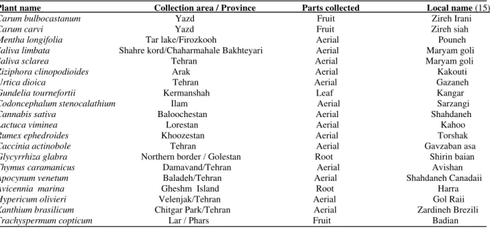
Table 1.Native Iranian plants chosen for the study, collection areas and plant parts collected.
Plant name Collection area / Province Parts collected Local name (15)
Carum bulbocastanum Yazd Fruit Zireh Irani
Carum carvi Yazd Fruit Zireh siah
Mentha longifolia Tar lake/Firozkooh Aerial Pouneh
Saliva limbata Shahre kord/Chaharmahale Bakhteyari Aerial Maryam goli
Saliva sclarea Tehran Aerial Maryam goli
Ziziphora clinopodioides Arak Aerial Kakouti
Urtica dioica Tehran Aerial Gazaneh
Gundelia tournefortii Kermanshah Leaf Kangar
Codoncephalum stenocalathium Ilam Aerial Sarzangi
Cannabis sativa Baloochestan Aerial Shahdaneh
Lactuca viminea Lorestan Aerial Kahoo
Rumex ephedroides Khoozestan Aerial Torshak
Caccinia actinobole Tehran Aerial Gavzaban asa
Glycyrrhiza glabra Northern border / Golestan Root Shirin baian
Thymus caramanicus Damavand/Tehran Aerial Avishan
Apocynum venetum Baladeh/Tehran Aerial Shahdaneh Canadaii
Avicennia marina Gheshm Island Root Harra
Hypericum olivieri Velenjak/Tehran Aerial Gol Raii
Xanthium brasilicum Chitgar Park/Tehran Aerial Zardineh Brezili
Ecology and Systematic, Research Institute of Applied Science, ACECR, Tehran, Iran) and Dr. Mozaffarian (Department of Botany, Research Institute of Forests and Rangelands, Tehran, Iran) and voucher numbers are kept at the Shahid Beheshti University Herbarium. To prepare organic extracts, dried plant parts were soaked in an equal mixture of methanol, diethyl ether and petroleum benzene (Merck, Darmstadt, Germany) at a ratio of 1:10 (w/v) at room temperature for 24 hr before filtration. The filtrates were concentrated, using a Rotavapor (Eyela, Tokyo, Japan) to about 5 ml and placed at –15 oC to remove heavy hydrocarbons and lipids. The extracts were then diluted with cold methanol, filtered, and the filtrates were evaporated to leave a solid pellet which was then reconstituted in methanol (10%, w/w) (14).
Anti- Helicobacter pylori activity measured by disc diffusion
Suspensions of fresh cultures were made in saline and turbidity was adjusted to 9 x 10⁸ or 1.5×10⁸ bacteria/ml (corresponding to McFarland standards 3 and 0.5 respectively).It has been reported that a McFarland standard of 3 or 4 yields actual counts of 5×10⁶ cells /ml for H. pylori (16). Hence, both inocula were
used to test anti-Helicobacter activity of the plant extracts. In each case, 200 µl of the appropriate microbial suspension was placed on a large (50 ml) Mueller Hinton agar plate (Merck, Darmstadt, Germany) containing 10% fetal calf serum (Gibco/BRL, Paisley, UK)and spread evenly in all directions.
Antibiotic discs including metronidazole (5 µg), amoxicillin (30 µg), ampicillin (10 µg), tetracycline (30 µg), furazolidone (50 µg), azithromycin (15 µg), erythromycin (15 µg) (HiMedia Laboratories, Pvt. Limited, Mumbai- 400 086 India) and clarithromycin
(15 µg) (Becton Dickinson, Abbot
Laboratories, Chicago, IL., USA) were placed on the bacterial lawns and the plates were incubated at 37 oC under microaerophilic conditions for 2-5 days. For the plant extracts, sterile blank discs (6 mm) were inoculated with 25 µl (2.5 mg) of each extract or solvent
controls and were dried at 30-35 oC for 12-24 hr before being placed on the surface of the bacterial lawns.
Measurement of minimum inhibitory concentrations
Minimum inhibitory concentrations (MIC) were measured within the range of 0.015 to 1000 µg/ml, using tube and agar dilution methods. Fresh bacterial suspensions were prepared in saline and adjusted to McFarland turbidity standard 3. For the tube assay, serial 2-fold dilutions were made from a stock of crude plant extract in dimethyl sulfoxide (2 mg/ml) (Merck, Darmstadt, Germany) in 0.5 ml of Mueller Hinton broth containing 10% fetal calf serum. Finally, 0.5 ml of a 1:200 dilution of bacteria adjusted to MacFarland standard 3 in Mueller Hinton broth was added to the tubes. For the agar dilution assay, the extracts were added in 2-fold decreasing concentrations to Mueller Hinton agar containing 10% serum and 2 µl of the undiluted bacterial suspensions were spot inoculated on each plate. Controls of bacteria without extract or extracts without bacteria were also included. The tubes and plates were incubated for 2-5 days, before recording minimum inhibitory concentrations.
Results
Ten isolates of H. pylori were recovered from
the 32 gastric biopsies and their susceptibility was determined to eight antibiotics and the crude organic plant extracts by disc diffusion and employing MacFarland standard 3. Table 2 shows that the highest degree of antibiotic resistance was observed for metronidazole (9/10 isolates) followed by amoxicillin, tetracycline, erythromycin (4/10 isolates) and finally, ampicillin, furazolidone, azithromycin, clarithromycin (3/10 isolates). For plant extracts, an inhibition zone of 15 mm was arbitrarily chosen as the cut-off point for bacterial susceptibility. All H. pylori isolates
brasilicum. Eight were sensitive to Urtica dioica extract, seven to the extracts of
Avicennia marina, Hypericum olivieri, Gundelia tournefortii and Codoncephalum stenocalathium and six were susceptible to
Cannabis sativa extract. The extracts of Lactuca viminea, Rumex ephedroides, Caccinia actinobole and Apocynum venetum
showed no antibacterial activity. In addition, using the 10 most effective plant extracts, the disc test was carried out against two bacterial inocula (1.5×10⁸ and 3×10⁹ cells/ml). As shown in Table 3, all extracts showed high anti-H. pylori activity, using both inocula.
Table 4 shows the results of minimum
inhibitory concentrations determined for the 10 effective plant extracts against H. pylori
isolates, using broth and agar dilution methods. The results were identical for both
assays and were within the range of 125-500 µg/ml for organic extracts of
S. limbata and S. sclarea, 62.5-250 µg/ml for
organic extracts of M. longifolia, T. caramanicus, G. glabra and X. brasilicum
and 31.25-125 µg/ml for organic extracts of
Z. clinopodioides, C. bulbocastanum, C. carvi
and T. copticum. Such values are significant given that the crude extracts were used.
Table 2. Anti- Helicobacter pylori activity of 8 antibiotics and organic extracts of 16 native Iranian plants against 10 clinical isolates of H. pylori shown by disc diffusion.
Numbers shown in the table represent zones of inhibition measured in mm. For plant extracts, zones of > 15 mm show sensitivity and R, indicates resistance. H. pylori ATCC 43504 was used as the susceptible control strain, nt, not tested.
Table 3. Comparison of anti- Helicobacter pylori activities of the organic extracts from ten Iranian plants using MacFarland standards 0.5 and 3, measured by disc diffusion.
Isolate number
C. bulbocastanum C. carvi M. longifolia S. limbata S. sclarea Z. clinopodioides G. glabra T. caramanicus X. brasilicum T. copticum
MF#0.5 MF#3 MF#0.5 MF#3 MF#0.5 MF#3 MF#0.5 MF#3 MF#0.5 MF#3 MF#0.5 MF#3 MF#0.5 MF#3 MF#0.5 MF#3 MF#0.5 MF#3 MF#0.5 MF#3
1 29 27 27 25 25 23 22 20 20 19 27 25 24 22 31 29 32 29 31 28
2 42 40 39 38 28 25 23 22 23 20 28 25 27 25 27 24 32 28 28 25
3 40 38 28 26 33 31 25 20 22 19 30 28 25 22 29 26 28 25 29 26
4 30 26 28 25 21 18 22 18 18 15 28 20 24 20 29 24 28 26 33 30
5 31 28 27 25 31 28 21 18 22 18 24 20 32 30 26 22 32 28 33 29
6 27 25 18 16 27 25 23 20 24 22 30 25 27 25 29 26 32 28 34 31
7 27 24 24 22 34 31 23 22 22 19 32 28 30 28 29 25 34 30 36 32
8 27 25 24 20 42 40 24 23 24 22 33 30 29 26 31 27 37 35 36 30
9 35 30 31 29 40 38 30 28 30 28 37 35 33 31 34 32 42 39 42 39
10 34 32 34 32 42 40 32 30 33 31 36 34 36 30 34 31 39 35 43 40
Control 36 35 31 30 36 35 29 28 31 30 40 37 44 42 45 42 36 35 42 40
Numbers shown in the table represent zones of inhibition measured in mm. For plant extracts, zones of > 15 mm were chosen as sensitivity cut off point. H. pylori ATCC 43504 was used as the susceptible control strain.
Table 4. Minimum inhibitory concentrations (MIC) measured for 10 organic extracts from native Iranian plants against clinical isolates of H. pylori.
Isolate number
Plant Extract 1 2 3 4 5 6 7 8 9 10 Control
C. bulbocastanum 125 31.25 31.25 62.5 62.5 62.5 125 125 62.5 62.5 62.5
C. carvi 62.5 31.25 62.5 62.5 62.5 125 125 125 62.5 31.25 125
M. longifolia 125 125 62.5 250 250 125 62.5 62.5 62.5 62.5 62.5
S. limbata 250 250 500 500 500 250 250 125 125 125 250
S. sclarea 250 500 500 500 250 500 250 125 125 125 250
Z. clinopodioides 125 125 125 125 125 62.5 62.5 31.25 31.25 31.25 31.25
G. glabra 250 125 125 125 62.5 125 62.5 62.5 62.5 62.5 15.6
T. caramanicus 125 250 125 125 250 125 62.5 62.5 62.5 62.5 15.6
X. brasilicum 125 250 250 125 62.5 62.5 62.5 62.5 62.5 62.5 125
T. copticum 125 125 125 62.5 62.5 62.5 31.25 62.5 31.25 31.25 62.5
MIC was determined by tube and agar dilution methods, using MacFarland standard # 3 and values are shown inµg/ml. H. pylori ATCC 43504 was used as control.
Discussion
Due to increasing antibiotic resistance in clinical isolates of H. pylori, the search for new and safe anti-H. pylori compounds are of
utmostimportance and native medicinal plants seem to be a logical source for seeking new agents.
We determined the biological activity of twenty native Iranian plants against 10 clinical isolates of H. pylori and found that sixteen had good anti- H. pylori activity. As a matter of
fact, inhibition zones for the plant extracts obtained by disc diffusion were equal or larger than those of the eight antibiotics commonly used to eradicate H. pylori infections. These
findings suggest the potential of natural agents for treatment of H. pylori infections. Minimum
inhibitory concentration values were also encouraging since only a fraction of the crude extract is expected to have antimicrobial activity. We have previously shown the antibacterial activity of Xanthium brasilicum
from Iran and identified Xanthatin (8-Epi-xanthatin) as its active antibacterial compound
(14, 17 and unpublished data). Antibacterial and anti-ulcerogenic activities of
Xanthanolides have also been reported by
other researchers (17-20). There are some reports on anti-H. pylori activity of a number of plant extracts including some species within the genera used in this study (21-29). However, in vitro susceptibility does not
necessarily mean success in vivo. Graham et al. showed that neither garlic nor red pepper containing capsaicin had any in vivo effect on H. pylori despite their anti- H. pylori activity in vitro (30). Therefore, a wide range of plants with anti- H. pylori activity must be screened before exploring the potential use of effective agents for in vivo use. More extensive experiments are also needed to determine the
in vivo activities of these edible medicinal
plants and furthermore, identifying the active components of each plant as well as their therapeutic values.
Conclusion
Majority of the plant extracts used in this study had considerable in vitro activity against clinical isolates of H. pylori. Considering that
these plants are edible and are traditionally used for treatment of a number of ailments, their anti-H. pylori activity is quite significant
and could present alternative treatments for
H. pylori infections.
Acknowledgment
References
1. Graham DY. Helicobacter pylori infection in the pathogenesis of duodenal ulcer and gastric cancer: a model. Gastroenterology 1997; 113:9183-9191.
2. Marshall BJ, Warren JR. Unidentified curved bacilli in the stomach of patients with gastritis and peptic ulceration. Lancet 1984; 1:1311-1315.
3. International Agency for Research on Cancer. IARC Monographs on the Evaluation of Carcinogenic Risks to Humans, Schistosomes, Liver Flukes and Helicobacter pylori. Lyon, France: World Health Organization; 1994. p.61
4. Nomura A, Stemmermann GN, Chyou PH, Kato I, Perez-Perez GL, Blaser MJ. Helicobacter pylori infection and gastric carcinoma among Japanese Americans in Hawaii. N Engl J Med 1991; 325:1127-1131.
5. Graham DY, Qureshi WA. Antibiotic resistant Helicobacter pylori infection and its treatment. Curr Pharm Des 2000; 6:1537-1544.
6. O’Morain C, Montague S. Challenges to therapy in the future. Helicobacter 2000; 5:23–26.
7. Megraud F. Resistance of Helicobacter pylori to antibiotics. Aliment Pharmacol Ther 1997; 11: 43-53.
8. Adamak RJ, Suerbaum S, Plaffenbach B, Opferkuch W. Primary and acquired Helicobacter pylori resistance to clarithromycin, metronidazole and amoxicillin-influence on treatment outcome. Am J Gastroenterol 1998; 93:386-389.
9. Falsafi T, Mobasheri F, Nariman F, Najafi M. Susceptibilities to different antibiotics of Helicobacter pylori strains isolated from patients at the pediatric medical center of Tehran, Iran. J Clin Microbiol 2004; 42:387-389. 10. Li Y, Xu C, Zhang Q, Liu JY, Tan RX. In vitro anti-Helicobacter pylori action of 30 Chinese herbal
medicines used to treat ulcer diseases. J Ethnopharmacol 2005; 98:329-333.
11. Ohta R, Yamada N, Kaneko H, Ishikawa K, Fukuda H, Fujino T, et al. In vitro inhibition of the growth of Helicobacter pylori by oil-macerated garlic constituents. Antimicrob Agents Chemother 1999; 43:1811–1812. 12. Ingolfsdottir K, Hjalmarspottir M, Slgurdsson A, Gudjonscottir G, Brynjolfsdottir A. In vitro susceptibility of
Helicobacter pylori to proto-lichenistic acid from the lichen Certaria islandica. Antimicrob Agents Chemother 1997; 41:215-217.
13. Yee YK, Wing-Leungkoo M. Anti Helicobacter pylori activity of Chinese tea: An in vitro study. Aliment Pharmacol Ther 2000; 4:635-638.
14. Nariman F, Eftekhar F, Habibi Z, Falsafi T. Anti-Helicobacter pylori activities of six Iranian plants. Helicobacter 2004; 9:146-151.
15. Mozaffarian V. Dictionary of Iranian plant names. Farhang Moaser, 1996.
16. DeCross AJ, Marshall BJ, McCallum RW, Hoffman SR, Barrett LJ, Guerrant RL. Metronidazol susceptibility testing for H. pylori: Comparison of disk, broth and agar dilution methods and their clinical relevance. J Clin Microbiol 1993; 31:1971-1974.
17. Eftekhar F, Nariman F, Habibi Z and Mohammadi N. Antibacterial activity of aerial extracts from Xanthium brasilicum. Iran J Pharm Res 2007; 6:65-68.
18. Sato Y, Oketani H, Yamada T, Singyouchi K, Ohtsubo T, Kihara M, et al. A xanthanolide with potent
antibacterial activity against methicillin-resistant Staphylococcus aureus. J Pharmacol 1997; 49:1042-1047. 19. Kim YS, Kim JS, Park SH, Choi HU, Lee CO, Kim SK, et al. Two cytotoxic sesquiterpene lactones from the
leaves of Xanthium strumarium and their in vitro inhibitory activity of n-farnesyltransferase. Planta Med 2003; 69:375-377.
20. Favier LS, María AOM, Wendel GH,. Borkowskia EJ, Giordano OS, Pelzerb L, et al. Anti-ulcerogenic activity of xanthanolide sesquiterpenes from Xanthium cavanillesii in rats. J Ethnopharmacol 2005;100:260-267.
21. Mahady GB, Pendland SL, Stoia A, Hamill FA, Fabricant D, Dietz BM, et al. In vitro susceptibility of Helicobacter pylori to botanical extracts used traditionally for the treatment of gastrointestinal disorders. Phytother Res 2005;19:988–991.
22. Nostro A, Cellini L, Di Bartolomeo S, Di Campli E, Grande R, Cannatelli MA, et al. Antibacterial effect of plant extracts against Helicobacter pylori. Phytother Res 2005; 19:198-202.
23. Deriu A, Branca G, Molicotti P, Pintore G, Chessa M, Tirillini B, et al. In vitro activity of essential oil of Myrtus communis L. against Helicobacter pylori. Int J Antimicrob Agents 2007; 30:562–565.
24. Yeilada E, Gürbüz I, Shibata H. Screening of Turkish anti-ulcerogenic folk remedies for anti-Helicobacter pylori activity. J Ethnopharmacol 1999; 66:289-293.
25. Wang YC, Huang TL. Screening of anti-Helicobacter pylori herbs deriving from Taiwanese folk medicinal plants. FEMS Immun Med Microbiol 2005; 43:295–300.
26. Stamatis G, Kyriazopoulos P, Golegou S, Basayiannis A, Skaltsas S, Skaltsa H. In vitro anti-Helicobacter pylori activity of Greek herbal medicines. J Ethnopharmacol 2003; 88:175-179.
27. Gürbüz I, Üstün, O, Yesilada E, Sezik E, Kutsal O. Anti-ulcerogenic activity of some plants used as folk remedy in Turkey. J Ethnopharmacol 2003; 88: 93-97.
29. Fukai T, Marumo A, Kaitou K, KandaT, Terada S, Nomura T. Anti-Helicobacter pylori flavonoids from licorice extract. Life Sci 2002; 71:1449-1463.