METHICILLIN-SENSITIVE STAPHYLOCOCCUS
Mattana, C.M.1*; Satorres, S.E.1; Sosa, A.2; Fusco, M.2; Alcaráz L.E.1
1
Area Microbiologia, Facultad de Química, Bioquímica y Farmacia. Universidad Nacional de San Luis, Chacabuco y Pedernera,
5700 San Luis, Argentina; 2Area de Farmacognosia. Facultad de Química, Bioquímica y Farmacia. Universidad Nacional de San
Luis, Chacabuco y Pedernera, 5700 San Luis, Argentina.
Submitted: June 23, 2009; Returned to authors for corrections: September 04, 2009; Approved: February 18, 2010.
ABSTRACT
Antibacterial activity of organic and aqueous extracts of Acacia aroma was evaluated against
methicillin-resistant Staphylococcus aureus (MRSA), methicillin sensitive Staphylococcus aureus (MSSA) and
methicillin-resistant Staphylococcus epidermidis. Inhibition of bacterial growth was determined using agar
diffusion and bioautographic methods. Among all assayed organic extracts only ethanolic and ethyl acetate
extracts presented highest activities against all tested Staphylococcus strains with minimal inhibitory
concentration (MIC) values ranging from 2.5 to 10 mg/ml and from 2.5 to 5 mg/ml respectively. The
aqueous extracts show little antibacterial activity against Staphylococcus strains. The bioautography assay
demonstrated well-defined growth inhibition zones against S. aureus in correspondence with flavonoids
and saponins. A. aroma would be an interesting topic for further study and possibly for an alternative
treatment for skin infections.
Key words:Acacia aroma; Antibacterial activity; Staphylococcus; Tusca.
INTRODUCTION
The indiscriminate use of commercial antimicrobial drugs
commonly employed in the treatment of infectious diseases,
and the bacterial genetic ability to transmit and acquire
resistance to drugs, which are utilized as therapeutic agents has
compromised the use of newer generations of antibiotics (13,
17). This situation is growing and the outlook for the use of
antimicrobial drugs in the future is still uncertain. Therefore,
actions must be taken to reduce this problem; for example,
controlling the use of antibiotics, developing research to better
understand the genetic mechanisms of resistance, and
continuing studies to develop new drugs, either synthetic or
natural.
Plants have been a valuable source of natural products for
human health care, and the use of plant compounds for
pharmaceutical purposes has gradually increased around the
world (7, 8, 16, 18).
Most studies on the antibacterial activity of plant extracts
have been restricted to analysis of their bacteriostactic and
bactericidal properties (6, 8, 18, 25). New assays investigating
other potential roles, such as the mediation of pathogenicity via
quorum sensing inhibition, have recently emerged (20).
The use of plants against skin disease is a common
practice in the popular medicine of most cultures, although the
precise cause of disease and mechanism of cure is not always
understood. Staphylococcus aureus and Staphylococcus
epidermidis are ubiquitous colonizer of the skin and mucous
membranes of humans and animals. Many strains of S. aureus
carry resistance genes for penicillin, tetracyclines, methicillin
and now, vancomycin. Methicillin-resistant Staphylococcus
aureus (MRSA) is a major pathogen causing nosocomial
infections, but it has emerged as a problematic pathogen in the
community setting as well. These strains called
Community-Associated Methicillin-Resistant Staphylococcus aureus
(CA-MRSA) cause infections in healthy individuals without
predisponent risk factor and outside the hospital setting. MRSA
and CA-MRSA presents a significant threat to public health
and are difficult to manage (14). Vancomycin and other
glycopeptides antibiotics are the current mainstay of therapy
for infections caused by MRSA. However, therapy with
glycopeptides is frequently accompanied by side effects and
possible risk of emergence of isolates with reduced
susceptibility to vancomycin, such as
Vancomycin-Intermediate Staphylococcus aureus (VISA) and heterogeneous
VISA (hVISA) (2); hence, there is a constant need for new and
effective therapeutic agents, being the phytomedicine a valid
alternative.
Acacia aroma, commonly known as tusca (4), a native
species of Argentina, is member of genus Acacia subgenus
Acacia, which grow widely in the provinces of Tucumán, Salta,
Santiago del Estero, Catamarca, La Rioja, Formosa, Chaco,
Córdoba, San Luis and Santa Fe (5). This plant is used in
Argentine folkloricmedicine for wound healing, antiseptic and
for the treatment of gastrointestinal disorders. Leaf and bark
infusions have diuretic, anti-inflammatory and cicatrizing uses.
The acceptance of traditional medicine as an alternative
form for health care and the development of microbial
resistance to the available antibiotics has led authors to
determine the antibacterial properties of A. aroma and validate
thepopular use of this plant.
The purpose of the presents study wasto investigate the
antibacterial activity of leaf extracts of A. aroma against
methicillin-resistant S. aureus (MRSA), methicillin-sensitive S.
aureus (MSSA) and methicillin-resistant S. epidermidis
isolates in infected skin wounds. The extracts with the highest
antibacterial effectiveness could be of therapeutic usefulness
for subsequent application in pharmaceutical formulations
against Staphylococcus spp.
MATERIALS AND METHODS
Plant material
Aerial parts of Acacia aroma were collected in
December-January 2007, in the Northwestern region of the Province of
San Luis, Argentina. Acacia aroma (Fabaceae) was
authenticated by Dr. Del Vitto, Botany Department, San Luis
National University (UNSL). A voucher specimen was
deposited in the herbarium of UNSL under the number 487.
The parts used were leaves.
Preparation of Acacia aroma extracts Aqueous extracts:
• Infusion: It was prepared according to Farmacopea Argentina 6th ed. 1978 (9), applying
constant actionof hot water during 20 min. • Decoction: Dried and powdered plant material
(5g) was boiled with 70 ml of water for 20 min.
After cooling to 40-45 ºC, the liquid was filtered
and the volume adjusted to 100 ml with distilled
water (9).
Organics extracts: A known amount of powdered plant material (100 g) was sequentially extracted at room
temperature with solvents of increasing polarity: petroleum
ether (60-80°C), dichloromethane, diethyl acetate, methanol
and ethanol. The extracts were filtered through Whatman nº 4
filter paper, dried under reduced pressure at 40ºC, and
weighed.
Microorganisms and media
followed: I-Local clinical isolates: methicillin resistant
Staphylococcus aureus (n=20), methicillin sensitive S. aureus
(n=18), methicillin resistant Staphylococcus epidermidis
(n=16) obtained from skin diseases. The written informed
consent was obtained from all subjects prior to the study.
II-Reference strains: Staphylococcus aureus ATCC 29213,
Staphylococcus aureus ATCC 43300 and Staphylococcus
epidermidis ATCC 12228.
The strains were identified by the use of Biochemical
profiles according to the recommendations of the Manual of
Clinical Microbiology (15). All organisms were maintained in
brain-heart infusion (BHI medium) containing 30% (v/v)
glycerol at −20°C. The inocula were prepared by adjusting the
turbidity of the suspension to match the 0.5 Mc Farland.
Antibacterial testing
Antibacterial activity of the crude organic extracts and
aqueous extract were determined by the modified agar well
diffusion method of Perez et al. (19). The different organic
extracts were dissolved in dimethylsulfoxide (DMSO) and the
aqueous extracts were dissolved in water. The extracts were
sterilizedby filtration through a 0.2 membrane filter.
Agar diffusion assay
The agar well diffusion method was employed.
Mueller-Hinton agar (MHA, Laboratorios Britania, Argentina) 25 ml
was poured into each petri plate. Once the agar solidified, the
microorganisms were inoculated on the surface of the plates (1
× 108 CFU/ml). Subsequently, the surface of the agar was
punched with a 6-mm-diameter wells. Each well was filled
with 50 l of each plant extract. The concentration of the
extracts employed was 20 mg/ml. Simultaneously, gentamicin
sulfate was used as positive control (1µg per well). Control
wells containing the same volume of DMSO, diethyl acetate,
ethanol, petroleum ether, dichloromethane, and distilled water
were made. After a 24-hourincubation at 35ºC, all plates were
observed for zones of growth inhibition, and the diameter of
these zones was measured in millimeters.
All tests were performed in duplicate and the antibacterial
activity was expressed as the mean of inhibition diameters
(mm) produced.
Minimal inhibitory concentration (MIC)
Serial agar macro-dilution method: The tests were performed in MHA medium. After cooling and drying, the
plates were inoculated with swabs containing each bacterial
cell suspension (1 × 10⁸ CFU/ml). Then, the surface of the agar
was punched with 6-mm-diameter wells. Serial two-fold
dilution of each extract were added in each well in a
concentration ranging from 20 mg/ml to 2.5 mg/ml. The plates
were incubated aerobically for 24 hours at 37°C. A growth
control of each tested strain was included.
MIC was defined as the lowest concentration of A. aroma
extract in which no colony was observed after incubation
Phytochemical screening
Characterization of chemical metabolites:
• Detection of glucides: Regular methods were carried out using developers: Molisch reactive (
-Naftol and H2SO4) for the detection and
differentiation of glucides. Positive result was
indicated by purple coloring.
• Detection of tannin: 5ml of plant extract was evaporated and theremains were dissolved in 10
ml of distilled water. The aqueous extract was
divided into three parts: 1-2 drops of 10% ferric
chloride solution were added to one of the parts, a
blue coloring indicated the possible presence of
hydrolysable tannin. The remaining parts were
poured with gelatin, and lead acetate solution at
10%. The occurrence of white precipitate
indicated a positive result for the tannin test. • Detection of flavonoids: For the visualization of
flavonoids UV light was used, previously
exposing the specimen to ammonia vapors
(ionization in basic medium) giving an intense
yellow coloring for positive results.
• performed in a test tube evaporating 5 ml of the extract, then it was shaken vigorously, and left to
settle for a period of 15 to 20 min. The presence
of saponins was classified by the height and
permanence of the foam formation, being
observed for 5 min.
• Detection of alkaloids: 30 ml of the plant extract was dried, and then, adding5 ml of hydrochloric
acid at 10% to it. After filtering, some drops of
alkaloid precipitation reactive were added. A
slight turbidity or precipitate colored proves the
possible presence of alkaloids.
The determinations were done according to the
recommendations of the Manual of Farmacognosis (12).
Thin-Layer chromatography (TLC)
Ethanolic and ethyl acetate extracts were analyzed for
TLC. The volume of injection of both extracts was fixed at 3µl
(20 mg/ml) which were spotted on chromatographic silica gel
60 F254 plates (Merck). To detect flavonoids three different
solvent systems were used: ethyl acetate:formic acid:methyl
ethyl acetone:water (5:1:3:1); ethyl acetate:formic
acid:water:methanol (10:2:2:1); ethanol: chloroform (1:3).
Spots were visualized by UV light. For the detection of
sapogenines, chloroform:methanol:water (70:30:4); buthanol:
acetic acid:water (4:1:1); chloroform:methanol:water
(65:35:10) were used as development solvents. Revealing with
a mix of 5% of sulfur in ethanol, 1% of vanillin in ethanol and
heat. Three micro liters of rutine, quercetin, oleanolic acid and
ß-amirin were used as standards. TLC plates were dried
overnight in a sterile room for complete removal of solvent.
Bioautography
Developed TLC plates were covered with 1-2 mm layer of
soft medium (BHI agar 0.6%) containing 0.1 % (w/v) 2,3,5
triphenyltetrazolium chloride (tetrazolium red) and a
suspension of S. aureus at a final concentration of 107
CFU/ml. The plates were placed in a sterile tray, sealed to
prevent the thin agar layer from drying, and incubated at 37°C
for 24 h. Where bacterial growth has been inhibited, an
uncolored area can be seen on the deep pink-red background.
The plates were run in duplicate.
RESULTS AND DISCUSSION
The antibacterial activity of leaves of Acacia aroma was
assayed in vitro conditions by agar well diffusion method
against Staphylococcus spp. The inhibition of bacterial growth
by seven extracts of A. aroma is summarized in Table 1. The
results showed that the ethyl acetate and ethanolic extracts
presented the highest activities (inhibition diameters of 14-16
mm). The petroleum ether, dichloromethane and methanolic
extracts presented low activity against the investigated bacteria
(inhibition diameters of 9-10 mm). On the other hand, the
infusion and decoction show little antibacterial activity against
all tested microorganisms (inhibition diameters of 7 mm).
Table 1. Antibacterial activity of organic and aqueous extracts of Acacia aroma by agar diffusion method.
Inhibition zone diameter (mm)
Microorganisms n A B C D E F G Gen
Methicillin-sensitive S.aureus* 18 7.3±0.3 7.2±0.2 9.5±0.4 9.5±0.4 14.9±0.8 15.0±0.8 9.1±0.3 18.0±1.0
Methicillin-resistant S. aureus* 20 7.2±0.3 7.1±0.3 9.5±0.4 9.5±0.5 15.1±0.7 15.2±0.8 9.2±0.2 17.0±0.2
S. aureus ATCC 43300 1 7.2±0.3 7.0±0.0 10.0±0.0 10.0±0.0 15.0±0.0 14.7±0.3 9.0±0.1 12.0±0.5
S. aureus ATCC 25923 1 6.9 ± 1.1 7.4±0.0 9.1±0.2 9.5±0.0 14.5±0.7 15.0±0.0 9.0±0.0 14.0±0.4
Methicillin-resistant S. epidermidis* 16 7.2±0.3 7.1±0.2 9.4±0.4 9.3±0.4 15.0±0.8 15.3±0.7 9.1±0.2 17.0±0.8
Alcohol extracts exhibited a higher degree of antibacterial
activity, compared to water extracts. This observation
confirmed the evidence from a previous study which reported
that alcohol is a better solvent for extraction of antimicrobial
substances from medicinal plants than water (22).
DMSO, ethanol, petroleum ether, dichloromethane, diethyl
acetate, and methanol blanks showed no inhibitory effect.
Whereas the in vitro results proved that aqueous extracts
showed little inhibitory growth activity against Staphylococcus;
it is of common knowledge the use of A. aroma leaves in form
of decoctions in popular medicine to clean wounds or skin
infections. Other components could trigger the immune system
helping to strengthen the antibacterial effect inherent to the
plant that has not been shown in conditions in-vitro. The ethyl
acetate and ethanolic extracts were selected because of their
higher antibacterial activity to determine the minimal
inhibitory concentration (MIC).
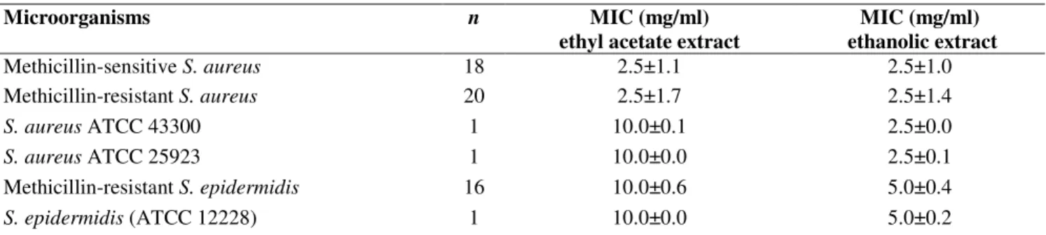
The data obtained, through the determination of MIC are
shown in Table 2. The results revealed variability in the
inhibitory concentrations of both extracts for Staphylococcus.
The MIC values of ethyl acetate and ethanolic extracts were in
the range of (concentrations) 2.5 to 10 mg/ml, and 2.5 to 5
mg/ml respectively. There is little or no scientific information
concerning the antibacterial activity of A. aroma in Argentina.
A study on A. aroma, performed in Tucumán, Argentina
reported MIC values inferior to our results (3). However, it is
difficult to compare the data because several variables
influence the results, such as the environmental and climate
conditions of the plant, and the choice of the extraction method
and antibacterial test. Moreover, there is no standard criteria
for the evaluation of the plant activity, therefore, the results
obtained by different researchers could be different (21).
Table 2. The MIC values of ethyl acetate and ethanolic extracts of A. aroma against strains of Staphylococcus aureus and
Staphylococcus epidermidis. Results are the mean of MIC values followed by the standard deviation.
Microorganisms n MIC (mg/ml)
ethyl acetate extract
MIC (mg/ml) ethanolic extract
Methicillin-sensitive S. aureus 18 2.5±1.1 2.5±1.0
Methicillin-resistant S. aureus 20 2.5±1.7 2.5±1.4
S. aureus ATCC 43300 1 10.0±0.1 2.5±0.0
S. aureus ATCC 25923 1 10.0±0.0 2.5±0.1
Methicillin-resistant S. epidermidis 16 10.0±0.6 5.0±0.4
S. epidermidis (ATCC 12228) 1 10.0±0.0 5.0±0.2
n: strain number; results are the mean of inhibition zone diameter followed by the standard deviation.
Methicillin resistant S. aureus and methicillin resistant S.
epidermidis strains which are already known to be
multirresistant to drugs were highly susceptible to ethyl acetate
and ethanolic extracts. Likewise, methicillin susceptible S.
aureus strains were also inhibited by both extracts; that is to
say, the Staphylococcus spp. susceptibility to the most active
extracts of A. aroma didnot correlate with the susceptibility or
resistance to oxacillin, in agreement with Arias et al.
To obtain some information on the active components,
ethyl acetate and ethanolic extracts were analyzed by TLC on
silica gel. TLC plates were run in duplicate and one set was
used as the reference chromatogram, and the other set was
assayed for bioautography. TLC analysis revealed the presence
of flavonoids: rutine (Rf of 0.71) and quercetin (Rf of 0.94) and
sapogenines: oleanolic acid (Rf of 0.87) and amirine (Rf of
0.50). This is in agreement with observations by other authors
(3). The bioautography assay for qualitative antibacterial
against S. aureus (Fig. 1) in correspondence with those
flavonoids and sapogenines bands, although the sapogenines
showed less visible zones of inhibition. Antimicrobial activity
of flavonoids and saponines has also been reported against
methicillin-resistant Staphylococcus aureus (1, 10, 11, 23, 24),
however studies regarding the mode of action for these
compounds in the bacterial cell should be done.
Figure 1. TLC of Acacia Aroma ethyl acetate extract. (A) Visual appearance; (B) Staphylococcus aureus (ATCC 43300)
bioautography overlay. Arrows indicate regions of inhibition
growth visualised with tetrazolium red. (1) Crude ethyl acetate
extract, (2) Rutine (flavonoids standard)
Our results suggest that Acacia aroma extracts possess
compounds with antibacterial properties which can be used as
bacterial growth inhibitory agents in new drugs for therapy
Staphylococcus diseases. Although this study investigated the
in vitro antibacterial activity, however, in vivo data may be
favorable fordetermining the potential usefulness of this plant.
Further work, including fractioning to isolate active
constituents and subsequent pharmacological evaluation, is
needed.
Investigations are in progress to determine the degree of
toxicity of these extracts. Acacia aroma is a promising plant as
its antibacterial activity showed effective results in treatment
against the MRSA. Our findings can set the basis for further
studies to obtain optimized preparations of the herbal extract
with activity against Staphylococcus.
ACKNOWLEDGEMENT
This work is part of the Project 8802 financed by
Universidad Nacional de San Luis, Argentina.
REFERENCES
1. Alcaraz, L.E.; Blanco, S.E.; Puig, O.N.; Tomas, F.; Ferreti, F.H. (2000). Antibacterial activity of flavonoids against methicillin-resistant
Staphylococcus aureus strains. J. Theor. Biol. 205, 231-240.
2. Appel Baum, P.C. (2007). Reduced glycopeptides susceptibility in methicillin-resistant Staphylococcus aureus (MRSA). Int. J. Antimicrob. Agents. 30, 398-408.
3. Arias, M.E.; Gomez, J.D.; Cudmani, N.M.; Vattuone, M.A.; Isla, M.I. (2004). Antibacterial activity of ethanolic and aqueous extracts of Acacia aroma Gill. Ex Hook et Arn. Life Sciences. 75, 191-202.
4. Burkart, A. (1952). Las leguminosas argentinas silvestres y cultivadas.
Acme Agency, Buenos Aires, 569 pp.
5. Cabrera, A.L. (1971). Fitogeografía de la República Argentina. Boletín de la Sociedad Argentina de Botánica XIV, (1-2), 15-16.
6. Chomnawang, T.M.; Surassmo, S.; Ukoolkarn V.S.; Gritsanapan, W. (2005). Antimicrobial effects of thaimedicinal plants against acne-inducing bacteria. J Ethnopharmacol. 101, 330-333.
7. Cowan, M.M. (1999). Plant products as antimicrobial agents. Clin. Microbiol. Rev. 12, 564-582.
8. Essawi, T.; Srour, M. (2000). Screening of some Palestinian medicinal plants for antibacterial activity. J Ethnopharmacol. 70, 343-349. 9. Farmacopea Nacional Argentina (1978). Codex Medicamentarius
Argentine. Sexta Edición. Bs.As. Editorial Codex, S.A.
10. Hernández, N.C.; Tereschuk, M.L.; Abdala, L.R. (2000). Antimicrobial activity of flavonoids in medicinal plants from Tafí del Valle (Tucumán, Argentina). J Ethnopharmacol. 73, 317-322.
11. Kannabiran, K.; Thanigaiarassu, R.; Khanna, V. (2008). Antibacterial activity of saponin isolated from the leaves of Solanum trilobatum Linn.
JABS. 2, 109-112.
12. Kuklinski, C. (2000). Farmacognosia. Ed. Omega S.A. España. 13. Levy, S.B. (2002). Factors impacting on the problem of antibiotic
resistance. J. Antimicrob. Chemother.. 49, 25-30.
14. Li, M.; Diep, B.M; Villaruz, A.; Braughton, K.; Jiang, X.; Deleo, F.; Chambers, H.; Lu yuan, O. (2009). Evolution of virulence in epidemic community-associated methicillin-resistant Staphylococcus aureus.
Proc Natl Acad Sci USA. 106, 5883-5888.
15. Murray, P.; Rosenthal, K.; Pfaller, M. (2006). Microbiología Médica. Quinta Edición. España. Editorial Elsevier S.A.
17. Pallecchi, L.;Lucchetti, C.; Bartoloni, A.; Bartalesi, F.; Mantella, A.; Gamboa, H.; Carattoli, A.; Paradisi, F.; Rossolini G. (2007). Population Structure and Resistance Genes in Antibiotic-Resistant Bacteria from a Remote Community with Minimal Antibiotic Exposure. Antimicrob. Agents Chemother. 51, 1179-1184.
18. Palombo, E.; Semple, S.J. (2002). Antibacterial activity of Australian plant extracts against methicillin-resistant Staphylococcus aureus
(MRSA) and Vancomycin resistant enterococci (VRE). J. Basic Microbiol. 42, 444-448.
19. Perez, C.; Pauli, M.; Bazevque, P. (1990). An antibiotic assay by the agar well diffusion method. Acta Biologiae et Medicinal Experimentalis. 15, 113-115.
20. Rasmussen, T.B.; Givskov, M. (2006). Quorum sensing inhibitors: a bargain of effects. Microbiology. 152, 895-904.
21. Rios, J.L.; Recio, M.C.; Villar, A. (1988). Screening methods for natural products with antimicrobial activity: a review of the literature. J
Ethnopharmacol. 23, 127-149.
22. Rojas, J.J.; Ochoa, V.J.; Ocampo, S.A.; Muñoz, J.F. (2006). Screening for antimicrobial activity of ten medicinal plants used in Colombian folkloric medicine: A possible alternative in the treatment of non-nosocomial infections. BMC Complementary and Alternative Medicine 6,
http://www.biomedcentral.com/1472-6882/635.
23. Soetan, K.O.; Oyekunle, M.A.; Aiyelaagbe, O.O.; Fafunso, M.A. (2006). Evaluation of the antimicrobial activity of saponins extract of Sorghum bicolor L. Moench. African J. Biotechnol.. 5, 2405-2407.
24. Tanaka, H.; Sato, M.; Fujiwara, S.; Hirata, M.; Etoh, H.; Takeuchi, H. (2007). Antibacterial activity of the flavonoids isolated for Erythrina variegate against methicillin-resistant Staphylococcus aureus.Lett. Appl. Microbiol. 35, 494-498.
25. Zampini, I.C.; Vattuone, M.A.; Isla, M.I. (2005). Antibacterial activity of