Hydroxyecdysone during
Bombyx
Metamorphosis
Enen Guo1,2, Qianyu He1, Shumin Liu1, Ling Tian1, Zhentao Sheng1, Qin Peng1, Jingmin Guan1, Mingan Shi1, Kang Li2, Lawrence I. Gilbert3, Jian Wang4, Yang Cao2*, Sheng Li1*
1From Key Laboratory of Insect Developmental and Evolutionary Biology, Institute of Plant Physiology and Ecology, Shanghai Institutes for Biological Sciences, Chinese Academy of Sciences, Shanghai, China,2Laboratory of Insect Molecular Biology and Biotechnology, Guangdong Provincial Key Laboratory of Agro-animal Genomics and Molecular Breeding, College of Animal Sciences, South China Agricultural University, Guangzhou, China,3Department of Biology, University of North Carolina, Chapel Hill, North Carolina, United States of America,4Department of Entomology, University of Maryland, College Park, Maryland, United States of America
Abstract
Little is known about how the putative juvenile hormone (JH) receptor, the bHLH-PAS transcription factor MET, is involved in 20-hydroxyecdysone (20E; the molting hormone) action. Here we report that two MET proteins found in the silkworm,
Bombyx mori, participate in 20E signal transduction.Metis 20E responsive and its expression peaks during molting and pupation, when the 20E titer is high. As found with results from RNAi knockdown ofEcR-USP(the ecdysone receptor genes), RNAi knockdown ofMetat the early wandering stage disrupts the 20E-triggered transcriptional cascade, preventing tissue remodeling (including autophagy, apoptosis and destruction of larval tissues and generation of adult structures) and causing lethality during the larval-pupal transition. MET physically interacts with EcR-USP. Moreover, MET, EcR-USP and the 20E-response element (EcRE) form a protein-DNA complex, implying that MET might modulate 20E-induced gene transcription by interacting with EcR-USP. In conclusion, the 20E induction of MET is required for the maximal action of 20E duringBombyxmetamorphosis.
Citation:Guo E, He Q, Liu S, Tian L, Sheng Z, et al. (2012) MET Is Required for the Maximal Action of 20-Hydroxyecdysone duringBombyxMetamorphosis. PLoS ONE 7(12): e53256. doi:10.1371/journal.pone.0053256
Editor:Didier Picard, University of Geneva, Switzerland
ReceivedJuly 12, 2012;AcceptedNovember 27, 2012;PublishedDecember 27, 2012
Copyright:ß2012 Guo et al. This is an open-access article distributed under the terms of the Creative Commons Attribution License, which permits unrestricted use, distribution, and reproduction in any medium, provided the original author and source are credited.
Funding:This study was supported by the 973 program (2012CB114605), the National Science Foundation of China (31125025) and the Science and Technology Commission of Shanghai Municipality (10JC1416700) to S. Li (corresponding author), the National Science Foundation of China (31101670) to LT, and the 973 program (2012CB114602) to YC. The funders had no role in study design, data collection and analysis, decision to publish, or preparation of the manuscript.
Competing Interests:The authors have declared that no competing interests exist.
* E-mail: [email protected] (YC); [email protected] (SL)
Introduction
The molting hormone, 20-hydroxyecdysone (20E), and juvenile hormone (JH) coordinately control insect molting and metamor-phosis. Overall, 20E orchestrates the molting process, whereas JH determines the nature of the molt. In the fruitfly, Drosophila melanogaster, Methoprene-tolerant (MET), a bHLH-PAS transcrip-tion factor [1], binds JH at physiological concentratranscrip-tionsin vitro[2] and is postulated to be the JH receptor [3]. MET forms homodimers or heterodimers with its paralog, germ-cell expressed (GCE), and JH reduces this dimerization [4]. AlthoughMetandgce null single mutants are fully viable, Met gce double mutants die during the larval-pupal transition [5,6], resembling what is seen in JH-deficient animals [7]. Functionally, MET/GCE mediates JH action to prevent 20E-triggered apoptosis of larval fat body [6,7] and differentiation of the optic lobe of the adult brain [8]. In the beetle Tribolium castaneum, MET plays a similar key role in JH action during the larval-pupal metamorphosis [9,10]. Recently, the ligand binding properties of MET were confirmed inTribolium, suggesting strongly that MET is the actual JH receptor [11].
A great deal more is known about the 20E signal transduction pathway. The 20E nuclear receptor complex is a heterodimer composed of ecdysone receptor (EcR) and ultraspiracle (USP) [12,13]. The heterodimeric EcR-USP is known as the ecdysone receptor and binds the 20E-response element (EcRE) with the assistance of a molecular chaperone complex [14]. In the absence
of 20E, the ecdysone receptor associates with transcriptional repressors. When 20E binds to the ecdysone receptor, the co-repressors dissociate [15,16]. The ligand-receptor complex (20E-ecdysone receptor complex) then recruits transcriptional co-activators to induce gene expression through the EcRE [17]. 20E triggers a transcriptional cascade, including transcription of the 20E primary-response genes (i.e. transcription factor genes Br-C, E74, E75, and E93) and, subsequently, the 20E secondary-response genes [18]. Moreover,Br-C, E74, E75, E93 and other 20E response genes positively impact 20E signaling. For example, E93 binds to many 20E response genes and cell death genes on polytene chromosomes. The expression of these genes is defective in E93 mutants, while E93 overexpression results in the upregulation of these genes [19].
competence factor for the 20E-ecdysone receptor complex [24], is also involved in JH action [25,26].
Previously, we performed RNAi knockdown studies of the ecdysone receptor (EcR-USP RNAi) during the early wandering stage in the silkworm,Bombyx mori.EcR-USPRNAi was shown to disrupt the 20E-triggered transcriptional cascade, preventing tissue remodeling and resulting in lethality during metamorphosis [27,28,29]. Surprisingly, RNAi knockdown of Met (Met RNAi) during this stage resembles the data resulting from EcR-USP RNAi. MET physically interacts with EcR-USP, which forms a protein-DNA complex with the 20E-response element (EcRE) supporting the conclusion that MET is required for the maximal action of 20E during metamorphosis inBombyx.
Results
The twoMetgenes are 20E responsive
There are twoMetgenes,Met1andMet2, in theBombyxgenome (GenBank accession numbers:Met1, EU249371;Met2, EU249372) (Figure S1A) [30]. Met1 and Met2 mRNA expression in the fat body was measured from day 2 of the 4thinstar to day 2 of the prepupal stage by quantitative real-time PCR (qPCR). The developmental profiles show thatMetmRNA levels reach a small peak during the 4thlarval molt and are very high during the larval-pupal transition (Figure 1A), suggesting that they are upregulated at stages when the 20E titer is high [31].Met1andMet2mRNA levels as well as the MET1 protein level were increased in the fat body 6 hr after 20E injection into day 2 of the 5th instar larvae (Figure 1B and S1B). They were also decreased 24 hr after EcR-USP RNAi at the initiation of the early wandering stage (Figure 1C). Furthermore, simultaneous addition of 20E and the protein synthesis inhibitor cycloheximide to the Bombyx DZNU-Bm-12 cells revealed thatMet1andMet2were 20E primary- and secondary-response genes, respectively (Figure 1D). In general, the Met1mRNA level in the fat body is much higher than theMet2 mRNA level. These data imply roles for the Met genes during metamorphosis. To further substantiate this, premise RNAi studies were conducted.
MetRNAi results in lethality
Met RNAi (10mg dsRNA per larva) was performed at the initiation of the early wandering stage. Met RNAi resulted in lethality during the larval-pupal-adult metamorphosis, with a higher percentage of lethality occurring fromMet2RNAi (,80%) compared toMet1RNAi (,50%) (Table 1). Although most of the MetRNAi treated silkworms were able to spin, their cocoons were much thinner (Figure S2A), and the larval-pupal transition was delayed significantly (,24 hr) (Figure 2A). Some Met RNAi treated silkworms died during the wandering stage (Figure 2A) or during pupation (Figure 2B), while some arrested during the mid-pupal stage lacked adult structures (Figure 2C). Overall,Met RNAi results in lethal phenotypes similar to EcR-USP RNAi treated animals [27], demonstrating that MET is functionally important duringBombyxmetamorphosis.
MetRNAi prevents tissue remodeling
Through the ecdysone receptor, the 20E-triggered transcrip-tional cascade is important in removing obsolete larval tissues via programmed cell death (PCD, mainly apoptosis and autophagy) and generating adult structures from progenitor cells during metamorphosis [18,32].
Since Met RNAi treated animals results in lethal phenotypes similar to those observed in EcR-USPRNAi treated animals, we investigated the effects of MetRNAi on larval tissue remodeling
duringBombyx metamorphosis to determine the possible role of MET in PCD. Eighteen hr after treatment,MetRNAi significantly prevented apoptotic events in the fat body as estimated by TUNEL labeling and quantification of caspase 3 activity [29] (Figure 3A). By 24 hr,MetRNAi nearly abolished autophagy, as estimated by LysoTracker staining [33,34] (Figure 3B). Twenty-four hr after pupation,MetRNAi dramatically inhibited fat body cell dissociation (Figure 3C). The inhibitory effects on fat body tissue remodeling by Met2 RNAi were stronger than for Met1 RNAi (Figure 3A–C). Similar to the fat body, silk gland lysis was also prevented byMetRNAi 24 hr after pupation. In this tissue, the inhibitory effects ofMet2RNAi were also stronger than for Met1RNAi (Figure S2B).
MetRNAi also affected adult structure formation. Most of the surviving Met RNAi pupae did not fully develop legs or wings during the late pupal stage (Figure 2D). Since half of the Met1 RNAi treated larvae survived to the adult stage, we closely examined developmental defects of their adult structures. Many of the survivingMet1RNAi adults failed to emerge normally (pupal cuticle remained attached to the head or abdomen) and they had shortened and distorted legs or unexpanded wings (Figure 2E and Figure S2C). These results demonstrate that MET is required for proper tissue remodeling duringBombyxmetamorphosis, including PCD of obsolete larval tissues and generation of adult structures. We next examined the question of the mechanism of MET action.
MetRNAi disrupts the 20E-triggered transcriptional cascade
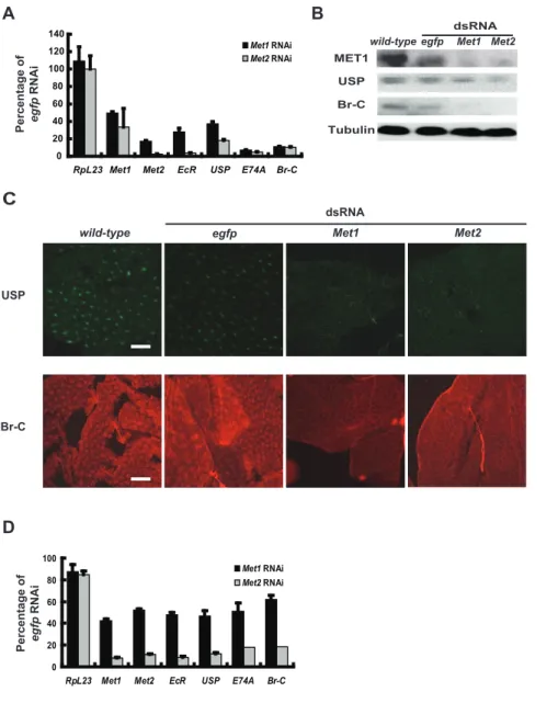
Since theMetRNAi effects resemble those ofEcR-USPRNAi at the phenotypic level, we investigated whether Met RNAi also disrupts the 20E-triggered transcriptional cascade in the fat body. As determined by qPCR, the 20E-response genesEcR,USP,Br-C, andE74Awere significantly downregulated 24 hr afterMetRNAi. Similar to the phenotypic effects of Met RNAi, the inhibitory effects on gene expression byMet2RNAi (70–95%) were stronger than forMet1 RNAi (50–90%) (Figure 4A). Moreover, Western blots for MET1, USP, and Br-C (Figure 4B) as well as immunohistochemistry for USP and Br-C (Figure 4C) revealed that protein levels were decreased by Met RNAi. To avoid the possibility of off-targeting, we generated two other sets of Met dsRNAs (Figure S1A) which exhibited similar but stronger inhibiting effects on gene expression when higher concentrations (30mg of dsRNA per larva) were used (Figure S3A). Since three different sets ofMetdsRNAs were used, the off-targeting problem should be largely minimized. To be consistent with the above experimental data (Figures 2, 3, 4 and S2), we still used the first set ofMetdsRNAs in the following experiments.
To verify the above results, samples of fat body were explanted 24 hr after RNAi treatment and cultured for an additional 6 hr in the presence of 20E. 20E treatment caused significant upregula-tion of all 6 genes in theegfpRNAi treated fat body, while this upregulation was dramatically decreased when Met2 RNAi was performed (Figure S3B). We therefore conclude thatMet RNAi disrupts the 20E-triggered transcriptional cascade in the fat body during larval-pupal metamorphosis.
To prevent effects of JH, Met RNAi experiments were performed,6 hr after pupation, a stage where JH is absent and 20E is present [30]. Surprisingly, bothEcR-USPRNAi and Met RNAi at this stage did not cause lethality. However, the 20E-response genes EcR, USP, Br-C, and E74A were significantly downregulated 24 hr afterMetRNAi treatment, and the inhibitory effects of Met2 RNAi were stronger than for Met1 RNAi (Figure 4D).
To further insure that the interference was not due to JH, we performedMetRNAi followed by the addition of 20E toBombyx DZNU-Bm-12 cells [35] which should lack JH.Met2mRNA levels were very low in these cells and the efficiency ofMet2RNAi was poor, butMet1RNAi decreasedMet1mRNA levels by about 90%. Six hr after 20E treatment, all the 20E-response genes were significantly upregulated in egfp RNAi treated cells, but this upregulation was significantly decreased in Met1 RNAi treated cells (Figure S3C). These data show conclusively that MET is required for the maximal ability of 20E to induce gene expression, and thus tissue remodeling, inBombyx.
In addition, RNAi knockdown of eitherEcRorMetinTribolium during the early quiescent stage resulted in lethality, delayed larval-pupal transition, and disrupted the 20E-triggered transcrip-tional cascade (Figure S4), demonstrating that MET is also required for the maximal action of 20E during metamorphosis in
Tribolium. We then turned to the question of transactivation of the 20E-ecdysone receptor complex by MET inBombyx.
MET, EcR-USP and EcRE are components of a protein-DNA complex
It has been reported that Drosophila MET physically interacts with EcR-USP [36,37]. A CytoTrap yeast two-hybrid experiment was carried out to investigate whether such direct associations among MET1, MET2, EcR and USP occur in Bombyx. As expected, EcR (the EcR-B1 isoform was used throughout the paper) and USP strongly associate with one another. Weak associations were observed between MET1 and MET2, MET1 and MET1, and MET2 and MET2, while intermediate associa-tions were formed between the two MET proteins and the ecdysone receptor (Figure 5A). To confirm the yeast two-hybrid Figure 1. TwoMetgenes in theBombyxgenome.Three biological replicates were used, one of which is represented. In each biological replicate, more than 10 larvae were used (A–C). (A) From day 2 of the 4thinstar to day 2 of the prepupal stage,Met1andMet2mRNA expression in fat body was
determined by qPCR. The developmental profiles show expression peaks during molting and pupation. 4L2D, day 2 of the fourth instar, and so on; M, molting; W, the wandering stage; PP, the prepupal stage. (B)Met1andMet2mRNA levels (left panel), and MET1 protein level (right panel) were increased by 20E treatmentin vivo. 20E (1mg per larva) was injected into selected larvae on day 2 of the fifth instar, and fat body was explanted for
qPCR analysis and Western blots 6 hr after 20E treatment. Tubulin was used as a loading control. (C)Met1andMet2mRNA levels (left panel), and MET1 protein level (right panel) were decreased byEcRRNAi andUSPRNAiin vivo. dsRNA (10mg per larva) was injected into larvae during the
initiation of the early wandering stage, and fat body was explanted for qPCR analysis and Western blots 24 hr after RNAi treatment. Tubulin was used as a loading control. (D) Simultaneous addition of 1mM 20E and 10mg/ml cycloheximide (CHX) toBombyxDZNU-Bm-12 cells for 2 hr revealed that
results that MET associates with EcR-USP, we performed immunoprecipitation experiments. When the HA-EcR, FLAG-USP, and V5-Met1 constructs were co-transfected into human HEK 293 cells, MET physically interacts with EcR-USP, while 20E treatment had little or no stimulating effects on the physical interactions between MET1 and EcR-USP (Figure 5B) confirming the results reported inDrosophila[36,37]. As negative controls, IgG was not able to pull down endogenous HA-EcR, FLAG-USP, or V5-MET1 (Figure S5A).
Since MET physically interacts with the ecdysone receptor, we investigated whether MET, ecdysone receptor and EcRE form a protein-DNA complex when the HA-EcR, FLAG-USP, V5-Met1, andcMyc-Met2constructs were co-transfected into HEK 293 cells. As expected, the overexpressed MET was not able to bind EcRE in the electrophoretic mobility shift assay (EMSA) but EcR-USP did (Figure S5B). Addition of the HA or FLAG antibody resulted in a shift of EcRE by HA-EcR or FLAG-USP (Figure 5C). When the V5 or cMyc antibody was added, binding of the ecdysone receptor-EcRE complex was also shifted by V5-MET1 or cMyc-MET2 (Figure 5D), demonstrating that MET, ecdysone receptor and EcRE form a protein-DNA complex.
Subsequently, Met1 RNAi and EcRE-Luc transfections were simultaneously conducted in DZNU-Bm-12 cells, followed by 20E treatment and measurements of EcRE-driven luciferase activity. Met1RNAi had no apparent effects on basal luciferase activity, but significantly decreased 20E-induced EcRE-driven luciferase activ-ity. Moreover, co-transfection withEcRandUSP increased 20E-induced EcRE-driven luciferase activity, and this induction was again significantly decreased byMet1RNAi (Figure 5F), suggesting that MET is required for the maximal action of 20E in inducing gene expression via physical interaction with the ecdysone receptor and EcRE.
Discussion
In this study, we demonstrate that MET is required for the maximal action of 20E inBombyx. Although theMet1mRNA level in the fat body is much higher than theMet2mRNA level, with the first set of dsRNAs, the inhibiting effects ofMet2RNAi on 20E-triggered gene expression and tissue remodeling were stronger than forMet1RNAi. However, results from the other two sets of dsRNAs showed thatMet1RNAi had higher inhibiting effects than Met2 RNAi. Met RNAi performed at different developmental stages and in cultured cells confirmed the major conclusion that MET is required for the maximal action of 20E in Bombyx. Unfortunately, it is difficult to determine with certainty which MET is more important and whether the two Met genes are functionally redundant in the 20E signal transduction pathway when only using RNAi methodology. One reason is that RNAi knockdown of one Met gene downregulates the otherMet gene, which is 20E responsive. Our preliminary data suggest that the Met1mRNA level in the midgut was more abundant than theMet2 mRNA level and that using the the first set of dsRNAs,Met1RNAi resulted in a more severe inhibitory effects on midgut remodeling thanMet2RNAi. Since the systemic RNAi approach might result in non-tissue-autonomous effects, we suppose that performing RNAi with the binary GAL4/UAS system in Bombyx [38,39] might be more useful for understanding which MET is more important in terms of different tissues or developmental stages. Mutation of the twoMetgenes, both separately and together, using Figure 2. Lethal and defective phenotypes caused byMetRNAi
in silkworms.dsRNA (10mg per larva) was injected into selected larva
during initiation of the early wandering stage.egfpdsRNA was used as a control. (A–C) TypicalMet1RNAi andMet2RNAi treated silkworms died during the wandering stage (A) or during pupation (B), while some were arrested at the mid-pupal stage (C). The pictures (A–C) show the dying animals afterMetRNAi. (D, E)MetRNAi affected adult structure formation. The survivingMet1RNAi andMet2RNAi treated pupae did not fully develop legs and wings during the late pupal stage (D). Many of the surviving Met1 RNAi adults failed to shed the pupal cuticle attached to their head or abdomen, exhibiting shortened and distorted legs or unexpanded wings (E).
doi:10.1371/journal.pone.0053256.g002
Table 1.MetRNAi results in lethality during the larval-pupal-adult metamorphosis.
dsRNA Treatment Injected larvae Larval lethality,% Prepupal lethality,% Pupal lethality,% Total lethality,%
egfp 10ug/larva 87 0 3.5 0 3.5
Met1 10ug/larva 92 15.2 13.1 21.7 50
Met2 10ug/larva 91 30.7 18.7 29.7 79.1
Met1+2 5+5ug/larva 97 8.2 19.6 23.7 52.5
dsRNA (10mg per larva) was injected into selected larvae during initiation of the early wandering stage. Lethality was scored at the larval, prepupal, and pupal stages to compare the effects ofegfp,Met1,Met2, andMet1+2dsRNAs.
doi:10.1371/journal.pone.0053256.t001
a gene-targeting method [40] should be able to eventually resolve the problem in the future.
BothBombyx Met genes are 20E responsive, exhibiting similar expression patterns to other 20E-response genes, includingBr-C, E74,E75andE93. RNAi knockdown of each of these genes also interrupts the 20E-triggered transcriptional cascade to different levels (unpublished data). Thus, it appears that the 20E induction of those 20E-response genes (including theMetgenes) is required for the maximal action of 20E to induce gene expression in Bombyx. In Drosophila, mutation of Br-C, E74, E75, or E93 interrupts the 20E signaling, with more pronounced effects in E93mutants. It has been well documented that the 20E induction of E93 determines a PCD response by positively impacting the 20E signaling [19]. The feedback regulation of 20E signaling should be common in insects.
Previously, we demonstrated that MET and GCE are functionally redundant in transducing JH signal to induceKr-h1 expression and to antagonize 20E-inducedBr-Cexpression [6,7]. However, our preliminary experiments suggest that MET and GCE might be not the same in modulating 20E signaling using the Met and gce mutants. It might be incomparable between the function ofMetandgceinDrosophilaand that ofMet1andMet2in Bombyx, since the duplication events in these two insect species are evolutionary independent [6,41]. Nevertheless, in Tribolium, Met RNAi during the early quiescent stage also disrupts the 20E-triggered transcriptional cascade, showing that the single MET protein in Tribolium and the two MET proteins in Bombyx have similar functions in modulating 20E signaling during metamor-phosis.
We have also tried to dissected out the molecular mechanism how MET is required for the maximal action of 20E inBombyx. Our preliminary results suggest that MET might bind
transcrip-tional co-activators, including CBP/p300 [42], which is important for transactivation of the 20E-ecdysone receptor complex via EcRE. The family of bHLH-PAS transcriptional regulators, consisting of transcription factors and co-activators, are critical components of gene expression networks that underlie essential developmental and environmental processes [43,44]. The mam-malian bHLH-PAS transcription factors, such as the dioxin receptor (DR), recruit many transcriptional co-activators, includ-ing CBP/p300, p160/SRC/NCoA, p140, and CARM1/PRMT1 for transactivation [45]. Ligand-activated nuclear receptors (i.e. estrogen receptor; ER) also recruit these transcriptional co-activators for transactivation [45]. Thus, it might be a common mechanism that a bHLH-PAS transcription factor recruits histone-modifying transcriptional co-activators to liganded nuclear receptors for transactivation [43,45].CBP/p300RNAi attenuated, but did not abolish, 20E-induced luciferase activity driven by EcRE, suggesting that the receptor complex may consist of other histone-modifying transcriptional co-activators. A good candidate is p160/SRC/NCoA, which has been demonstrated in Aedes, Drosophila, andTribolium[20,21,22,23].
Very recently, it has been documented that theBombyxMET2 might act as a JH receptor. In the presence of JH, MET2 associates with SRC, the p160/SRC/NCoA-like molecule in Bombyx, to interact the JHRE in inducingKr-h1expression [31]. Considering the MET function in both JH and 20E actions, we propose that MET plays a role mediating JH-20E crosstalk, and that the detailed molecular mechanism is surely worthy of further investigation.
Figure 3.MetRNAi prevents fat body remodeling.The inhibitory effects on fat body remodeling byMet2RNAi during the initiation of the early wandering stage were stronger than forMet1RNAi (A–C).egfpdsRNA was used as a control. (A)MetRNAi prevented the apoptotic events, estimated by TUNEL (left panel, green) and measured by caspase 3 activity (right panel) 18 hr after RNAi treatment. The inset shows that TUNEL (green) and DAPI (blue) co-localize in nuclei (Bar: 50mm). (B)MetRNAi prevented autophagy, estimated by LysoTracker (red) 24 hr after RNAi treatment. The inset shows that the LysoTracker (red) and DAPI (blue) stain the cytoplasm and the nuclei, respectively (Bar: 50mm). (C)MetRNAi dramatically prevented
cell dissociation of the fat body 24 hr after pupation (Bar: 50mm).
Materials and Methods
Insects and cell lines
Bombyx (P50) [28,29], Tribolium [9] were reared as previously described. Bombyx DZNU-Bm-12 cells [35] were maintained in TNM-FH (Sigma) medium supplemented with 10% heat-inacti-vated fetal bovine serum (Hyclone) at 27uC. And human HEK 293 cells were maintained in Dulbecco’s Modified Eagle Medium (Hyclone) supplemented with 10% heat-inactivated fetal bovine serum (Hyclone).
Conventional molecular, biochemical, and cellular approaches
The full-length Met1 and Met2 cDNA sequences (GenBank accession numbers: EU249371 and EU249372) were cloned using RACE. Details of qPCR and Western blotting were previously described [7,27,28,46]. Caspase 3 activity was determined according to the manufacturer’s instructions (Beyotime, Shanghai, China). TUNEL labeling (Beyotime) and LysoTracker staining (Invitrogen) were used to estimate apoptosis and autophagy, respectively, and monitored with an Olympus Fluoview FV1000 confocal microscope. Primers used here and elsewhere are listed in Table S1.
Figure 4.MetRNAi disrupts the 20E-triggered transcriptional cascade.RNAi was performed during initiation of the early wandering stage (A–C) and,6 hr after pupation (D). The RNAi knockdown efficiency byMet2RNAi is higher than forMet1RNAi, and the downregulation rate ofMet1
byMet2RNAi is higher than forMet2byMet1RNAi. The#1 set ofMetdsRNA was used.egfpdsRNA was used as a control. (A)Met1andMet2, and the 20E-response genesEcR,USP,E74AandBr-C, as determined by qPCR, were significantly downregulated 24 hr afterMetRNAi.RpL23is used as a negative control of 20E-response gene. (B) MET1, USP and Br-C protein levels, as determined by Western blots, significantly decreased 24 hr afterMet
RNAi. Tubulin was used as a loading control. (C) USP and Br-C protein levels, as estimated by immunohistochemistry, significantly decreased 24 hr afterMetRNAi. Localization of USP (green) and Br-C (red) were restricted to nuclei (Bar: 50mm). (D) RNAi was performed,6 hr after pupation. The
rest is as in (A).
doi:10.1371/journal.pone.0053256.g004
RNAi and hormone treatment
dsRNAs were generated using the T7 RiboMAXTM Express RNAi System (Promega). Preliminary data showed that the P50
strain ofBombyx (the Chinese strain variation, Dazao) was more sensitive to RNAi treatments than the other tested strains [47], and the P50 strain ofBombyx was used throughout this study. RNAi Figure 5. Physical interaction between MET and EcR-USP.(A) The CytoTrap yeast two-hybrid analyses revealed direct associations among MET1, MET2, EcR and USP. Strong associations between bait and prey proteins led to more yeast colonies. (B) When theHA-EcR,FLAG-USP, and V5-Met1constructs were co-transfected into human HEK 293 cells, 20E treatment for 6 hr at a final concentration of 1mM had little or no stimulating
effects on the physical interactions between MET and EcR-USP. In the immunoprecipation experiments, the bottom Western blot is input. IP, immunoprecipitate; Blot, Western blot. (C) TheHA-EcR,FLAG-USP,V5-Met1, andcMyc-Met2constructs were co-transfected into the human HEK 293 cells. After nuclear extracts were bound with biotin-labeled EcRE, the protein-DNA complexes were separated on a 5% native PAGE gel followed by EMSA. Addition of the HA or FLAG antibody resulted in a shift of EcRE. In (C) and (D), the shift was indicated by a black arrow in comparison with a gray arrow. (D) TheHA-EcR,FLAG-USP,V5-Met1, andcMyc-Met2constructs were co-transfected into human HEK 293 cells. After nuclear extracts were bound with biotin-labeled EcRE, the protein-DNA complexes were separated 5% native PAGE followed by EMSA. When the V5 or cMyc antibody was added, binding of EcR-USP-EcRE was shifted by MET1 and MET2 in EMSA showing that MET, EcR-USP and EcRE form a protein-DNA complex. (E)Met1
RNAi and transfection were simultaneously conducted in BombyxDZNU-Bm-12 cells for 48 hr, followed by 20E treatment for 6 hr at a final concentration of 1mM, and measurements of EcRE-driven luciferase activity were done. MET is required for 20E function to induce gene expression
knockdown was performed at two developmental stages, including initiation of the early wandering stage and 6 hr after pupation. After the RNAi treatment (10 or 30mg of dsRNA per animal), fat body from the abdominal segments was collected for bioassays. Three biological replicates were used, each of which consisted of 10 silkworms. The details of hormone treatmentin vivo(1mg 20E per animal; Sigma Aldrich) were previously described [28,29,48]. RNAi knockdown in DZNU-Bm-12 cells was performed using the Effectene transfection reagent (Qiagen) for 48 hr at a final concentration of 2mg/ml dsRNA. To determine the 20E primary-response genes, 1mM 20E and 10mg/ml cycloheximide (Sigma Aldrich) were used [48].
Antibodies and immunohistochemistry
TheBombyxMET1 and Br-C antibodies were produced by the Abmart Company (Shanghai). A cDNA fragment encoding amino acids 151M to 350Q of MET1 and the full-lengthBr-CZ4 cDNA were expressed inE. coliand their protein products were purified. Antigen-purified rabbit polyclonal antibodies against MET1 and Br-C were generated. The AB11 USP-specific monoclonal antibody was provided by Dr. K.F. Kafatos (Harvard University). The monoclonal antibodies against the V5 tag (Sigma Aldrich), cMyc tag (Santa Cruz), and Tubulin (Invitrogen) were also used. USP and Br-C were detected in explanted fat body from the 5th abdominal segment by immunohistochemistry with the above primary antibodies. The fluorescein-conjugated secondary anti-bodies (Jackson ImmunoResearch) were FITC-conjugated Affini-pure Goat Anti-Mouse IgG for USP and Cy3-conjugated Affinipure Goat Anti-Rabbit IgG for Br-C. Fluorescence signals were detected using the Olympus Fluoview FV1000 confocal microscope.
Yeast two-hybrid assay
Yeast two-hybrid assays were carried out using the CytoTrap system (Stratagene), which is based on the ability of human Sos to complement a temperature-sensitivecdc25allele (cdc25H) in yeast when Sos is targeted to the plasma membrane through bait-prey interactions. This system has been well characterized for protein-protein interaction studies between transcription factors and their associated proteins (SR6). First, the full lengthMET1,MET2, EcR-B1(EcR) andUSP1(USP) were amplified from silkworm genome, then these genes were cloned into bait or prey vector. MET1, MET2, EcR or USP was expressed as a fusion protein with human Sos as the bait protein. On the other hand, MET1, MET2, EcR and USP were expressed as prey proteins fused with a myristoylation (Myr) signal, targeting the proteins to the cell membrane. Expression of the prey is controlled by the GAL1 promoter, which is induced on galactose, and repressed on glucose medium. When bait and prey are co-transformed into thecdc25H strain, the only cells capable of growing at restrictive temperatures on galactose medium are those that have been rescued by the bait-prey interactions that recruit Sos to the cell membrane.
Transient transfection assay
Transient transfection assay in DZNU-Bm-12 cells was carried out for 48 hr using Effectene according to the manufacturer’s instructions. The final DNA concentration was 2mg/ml, and the DNA:Effectene ratio was 1:25. The vector used to overexpress V5-Met1andcMyc-Met2was pEGFP-N1 (Clontech) under the control of the ie1 promoter. After transfection, cells were treated with 1mM 20E, followed by immunoprecipitation, qPCR, and luciferase assay.
Transient transfection assays in HEK 293 cells were performed using Lipofectamine 2000 (Invitrogen). The full length MET1,
MET2,EcR-B1(EcR) andUSPwere amplified from the silkworm genome, and theHA,FLAG,V5,cMyctags were fused in their 5-ends, respectively and cloned into the pcDNA 3.1(+) vector (Invitrogen). After transfection, the cells were harvested for immunopreciptation, luciferase assay and EMSA.
Immunoprecipitation
After treatment, DZNU-Bm-12 cells and HEK 293 cells were harvested and lysed in ice-cold NP-40 lysis buffer (Beyotime). Lysates were incubated with FLAG, V5, or cMyc antibody or IgG for 4 hr, followed by incubation with protein G (GE Healthcare) overnight at 4uC. After extensive washing with cold NP-40 buffer, the samples were treated with RIPA lysis buffer (Beyotime) about 15min on the ice. Then immunoprecipitates were separated by SDS-PAGE and analyzed by Western blots after measured the protein concentration by the enhanced BCA protein assay kit (Beyotime).
Luciferase assay
Luciferase assays were carried out using the Dual Luciferase Assay System (Promega) and a Modulus Luminometer (Turner BioSystems). The reporter pGL3 vector (Promega) containing four repeated EcRE sequences (GACAAGGGTTCAATG-CACTTGTC) and a hsp70 mini promoter was used for the luciferase reporter. And the reference pRL vector (Promega) carrying Renila-luciferase driven by actin3 promoter was co-transfected into the cell with the reporter vector. The dual luciferase double reporter assay system and substrates were purchased from Promega.
EMSA
The HA-EcR, FLAG-USP, V5-Met1, and cMyc-Met2constructs were co-transfected into HEK 293 cells and nuclear extracts were prepared by the NE-PER Nuclear and Cytoplasmic Extraction Reagents (Thermo). The minimal EcRE (sence: AGTT-CAATGGCCT; anti-sense: AGGCCATTGAACT) was biotin-labeled as a probe using the Biotin 39 End DNA Labeling Kit (Pierce). After binding, the nuclear extract (15mg) containing the biotin-labeled EcRE and the protein-DNA complexes were separated on 5% nondenaturing PAGE gel. HA, FLAG, V5, and cMyc antibodies were added to the nuclear extract to detect the shift of EcRE. EMSA was performed using the LightShift Chemiluminescent EMSA Kit (Pierce).
Supporting Information
Figure S1 The diagram of the three sets ofMetdsRNA and confirmation of the MET and Br-C antibodies. (A) The diagram illustrates the three sets ofMetdsRNA. Red bar:#1 set ofMet1(491–916) andMet2(491–916) dsRNA; green bar:#2 set ofMet1(141–586) andMet2(1925–2336) dsRNA; yellow bar: #3 set ofMet1(948–1348) andMet2(245–669) dsRNA. (B and C) Western blotting confirmation of the MET1 and Br-C antibody afterMet1andBr-CRNAi. The arrow points to the MET1 protein and the Br-C protein isoforms with ideal molecular weights.efgp RNAi was used as a control. Tubulin was used as a loading control.
(PDF)
Figure S2 MetRNAi prevents removal of obsolete larval tissues and generation of adult structures.dsRNA (10mg per larva) was injected into larvae during initiation of the early wandering stage. More than 30 silkworms were used in each group. egfpdsRNA was used as a control. (A) Met RNAi larvae
form thinner cocoons. Cocoon images were collected after the silkworms stopped spinning. (B) Met RNAi prevented silk gland lysis 24 hr after pupation. The inhibiting effects, particularly on the middle silk gland, by Met2 RNAi were stronger than Met1 RNAi. (C)Met1RNAi affected adult structure formation. Many of the survivingMet1RNAi treated adults exhibited shortened and distorted legs (left panel) or unexpanded wings (right panel). (PDF)
Figure S3 Met RNAi disrupts the 20E-triggered tran-scriptional cascade during the early wandering stage and in DZNU-Bm-12 Cells.Three biological replicates were used and one was represented (A–C). In each biological replicate, more than 10 larvae were used (A and B).egfpdsRNA was used as a control. (A) The other two sets (#2 and #3) of Met dsRNA (30mg per larva) also disrupt the 20E-triggered transcriptional cascade during initiation of the early wandering stage. See Figure S1A for the locations of the three sets of Met dsRNA. (B) 20E treatment fails to induce expression of 20E-response genes in fat body explanted from theMet2RNAi silkworms during the early wandering stage. (C) Met1 RNAi disrupts the 20E-triggered transcriptional cascade, except Met2 whose expression level is extremely low, inBombyxDZNU-Bm-12 cells. RNAi knockdown was performed using the Effectene transfection reagent (Qiagen) for 48 hr at a final concentration of 2mg/ml dsRNA. The cells were treated with 20E for 6 hr at a final concentration of 1mM. (PDF)
Figure S4 Met is required for 20E action inTribolium. RNAi knockdown of either EcR or Met (,4 ng per larva) in Triboliumduring the early quiescent stage resulted in lethality (A), significantly delayed the larval-pupal transition (A and B), and disrupted the 20E-triggered transcriptional cascade (C). egfp dsRNA was used as a control. (A) Larval, prepupal, and pupal
numbers were counted 24 and 48 hr after RNAi treatment. Total lethality caused byegfp,MetandEcRdsRNAs was compared. (B) Phenotypic images were collected from the above experimental animals 24 (left) and 48 hr (right) after RNAi treatment. (C)Met, EcR,USP,E74, andBr-CmRNA levels, as determined by qPCR, were significantly down-regulated 24 hr afterMetRNAi. (PDF)
Figure S5 The negative controls for the IP and EMSA experiments. (A) The HA-EcR, FLAG-USP, and V5-Met1 constructs were co-transfected into human HEK 293 cells, the cells were treated by 20E for 6 hr at a final concentration of 1mM. The negative control IgG was not able to pull down HA-EcR, FLAG-USP, and V5-Met1. IP, immunoprecipitate; Blot, Western blot. (B) TheHA-EcRandFLAG-USPorV5-Met1andcMyc-Met2 constructs were co-transfected into the human HEK 293 cells. After nuclear extracts were bound with biotin-labeled EcRE, the protein-DNA complexes were separated on a 5% native PAGE gel followed by EMSA. The shift was indicated by a black arrow in comparison with a gray arrow.
(PDF)
Table S1 A list of all primers used in this paper. (DOC)
Acknowledgments
We sincerely thank Drs. Thomas G. Wilson and David Schooley for critical reading of the manuscript.
Author Contributions
Conceived and designed the experiments: S. Li YC JW. Performed the experiments: EG QH S. Liu LT ZS QP JG MS KL. Analyzed the data: EG S. Li. Wrote the paper: S. Li EG YC JW LG.
References
1. Ashok M, Turner C, Wilson TG (1998) Insect juvenile hormone resistance gene homology with the bHLH-PAS family of transcriptional regulators. Proc Natl Acad Sci U S A 95: 2761–2766.
2. Miura K, Oda M, Makita S, Chinzei Y (2005) Characterization of theDrosophila Methoprene -tolerant gene product. Juvenile hormone binding and ligand-dependent gene regulation. FEBS J 272: 1169–1178.
3. Riddiford LM (2008) Juvenile hormone action: a 2007 perspective. J Insect Physiol 54: 895–901.
4. Godlewski J, Wang S, Wilson TG (2006) Interaction of bHLH-PAS proteins involved in juvenile hormone reception inDrosophila. Biochem Biophys Res Commun 342: 1305–1311.
5. Wilson TG, Ashok M (1998) Insecticide resistance resulting from an absence of target-site gene product. Proc Natl Acad Sci U S A 95: 14040–14044. 6. Abdou MA, He Q, Wen D, Zyaan O, Wang J, et al. (2011)DrosophilaMet and
Gce are partially redundant in transducing juvenile hormone action. Insect Biochem Mol Biol 41: 938–945.
7. Liu Y, Sheng Z, Liu H, Wen D, He Q, et al. (2009) Juvenile hormone counteracts the bHLH-PAS transcription factors MET and GCE to prevent caspase-dependent programmed cell death in Drosophila. Development 136: 2015–2025.
8. Riddiford LM, Truman JW, Mirth CK, Shen YC (2010) A role for juvenile hormone in the prepupal development ofDrosophila melanogaster. Development 137: 1117–1126.
9. Konopova B, Jindra M (2007) Juvenile hormone resistance gene Methoprene-tolerant controls entry into metamorphosis in the beetleTribolium castaneum. Proc Natl Acad Sci U S A 104: 10488–10493.
10. Parthasarathy R, Tan A, Palli SR (2008) bHLH-PAS family transcription factor methoprene-tolerant plays a key role in JH action in preventing the premature development of adult structures during larval-pupal metamorphosis. Mech Dev 125: 601–616.
11. Charles JP, Iwema T, Epa VC, Takaki K, Rynes J, et al. (2011) Ligand-binding properties of a juvenile hormone receptor, Methoprene-tolerant. Proc Natl Acad Sci U S A 108: 21128–21133.
12. Koelle MR, Talbot WS, Segraves WA, Bender MT, Cherbas P, et al. (1991) The Drosophila EcRgene encodes an ecdysone receptor, a new member of the steroid receptor superfamily. Cell 67: 59–77.
13. Yao TP, Forman BM, Jiang Z, Cherbas L, Chen JD, et al. (1993) Functional ecdysone receptor is the product of EcR and Ultraspiracle genes. Nature 366: 476–479.
14. Arbeitman MN, Hogness DS (2000) Molecular chaperones activate the Drosophila ecdysone receptor, an RXR heterodimer. Cell 101: 67–77. 15. Tsai CC, Kao HY, Yao TP, McKeown M, Evans RM (1999) SMRTER, a
Drosophilanuclear receptor coregulator, reveals that EcR-mediated repression is critical for development. Mol Cell 4: 175–186.
16. Kimura S, Sawatsubashi S, Ito S, Kouzmenko A, Suzuki E, et al. (2008) Drosophilaarginine methyltransferase 1 (DART1) is an ecdysone receptor co-repressor. Biochem Biophys Res Commun 371: 889–893.
17. Henrich VC (2012) The ecdysteroid receptor. Insect Endocrinology, Academic Press, San Diego, CA, U S A pp.177–218.
18. Yin VP, Thummel CS (2005) Mechanisms of steroid-triggered programmed cell death inDrosophila. Semin Cell Dev Biol 16: 237–243.
19. Lee CY, Wendel DP, Reid P, Lam G, Thummel CS, et al. (2000) E93 directs steroid-triggered programmed cell death inDrosophila. Mol Cell 6: 433–443. 20. Bai J, Uehara Y, Montell DJ (2000) Regulation of invasive cell behavior by
taiman, a Drosophila protein related to AIB1, a steroid receptor coactivator amplified in breast cancer. Cell 103: 1047–1058.
21. Zhu J, Chen L, Sun G, Raikhel AS (2006) The competence factor beta Ftz-F1 potentiates ecdysone receptor activity via recruiting a p160/SRC coactivator. Mol Cell Biol 26: 9402–9412.
22. Li M, Mead EA, Zhu J (2011) Heterodimer of two bHLH-PAS proteins mediates juvenile hormone-induced gene expression. Proc Natl Acad Sci U S A 108: 638–643.
23. Zhang Z, Xu J, Sheng Z, Sui Y, Palli SR (2011) Steroid receptor co-activator is required for juvenile hormone signal transduction through a bHLH-PAS transcription factor, methoprene tolerant. J Biol Chem 286: 8437–8447. 24. Broadus J, McCabe JR, Endrizzi B, Thummel CS, Woodard CT (1999) The
Drosophilabeta FTZ-F1 orphan nuclear receptor provides competence for stage-specific responses to the steroid hormone ecdysone. Mol Cell 3: 143–149. 25. Bernardo TJ, Dubrovsky EB (2012) TheDrosophilaJuvenile Hormone Receptor
26. Dubrovsky EB, Dubrovskaya VA, Bernardo T, Otte V, DiFilippo R, et al. (2011) TheDrosophilaFTZ-F1 nuclear receptor mediates juvenile hormone activation of E75A gene expression through an intracellular pathway. J Biol Chem 286: 33689–33700.
27. Tian L, Guo E, Diao Y, Zhou S, Peng Q, et al. (2010) Genome-wide regulation of innate immunity by juvenile hormone and 20-hydroxyecdysone in theBombyx fat body. BMC Genomics 11: 549.
28. Tian L, Guo E, Wang S, Liu S, Jiang RJ, et al. (2010) Developmental regulation of glycolysis by 20-hydroxyecdysone and juvenile hormone in fat body tissues of the silkworm,Bombyx mori. J Mol Cell Biol 2: 255–263.
29. Tian L, Liu S, Liu H, Li S (2012) 20-hydroxyecdysone upregulates apoptotic genes and induces apoptosis in theBombyxfat body. Arch Insect Biochem Physiol 79:207–219.
30. Kayukawa T, Minakuchi C, Namiki T, Togawa T, Yoshiyama M, et al. (2012) Transcriptional regulation of juvenile hormone-mediated induction ofKruppel homolog 1, a repressor of insect metamorphosis. Proc Natl Acad Sci U S A 109: 11729–11734.
31. Muramatsu D, Kinjoh T, Shinoda T, Hiruma K (2008) The role of 20-hydroxyecdysone and juvenile hormone in pupal commitment of the epidermis of the silkworm,Bombyx mori. Mech Dev 125: 411–420.
32. Ryoo HD, Baehrecke EH (2010) Distinct death mechanisms in Drosophila development. Curr Opin Cell Biol 22: 889–895.
33. Rusten TE, Lindmo K, Juha´sz G, Sass M, Seglen PO, et al. (2004) Programmed autophagy in theDrosophilafat body is induced by ecdysone through regulation of the PI3K pathway. Dev Cell 7: 179–192.
34. Scott RC, Schuldiner O, Neufeld TP (2004) Role and regulation of starvation-induced autophagy in theDrosophilafat body. Dev Cell 7: 167–178. 35. Khurad AM, Zhang MJ, Deshmukh CG, Bahekar RS, Tiple AD, et al. (2009) A
new continuous cell line from larval ovaries of silkworm,Bombyx mori. In Vitro Cell Dev Biol Anim 45: 414–419.
36. Li Y, Zhang Z, Robinson GE, Palli SR (2007) Identification and characteriza-tion of a juvenile hormone response element and its binding proteins. J Biol Chem 282: 37605–37617.
37. Bitra K, Palli SR (2009) Interaction of proteins involved in ecdysone and juvenile hormone signal transduction. Arch Insect Biochem Physiol 70: 90–105. 38. Imamura M, Nakai J, Inoue S, Quan GX, Kanda T, et al. (2003) Targeted gene
expression using the GAL4/UAS system in the silkwormBombyx mori. Genetics 165: 1329–1340.
39. Ma L, Xu H, Zhu J, Ma S, Liu Y, et al. (2011)Ras1CA
overexpression in the posterior silk gland improves silk yield. Cell Res 21: 934–943.
40. Takasu Y, Kobayashi I, Beumer K, Uchino K, Sezutsu H, et al. (2010) Targeted mutagenesis in the silkworm Bombyx moriusing zinc finger nuclease mRNA injection. Insect Biochem Mol Biol 40:759–765.
41. Baumann A, Fujiwara Y, Wilson TG (2010) Evolutionary divergence of the paralogs Methoprene tolerant (Met) and germ cell expressed (gce) within the genusDrosophila. J Insect Physiol 56: 1445–1455.
42. Chan HM, La Thangue NB (2001) p300/CBP proteins: HATs for transcrip-tional bridges and scaffolds. J Cell Sci 114: 2363–2373.
43. Partch CL, Gardner KH (2010) Coactivator recruitment: a new role for PAS domains in transcriptional regulation by the bHLH-PAS family. J Cell Physiol 223: 553–557.
44. Kewley RJ, Whitelaw ML, Chapman-Smith A (2004) The mammalian basic helix-loop-helix/PAS family of transcriptional regulators. Int J Biochem Cell Biol 36: 189–204.
45. Perissi V, Rosenfeld MG (2005) Controlling nuclear receptors: the circular logic of cofactor cycles. Nat Rev Mol Cell Biol 6: 542–554.
46. Liu Y, Zhou S, Ma L, Tian L, Wang S, et al. (2010) Transcriptional regulation of the insulin signaling pathway genes by starvation and 20-hydroxyecdysone in theBombyxfat body. J Insect Physiol 56: 1436–1444.
47. Huang J, Zhang Y, Li M, Wang S, Liu W, et al. (2007) RNA interference-mediated silencing of the bursicon gene induces defects in wing expansion of silkworm. FEBS Lett 581: 697–701.
48. Zhou S, Zhou Q, Liu Y, Wang S, Wen D, et al. (2010) Two Tor genes in the silkwormBombyx mori. Insect Mol Biol 19: 727–735.