Transcriptional Profiling of Bergmann Glial Cells from
Mouse Cerebellum
Samir Koirala1,2, Gabriel Corfas1,2,3*
1F.M. Kirby Neurobiology Center, Children’s Hospital Boston, Boston, Massachusetts, United States of America,2Department of Neurology, Harvard Medical School, Boston, Massachusetts, United States of America,3Department of Otolaryngology, Harvard Medical School, Boston, Massachusetts, United States of America
Abstract
Bergmann glial cells play critical roles in the structure and function of the cerebellum. During development, their radial processes serve as guides for migrating granule neurons and their terminal endfeet tile to form the glia limitans. As the cerebellum matures, Bergmann glia perform important roles in synaptic transmission and synapse maintenance, while continuing to serve as essential structural elements. Despite growing evidence of the diverse functions of Bergmann glia, the molecular mechanisms that mediate these functions have remained largely unknown. As a step toward identifying the molecular repertoire underlying Bergmann glial function, here we examine global gene expression in individual Bergmann glia from developing (P6) and mature (P30) mouse cerebellum. When we select for developmentally regulated genes, we find that transcription factors and ribosomal genes are particularly enriched at P6 relative to P30; whereas synapse associated molecules are enriched at P30 relative to P6. We also analyze genes expressed at high levels at both ages. In all these categories, we find genes that were not previously known to be expressed in glial cells, and discuss novel functions some of these genes may potentially play in Bergmann glia. We also show that Bergmann glia, even in the adult, express a large set of genes thought to be specific to stem cells, suggesting that Bergmann glia may retain neural precursor potential as has been proposed. Finally, we highlight several genes that in the cerebellum are expressed in Bergmann glia but not astrocytes, and may therefore serve as new, specific markers for Bergmann glia.
Citation:Koirala S, Corfas G (2010) Identification of Novel Glial Genes by Single-Cell Transcriptional Profiling of Bergmann Glial Cells from Mouse Cerebellum. PLoS ONE 5(2): e9198. doi:10.1371/journal.pone.0009198
Editor:Thomas A. Reh, University of Washington, United States of America
ReceivedOctober 6, 2009;AcceptedJanuary 22, 2010;PublishedFebruary 12, 2010
Copyright:ß2010 Koirala, Corfas. This is an open-access article distributed under the terms of the Creative Commons Attribution License, which permits unrestricted use, distribution, and reproduction in any medium, provided the original author and source are credited.
Funding:This work was supported by National Institute of Neurological Disorders and Stroke (NINDS) grant R01 NS35884 (to G.C.), National Institutes of Health (NIH) Mental Retardation Research Center grant P30-HD 18655 (to G.C.), and a Fundamental Neurobiology National Institutes of Health (NIH) training grant T32 NS007484-06 (to S.K.). The funders had no role in study design, data collection and analysis, decision to publish, or preparation of the manuscript.
Competing Interests:The authors have declared that no competing interests exist. * E-mail: gabriel.corfas@childrens.harvard.edu
Introduction
The Bergmann glial cell is a type of astroglia that performs a range of important functions in the cerebellum throughout the life of the animal. During development, the radial processes of Bergmann glia provide structural support to the expanding cerebellar plate, and endfeet of these cells adhere together to form a continuous glia limitans overlying the cerebellum [1,2,3]. The radial fibers of Bergmann glia also act as essential guide rails for the migration of cerebellar granule cells [4,5]. Mice with Bergmann glial defects during development have severe abnor-malities including pial rupture, disrupted neuronal migration and layering, and altered connectivity [6,7]. In addition, it has been proposed that Bergmann glia also contribute to the elaboration of Purkinje cell dendrites [8,9,10] and the stabilization of synaptic connections onto these neurons [11].
After completion of cerebellar morphogenesis, Bergmann glia remain important for structural support [12,13], but also serve additional roles in synapse maintenance, function and plasticity [14,15,16,17]. Bergmann glial processes ensheathe synapses on Purkinje cells, and play a supportive role in normal transmission by maintaining a physiological synaptic microenvironment. This involves buffering of ions, uptake of neurotransmitters, and
production of glutamine, which neurons then convert to glutamate [16,18,19]. Recent studies suggest that Bergmann glia may also play more active roles at synapses. These cells respond to synaptic activity and locomotor behavior with Ca++
elevationsin vivo[20], and may in turn modulate synaptic transmission, synaptic plasticity, and blood perfusion changes around synapses [20,21,22].
addition, we identify a number of genes as potential candidates to mediate Bergmann glial roles in maintenance of cerebellar morphology, and in synaptic structure and function. Surprisingly, we also find that Bergmann glia express a large set of genes thought to be expressed specifically in stem cells, suggesting that this glial type may harbor progenitor potential. Together, this information should be useful to future studies of Bergmann glia and glial cells in general.
Results
Single Cell Isolation and cDNA Synthesis from Bergmann Glia
To examine gene expression in Bergmann glia, we chose a single-cell cDNA library approach [25] using acutely isolated single-cells from
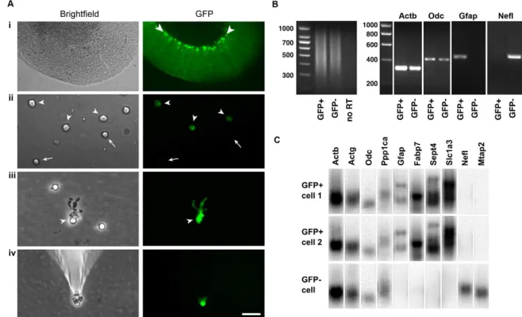
the cerebella of mice expressing GFP under the Glial fibrillary acidic protein (Gfap) promoter [26]. As shown Fig. 1Ai, Bergmann glial cell bodies could be easily visualized by their robust GFP expression in a live slice from a GFAP-GFP mouse cerebellum. After gentle dissociation of the tissue, four main types of cells were seen. Most cells were small with round cell bodies and lacked GFP signal (arrows in Fig. 1Aii), and most likely represent granule cells. Another frequent cell type had round cell bodies and relatively weak GFP fluorescence (arrowheads in Fig. 1Aii), almost certainly representing astrocytes. A third cell type, with large cell bodies and no fluorescence, we identified as Purkinje cells (not shown). Finally, a rather infrequent type (,1% of cells) could be readily distinguished from the others by a distinctive ‘‘bushy’’ unipolar morphology and strong GFP expression (arrowheads in Fig. 1Aiii). These putative Bergmann glia and some GFP- cells as controls were harvested
Figure 1. Harvesting of individual Bergmann glia and quality control of single-cell cDNA. Ai, a live cerebellar slice obtained from an adult GFAP-GFP transgenic mouse imaged under phase contrast optics (left) and fluorescence illumination (right). The Bergmann glia (see arrowheads) are the cells with the most fluorescence.Aii, freshly dissociated cells include putative astrocytes, which are devoid of processes and show relatively weak GFP fluorescence (arrowheads), and putative granule neurons, which have small, round, GFP-negative cell bodies (arrows).Aiii, a freshly dissociated Bergmann glia (arrowhead in left panel) can be distinguished from other cells by the bushy processes that emanate from one side of the soma–these are the long Bergmann glial processes that have partially retracted or been sheared off during tissue dissociation. In addition, Bergmann glia display strong GFP fluorescence (arrowhead in right panel), with the mean GFP intensity of their cell bodies 2.960.8 fold that of astrocytes; n = 17 cells).Aiv, a single Bergmann glia being washed by placement in a new dish containing fresh buffer, before being picked again with a new microelectrode. This step is performed to exclude contaminating cells or mRNAs. Scale bar, 70mm inAi, 25mm inAiiandAiii, 40mm inAiv.B, left panel, agarose gel
electrophoresis of cDNAs generated from single GFP+and GFP2cells. The gels show that most of the cDNA lies between 300 and 1000 bases. Right panel, agarose gels showing PCR with primer pairs directed towardsb-actin (Actb), ornithine decarboxylase (Odc),Gfap, and neurofilament light chain (Nefl). The results show that the single cell cDNAs from Bergmann glia (GFP+) and neurons (GFP2) contain both high and low abundance transcripts (ActbandOdc, respectively). Bergmann glia are positive for the astroglial markerGfapand negative for the neuronal markerNefl, whereas neurons are negative forGfapand positive forNefl.C. Southern blot analysis of cDNAs from two putative Bergmann glial cells (GFP+) and a putative neuron
(GFP2) shows presence of the high, medium, and two low abundance markers (Actb,Actg, andOdcandPpp1ca, respectively). In addition, the GFP+
cells are positive forGfap,Fabp7(BLBP),Sept4andSlc1a3(GLAST), confirming their glial identity, whereas the GFP2cell lacks all these markers. Conversely, the GFP+cells are absent for the neuronal markers NeflandMtap2 whereas the GFP2cell is positive. These results confirm the preservation of low to high abundance transcripts after the single-cell RT-PCR amplification, and also confirm the cell identity of the Bergmann glia used for microarray analysis.
individually using glass microelectrodes (Fig. 1Aiv). To minimize the possibility of contamination from other mRNAs, each cell was subjected to a rinse in a new dish with fresh buffer and picked with a new microelectrode before cDNAs were generated using protocols described before [25,27,28]. In line with these protocols, single cell RT-PCR amplification generated cDNAs of 300–1000 base pairs (Fig. 1B, left panel). The single cell cDNAs were then subjected to rigorous quality control using PCR (Fig. 1B, right panels). Single cell cDNA libraries were considered to be of good quality if they were positive for high (b-actin or Actb) and low abundance (ornithine decarboxylase orOdc) markers, confirming that mRNAs of widely varying abundance were preserved during the RT-PCR amplifica-tion. High quality cDNAs of GFP+ and GFP2 cells were then further characterized. GFP+cells were positive forGfapbut negative for neurofilament light chain (Nefl), confirming that they were glia and that the samples were free of contaminating neuronal mRNA. GFP2cells were negative forGfapand positive forNefl, indicating they were most likely neurons. Finally, an additional quality control step was performed using Southern blot analysis (Fig. 1C). GFP+ and GFP2 cells were positive for high, medium, and two low abundance transcripts (Actb, high;c-actin orActg, medium;Odcand protein phosphatase 1ca orPpp1ca, low), indicating good amplifi-cation. GFP+cells were positive forGfap, fatty acid binding protein 7 orFabp7(also called brain lipid binding protein or BLBP),Sept4
(Septin 4) and the glutamate transporter, Slc1a3 (GLAST), confirming that they were astroglial cells. They were negative for the neuronal markers Nefl and microtubule-associated protein 2 (Mtap2). Conversely, GFP2cells were negative for glial markers but positive for neuronal ones, confirming their identity as neurons.
Purity and Accuracy of Expression Profiles of Individual Bergmann Glia
To examine the global transcriptional profiles of Bergmann glia, the amplified cDNA generated from five P6 and five P30 cells were individually hybridized to Affymetrix 430 2.0 Mouse Expression Arrays. On average, 31.761.5% of the 45101 probe sets per array showed positive expression at P6, and 27.562.3% at P30.
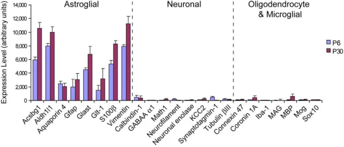
Expression profiles of all the Bergmann glia demonstrated high levels of expression of known astrocyte-specific genes [23], consistent with their long-held classification as specialized astroglia (Fig. 2). In contrast, the expression of several genes considered to be markers for neurons, oligodendrocytes or microglia [23] was low or absent (Fig. 2), confirming that the samples were indeed free from contaminating mRNAs from other cell types.
To determine the level of heterogeneity between samples of the same and different ages, we performed two analyses. Pair-wise comparisons of all individual samples showed that cells from the same age are more similar than between ages (same age: mean correlation coefficient = 0.8 for P6, 0.82 for P30; between ages: mean correlation coefficient = 0.66; representative samples shown in Fig. 3A). The similarity between cells of the same age is comparable to that reported between individual cells of a glioblastoma cell line (mean correlation coefficient = 0.86; [25], using very similar techniques, suggesting that Bergmann glia from a particular age and sagittal location (vermis) are quite homogeneous. The similarity between the two ages (mean correlation coefficient = 0.66) is significantly higher than for disparate cell types (for example, olfactory epithelium neurons vs. heart cells, mean correlation coefficient = 0.42; [25], as expected for cells of the same type. Unsupervised hierarchical clustering of the samples indicated that cells were more distinct between ages than within each age (Fig. 3B). Taken as a whole, these results affirm the cell-type specificity and reproducibility of single cell expression profiling, and the validity of comparisons between ages.
Identification of Genes that Are Developmentally Regulated in Bergmann Glia
Genes expressed differentially between P6 and P30 were identified using two criteria: those with a greater than 3-fold difference in normalized expression between ages, and those that were flagged ‘‘present’’ at one age and ‘‘absent’’ at the other. In total, 435 genes were found to fit the criteria for P6.P30 expression (Table S1), and 137 fit the criteria for P30.P6
Figure 2. Expression profiles of cell type control genes confirm purity of Bergmann glial cDNA generated by single cell RT-PCR.
Mean expression levels of well established markers for astroglia, neurons, oligodendrocytes and microglia were analyzed in the transcriptional profiles of ten Bergmann glia (five each from P6 and P30) using GeneSpring GX 7.3 software. For genes represented by multiple probe sets, the averaged expression of all probe sets were used. Astroglial genes were robustly expressed whereas markers of other cell types were absent or extremely low, confirming the astroglial identity of Bergmann glia and the absence of contaminating mRNAs from other cell types during cell harvesting. Error bars represent6SEM.
expression (Table S2). Of these, the top twenty most differentially expressed probe sets determined using the two approaches are shown in Tables 1 and 2 for P6.P30 and in Tables 3 and 4 for P30.P6. Genes in all tables were categorized and annotated based on functional information obtained in online databases and/ or previous studies.
We found that the set of genes expressed more highly at P6 is enriched in molecules known or predicted to be involved in cell growth and/or proliferation, cell cycle regulation, protein biosynthesis and other metabolic pathways, RNA processing and transport, and transcriptional regulation (including a large number of transcription factors) (Table S1). This suggests that at P6 Bergmann glia are in a state of active metabolism and growth, and some of them may potentially still be undergoing proliferation. The higher expression levels of ribosomal genes is also indicative of cells in a state of growth [29,30], consistent with the postnatal extension and elaboration of Bergmann glial processes.
On the other hand, the genes enriched at P30 include a different set of functional categories, namely molecules known or predicted to be involved in maintaining synapse structure and function, regulating exocytosis, forming gap junctions, and mediating molecular transport (including ion, protein, and carbohydrate transporters) (Table S2). This result suggests that a key role of
Bergmann glia in the adult cerebellum is to support/modulate synaptic function, and identifies some potential molecular players that may mediate this role. Interestingly, many of the differentially expressed genes in both sets (P6.P30 and P30.P6) have not previously been reported to be present in astroglial cells (Table 5), based on a search of the literature and exclusion of astrocyte-enriched genes listed by Cahoy and colleagues [23]. Further study of these genes, some of which are discussed below, may provide new insight into molecular mechanisms underlying Bergmann glial functions.
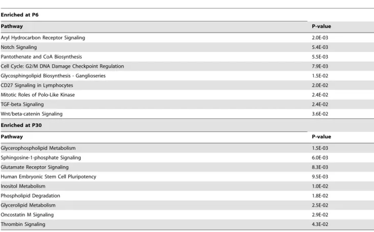
In addition to individual genes, we also sought to identify signaling and metabolic pathways that are preferentially active in Bergmann glia at P6 or at P30. To do this, we utilized the Ingenuity Pathway Analysis (IPA) tool from Ingenuity Systems, a resource based on a curated list of all canonical signaling and metabolic pathways [31]. IPA analysis of the differentially expressed gene sets in our data identified nine signaling or metabolic pathways statistically enriched at P6 and nine enriched at P30 (Table 6). The pathways enriched at P6 include the Notch, TGF-b and Wnt/b-catenin signaling pathways, which have key developmental roles in cell fate determination, cell growth, proliferation and maturation. The Notch pathway has been shown to be important for Bergmann glial specification and Figure 3. Single cell RT-PCR of individual Bergmann glia is sufficiently accurate for comparison of expression profiles by age. A, Scatter plots of raw gene expression level compared between two cells from the same age (P6-1 vs. P6-2; left panel) and between ages (P6-1 vs. P30-1; right panel) (a.u.: arbitrary units). The samples from the same age show high similarity (mean within-age correlation coefficient = 0.81), which is comparable to what is reported between individual cells of a glioblastoma cell line (0.86) [25]. The high level of concordance between samples of the same age increases the reliability of comparisons between ages. Samples of different ages are significantly more divergent (mean P6 vs. P30 correlation coefficient = 0.66).B, Dendrogram and sample clustering of individual Bergmann glia. Unsupervised hierarchical clustering based on overall gene expression profiles reveals two distinct clusters corresponding to the two ages, P6 and P30. This suggests that the samples from the two ages do indeed represent two statistically distinct populations suitable for valid comparison.
Table 2.Top 20 probe-sets present at P6 and absent at P30 (ranked by P values).
Probe ID Gene Symbol P value Description Functional classification
1448842_at Cdo1 5.28E-06 cysteine dioxygenase 1, cytosolic metabolic functions 1454778_x_at Rps28 2.89E-05 Ribosomal protein S28, mRNA component of ribosome 1448182_a_at Cd24a 6.02E-05 CD24a antigen cell cycle regulation 1433928_a_at Rpl13a 6.02E-05 ribosomal protein L13a component of ribosome 1435612_at Opcml 0.0001 opioid binding protein/cell adhesion molecule-like cell adhesion or repulsion 1460011_at Cyp26b1 0.00012 cytochrome P450, family 26, b1 morphogenesis/patterning 1418310_a_at Rlbp1 0.00012 retinaldehyde binding protein 1 transporter activity 1455085_at 1700086L19Rik 0.00012 RIKEN cDNA 1700086L19 gene function unknown 1433935_at AU020206 0.00016 expressed sequence AU020206 function unknown 1419123_a_at Pdgfc 0.00016 platelet-derived growth factor C growth factor signaling 1449013_at Eef2k 0.00016 eukaryotic elongation factor-2 kinase protein biosynthesis 1452499_a_at Kif2a 0.00021 kinesin family member 2A microtubule based motor
1454867_at Mn1 0.00025 meningioma 1 cell growth and proliferation
1416200_at Il33 0.00035 interleukin 33 cytokine signaling
1420329_at 4930455C21Rik 0.00036 RIKEN cDNA 4930455C21 gene transporter activity 1416107_at Nsg2 0.00036 neuron specific gene family member 2 intracellular signaling
1417038_at Sept9 0.00045 septin 9 cell cycle regulation
1421100_a_at Dab1 0.00053 disabled homolog 1 regulation of cell adhesion 1456010_x_at Hes5 0.00063 hairy and enhancer of split 5 regulation of transcription 1424275_s_at Trim41 0.00085 tripartite motif-containing 41 function unknown
doi:10.1371/journal.pone.0009198.t002
Table 1.Top 20 probe-sets showing largest fold-difference in expression (P6.P30).
Probe ID Gene Symbol Fold Change Description Functional Classification
1448182_a_at Cd24a 317.5 CD24a antigen cell cycle regulation
1456010_x_at Hes5 264.3 hairy and enhancer of split 5 (Drosophila) regulation of transcription
1416200_at Il33 248.6 interleukin 33 cytokine signaling
1435612_at Opcml 233 opioid binding protein/cell adhesion molecule-like cell adhesion or repulsion 1454778_x_at Rps28 200.7 Ribosomal protein S28, mRNA component of ribosome 1421100_a_at Dab1 181.8 disabled homolog 1 (Drosophila) regulation of cell adhesion 1455085_at 1700086L19Rik 177.9 RIKEN cDNA 1700086L19 gene function unknown 1448842_at Cdo1 170.9 cysteine dioxygenase 1, cytosolic metabolic functions 1415945_at Mcm5 164.7 minichromosome maintenance deficient 5 cell cycle regulation 1436808_x_at Mcm5 156.2 minichromosome maintenance deficient 5 cell cycle regulation 1419123_a_at Pdgfc 143.6 platelet-derived growth factor C growth factor signaling 1429372_at Sox11 140.7 SRY-box containing gene 11 regulation of transcription 1418310_a_at Rlbp1 140 retinaldehyde binding protein 1 transporter activity 1436505_at Ppig 137 peptidyl-prolyl isomerase G (cyclophilin G) protein folding
1460011_at Cyp26b1 135.1 cytochrome P450, family 26, b1 morphogenesis/patterning 1426307_at Cyb5r4 129.2 cytochrome b5 reductase 4 metabolic functions 1424010_at Mfap4 121.3 microfibrillar-associated protein 4 cell adhesion or repulsion 1423763_x_at Rps28 117.7 Ribosomal protein S28, mRNA component of ribosome 1428466_at Chd3 114.9 chromodomain helicase DNA binding protein 3 chromatin remodeling 1425052_at Isoc1 107.8 isochorismatase domain containing 1 metabolic functions
Table 3.Top 20 probe-sets showing largest fold-difference in expression (P30.P6).
Probe ID Gene Symbol Fold change Description Functional classification
1438044_at 1700047M11Rik 293.1 RIKEN cDNA 1700047M11 gene function unknown
1434264_at Ank2 266.8 ankyrin 2, brain synapse structure and function
1418288_at Lpin1 169.8 lipin 1 metabolic functions
1439568_at Greb1 159.4 gene regulated by estrogen in breast cancer protein electron transport 1430268_at 9630005C17Rik 154.6 RIKEN cDNA 9630005C17 gene function unknown 1448428_at Nbl1 143.3 neuroblastoma, suppression of tumorigenicity 1 cell cycle regulation 1427284_a_at Ttpa 137.4 tocopherol (alpha) transfer protein transporter activity 1434121_at Lgi4 126.1 leucine-rich repeat LGI family, 4 cell-cell interaction
1434265_s_at Ank2 108.7 ankyrin 2, brain synapse structure and function 1435436_at Epas1 101.2 endothelial PAS domain protein 1 regulation of transcription 1436470_at Rims2 97.07 regulating synaptic membrane exocytosis 2 exocytosis
1456642_x_at S100a10 95.89 S100 calcium binding protein A10 (calpactin) intracellular signaling 1421841_at Fgfr3 95.79 fibroblast growth factor receptor 3 growth factor signaling 1456523_at C77713 95.74 expressed sequence C77713 function unknown 1416762_at S100a10 85.57 S100 calcium binding protein A10 (calpactin) intracellular signaling 1451718_at Plp1 80.05 proteolipid protein (myelin) 1 myelin-associated
1438193_at Nrxn3 72.57 neurexin III synapse structure and function
1433788_at Nrxn3 69.35 neurexin III synapse structure and function
1423602_at Traf1 67.72 Tnf receptor-associated factor 1 intracellular signaling 1417220_at Fah 65.95 fumarylacetoacetate hydrolase hydrolase activity
doi:10.1371/journal.pone.0009198.t003
Table 4.Top 20 probe-sets present at P30 and absent at P6 (ranked by P values).
Probe ID Gene Symbol P value Description Functional classification
1434265_s_at Ank2 3.60E-06 ankyrin 2, brain synapse structure and function 1434264_at Ank2 3.60E-06 ankyrin 2, brain synapse structure and function 1438044_at 1700047M11Rik 7.65E-05 RIKEN cDNA 1700047M11 gene function unknown
1436173_at Dlc1 0.00087 Deleted in liver cancer 1 (Dlc-1) intracellular signaling 1451784_x_at H2-D1 0.00087 histocompatibility 2, D region locus 1 MHC class I receptor 1439568_at Greb1 0.00087 gene regulated by estrogen in breast cancer protein electron transport 1423523_at Aass 0.0011 aminoadipate-semialdehyde synthase metabolic functions
1438193_at Nrxn3 0.0011 neurexin III synapse structure and function
1420709_s_at Dao1 0.0011 D-amino acid oxidase 1 D-amino acid pathway 1425545_x_at H2-D1 0.00151 histocompatibility 2, D region locus 1 MHC class I receptor
1436205_at Nfasc 0.00186 neurofascin synapse structure and function
1417220_at Fah 0.00186 fumarylacetoacetate hydrolase hydrolase activity
1425567_a_at Anxa5 0.00189 annexin A5 intracellular signaling
1434121_at Lgi4 0.00191 leucine-rich repeat LGI family, 4 intracellular signaling 1417629_at Prodh 0.00194 proline dehydrogenase metabolic functions 1420545_a_at Chn1 0.00346 chimerin (chimaerin) 1 intracellular signaling 1427284_a_at Ttpa 0.00346 tocopherol (alpha) transfer protein ion transport 1436470_at Rims2 0.00346 regulating synaptic membrane exocytosis 2 exocytosis
1418288_at Lpin1 0.00346 lipin 1 metabolic functions
1421841_at Fgfr3 0.00346 fibroblast growth factor receptor 3 growth factor signaling
maturation [32,33,34], but the potential roles of the other pathways we have identified remain untested. At P30, the statistically enriched signaling pathways included the glutamate receptor signaling pathway, underscoring a role for Bergmann glia in synapse function/modulation. Unexpectedly, another enriched pathway is the embryonic stem cell pluripotency pathway, a finding that we discuss in greater detail below.
We were also surprised to find that at P6 Bergmann glia express a number of genes that have been traditionally thought of as neuron-specific (see Table S3). While we cannot completely exclude the possibility of contamination by some neuronal mRNAs, our quality control analysis (Fig. 2) argues against this, as does the finding that some of these are also enriched in cortical astrocytes [23]. We therefore believe that these genes are expressed by Bergmann glia in addition to neurons, but that the level of expression in glia may be significantly lower than in neurons, causing them not to have been detected in the glia by other techniques such as in situhybridization. We believe one of the strengths of our analysis is that we have not subtracted out
genes thought to be specific to other cell types from our microarray data, and therefore we can detect genes that may be present in multiple cell types, even if they are more abundant in other cells than in Bergmann glia.
Analysis of Genes Abundantly Expressed in Postnatal Bergmann Glia (P6 and P30) and Identification of Novel Cell-Specific Markers
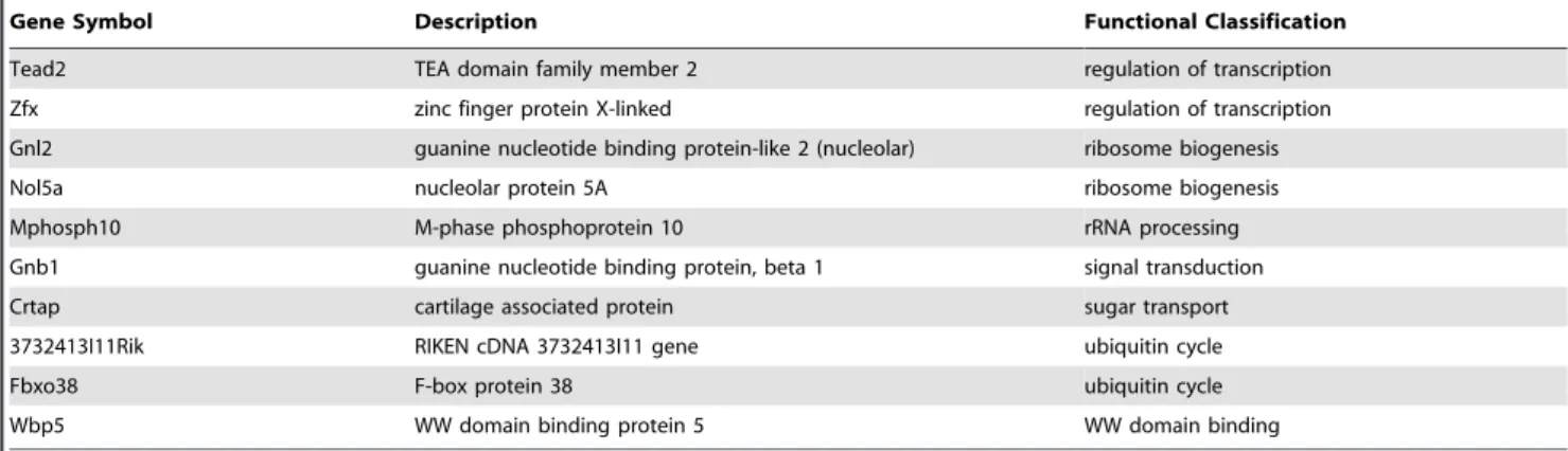
To gain further insight into the molecular makeup of Bergmann glia in the postnatal cerebellum, we also searched for genes that are expressed at both P6 and P30 at moderate to high levels (raw signal values .2000) (Table S4). As expected, this extensive list includes many astroglial markers such as vimentin, S100b, aquaporin 4 and aldehyde dehydrogenase 1 family, member L1 (Aldh1L1) [23]. It also includes genes not previously known to be expressed in astroglia, some of which are listed in Table 7.
While examining the set of abundantly expressed genes, we found, surprisingly, that Bergmann glia express mRNA for the Table 5.Differentially expressed (P6 vs. P30) genes in Bergmann glia not previously known to be present in astroglia.
Gene Symbol Description Functional Classification Age
Hs6st1 heparan sulfate 6-O-sulfotransferase 1 cell adhesion or guidance P6
Sema6a semaphorin 6A cell adhesion or guidance P6
Cd24a CD24a antigen cell cycle regulation P6
Atxn2 ataxin 2 cell growth and/or proliferation P6
Asxl1 additional sex combs like 1 (Drosophila) chromatin remodeling P6
Nnat neuronatin function unknown P6
Neurl2 neuralized-like 2 intracellular signaling (notch) P6
Shroom3 shroom family member 3 morphogenesis/pattern formation P6
Shfm1 split hand/foot malformation (ectrodactyly) type 1 protein processing P6 Adamts12 a disintegrin-like metallopeptidase, thrombospondin 1 motif, 12 proteolysis and/or cell-ECM interaction P6 Solh small optic lobes homolog proteolysis and/or cell-ECM interaction P6 Bmi1 Bmi1 polycomb ring finger oncogene regulation of transcription P6 Myef2 myelin basic protein expression factor 2, repressor regulation of transcription P6
Zfp131 zinc finger protein 131 regulation of transcription P6
Zfp260 zinc finger protein 260 regulation of transcription P6
Zfp414 zinc finger protein 414 regulation of transcription P6
Zfp532 zinc finger protein 532 regulation of transcription P6
Zfp560 zinc finger protein 560 regulation of transcription P6
Zfp651 zinc finger protein 651 regulation of transcription P6
Zfp704 zinc finger protein 704 regulation of transcription P6
Cyp26b1 cytochrome P450, family 26, subfamily b, polypeptide 1 retinoic acid signaling P6
Smn1 survival motor neuron 1 RNA synthesis or processing P6
Snapap SNAP-associated protein vesicle exocytosis P6
Syt16 synaptotagmin XVI vesicle exocytosis P6
Hap1 huntingtin-associated protein 1 vesicle transport P6
Dlc1 Deleted in liver cancer 1 (Dlc-1) cell adhesion or repulsion P30
Gpr89 G protein-coupled receptor 89 GPCR signaling P30
Chn1 chimerin (chimaerin) 1 intracellular signaling P30
Plp1 proteolipid protein (myelin) 1 myelin-associated P30
Nfasc neurofascin synapse structure and function P30
Nrxn3 neurexin III synapse structure and function P30
Rims2 regulating synaptic membrane exocytosis 2 transporter activity/exocytosis P30
myelin protein peripheral myelin protein (Pmp22) and for myelin protein zero-like 1 (Mpzl1), which may be important for myelination [35] (Table S4). Furthermore, in the adult, Bergmann glia also express mRNAs for proteolipid protein 1 (Plp1), the predominant component of CNS myelin, and leucine-rich repeat LGI family, member 4 (Lgi4), which is important for myelination in the PNS [36] (Table S2). These findings were supported byin situ hybridization data from the Allen Brain Atlas (Fig. S1):
although the strongest expression ofPmp22andPlp1in the adult is in putative oligodendrocytes (arrows in Fig. S1, top and second row), there is less intense but still significant staining in the Purkinje cell layer (PCL) consistent with expression in Bergmann glia (arrowheads).Lgi4(Fig. S1, third row) andMpzl1(arrowheads in Fig. S1, bottom row) also show labeling consistent with expression in Bergmann glia. Since Bergmann glia play no known role in myelination, it is unknown what alternate function these Table 6.Signaling and metabolic pathways enriched in Bergmann glia.
Enriched at P6
Pathway P-value
Aryl Hydrocarbon Receptor Signaling 2.0E-03
Notch Signaling 5.4E-03
Pantothenate and CoA Biosynthesis 5.5E-03
Cell Cycle: G2/M DNA Damage Checkpoint Regulation 7.9E-03
Glycosphingolipid Biosynthesis - Ganglioseries 1.5E-02
CD27 Signaling in Lymphocytes 2.0E-02
Mitotic Roles of Polo-Like Kinase 2.4E-02
TGF-beta Signaling 2.4E-02
Wnt/beta-catenin Signaling 3.6E-02
Enriched at P30
Pathway P-value
Glycerophospholipid Metabolism 1.5E-03
Sphingosine-1-phosphate Signaling 6.0E-03
Glutamate Receptor Signaling 8.3E-03
Human Embryonic Stem Cell Pluripotency 9.5E-03
Inositol Metabolism 1.0E-02
Phospholipid Degradation 1.8E-02
Glycerolipid Metabolism 2.5E-02
Oncostatin M Signaling 2.9E-02
Thrombin Signaling 4.3E-02
doi:10.1371/journal.pone.0009198.t006
Table 7.Genes with abundant Bergmann glial expression (at both P6 and P30) that were not previously known to be present in astroglia.
Gene Symbol Description Functional Classification Age
Cdh22 cadherin 22 cell adhesion or ECM binding P6, P30
Celsr2 cadherin EGF LAG seven-pass G-type receptor 2 cell adhesion or ECM binding P6, P30
Cbx3 chromobox homolog 3 chromatin remodeling P6, P30
Gpr153 G protein-coupled receptor 153 GPCR signaling P6, P30
Gpsm1 G-protein signalling modulator 1 (AGS3-like, C. elegans) GPCR signaling P6, P30 Kcnb1 potassium voltage gated channel, Shab-related subfamily ion channel or receptor activity P6, P30 Edf1 endothelial differentiation-related factor 1 regulation of transcription P6, P30 Tcfl5 transcription factor-like 5 (basic helix-loop-helix) regulation of transcription P6, P30
Stx5a syntaxin 5A SNARE receptor activity P6, P30
Stx8 syntaxin 8 SNARE receptor activity P6, P30
Kcnb1 potassium voltage gated channel, Shab-related subfamily ion channel or receptor activity P6, P30
Cbx3 chromobox homolog 3 chromatin remodeling P6, P30
genes may serve in these cells. Our data also indicated that Bergmann glia express agrin in vivo at P6 and P30 (Table S4), similar to what been shown in astrocytes in vitro [37]. Whether agrin synthesized by Bergmann glia plays any role in synapse formation or maintenancein vivomerits further investigation.
Analysis of in situ hybridization data for genes we found expressed in adult Bergmann glia identified several candidates that we believe may serve as selective markers for Bergmann glia. Within the adult cerebellum, the expression of four genes, leucine zipper protein 2 (Luzp2), G protein-coupled receptor 89 (Gpr89), leucine-rich repeat LGI family, member 4 (Lgi4), and Growth and differentiation factor 10 (Gdf10) appears to be restricted solely to the Purkinje cell layer (Fig. S2); and the cellular expression patterns of these genes within the PCL closely match those of well established astroglial markers expressed by Bergmann glia (Fig. S3). However, unlike most currently used markers that also label cerebellar astrocytes, these four genes appear to be completely specific to Bergmann glia. Gdf10 is particularly noteworthy because its specific expression in the Purkinje cell layer had been reported before [38], but was thought, we believe in error, to be in Lugaro cells rather than in Bergmann glia.
Gpr126, an Adhesion GPCR Expressed Specifically in Developing Bergmann Glia
GPCRs were one of the gene families of particular interest to us in light of our recent finding thatGpr56, an adhesion GPCR, is essential for cortical and cerebellar development [39,40]. More-over, few GPCRs have been studied in the context of glial function. Among the many GPCRs we found expressed in Bergmann glia, we were especially intrigued by the very specific spatiotemporal pattern of expression of Gpr126, another orphan receptor that is a close relative of Gpr56. At P6, Gpr126 was expressed specifically in the Purkinje cell layer (Fig. 4, center panels). Since RT-PCR analysis of single-cell cDNAs from
Purkinje cells showed that Gpr126 was absent in these neurons (data not shown), the pattern ofGPR126mRNA is consistent with expression by Bergmann glial cells. As predicted by the microarray data, in situ hybridization signal for Gpr126 was no longer detectable in the adult cerebellum (Fig. 4, right panels). At E15,
Gpr126was present in the cerebellar ventricular zone (arrowheads in Fig. 4, left panels), where precursors of cerebellar neurons and glia–including Bergmann glia–are located. In contrast,Gpr126was absent in the forebrain ventricular zone at E15 (Fig. 4, arrow in left panel), indicating that it does not play a role in cortical radial glia at this age. Based on a recent study in zebrafish, regulation of cyclic AMP by Gpr126 signaling plays a critical role in the initiation of myelination by Schwann cells [41]. WhetherGpr126
in Bergmann glia also regulates second messenger pathways involved in cell differentiation, or instead regulates cell adhesion similar to other adhesion GPCRs such asGpr56,Celsr2andCelsr3
[39,42,43] remains to be examined.
Bergmann Glia Express Genes Typical of Neural Stem Cells
A striking finding of our microarray analysis is that Bergmann glia, even in the adult, express numerous genes thought to be expressed specifically by stem cells (Table 8), and show enrichment of the embryonic stem cell pluripotency pathway (Table 6). Of the 220 genes that [24] identified as a core set of ‘‘stemness’’ genes common to multiple types of stem cells but not found in differentiated cells, 26.8% (60/220) are expressed by Bergmann glia at P6 (Table 8). Remarkably, 18.3% (41/220) remain expressed at P30. In addition, 26.3% (647/2458) of neural stem cell genes identified in the same study are also present in Bergmann glia at one or both ages (data not shown). These include transcription factors such asSox1,Sox2,Sox9,Hes1andHes5, which play important roles in cell proliferation and in maintaining neural stem cell identity. These findings support the intriguing hypothesis Figure 4. Identification of a developmentally regulated GPCR that is Bergmann glia-specific in the cerebellum.in situhybridization with a33P-labeled probe forGpr126, a little known GPCR of the adhesion family, reveals signal specifically in the Purkinje cell layer at P7 (arrows, middle panels), consistent with expression in Bergmann glia.Gpr126expression is developmentally regulated, and becomes undetectable in the adult (right panel). Unlike most classic Bergmann glial markers, which are also expressed by cortical radial glia,Gpr126is specific to Bergmann glia and not detected in cortical radial glia at E15 (arrow in top left panel). Labeling is seen in the ventricular zone of the developing cerebellar anlage at E15 (arrowheads in left panels), suggesting thatGpr126may be expressed in progenitors of Bergmann glia. All sections are oriented with rostral to the right. Scale bar, upper panels: left, 3 mm; center, 2 mm; right, 2.5 mm; lower panels: left, 100mm; center, 1 mm; right, 1.4 mm.
Table 8.Stem cell-enriched genes expressed in Bergmann glia.
Gene Symbol Description Functional Classification
Pls3 plastin 3 (T-isoform) actin binding
Aldh7a1 aldehyde dehydrogenase family 7, member A1 aldehyde metabolism
Pdcd2 programmed cell death 2 apoptosis
Cbr3 carbonyl reductase 3 Arachidonic acid metabolism
Rcn1 reticulocalbin 1 calcium ion binding
Tbrg1 transforming growth factor beta regulated gene 1 cell cycle regulation Fhl1 four and a half LIM domains 1 cell growth and differentiation
Msh2 mutS homolog 2 DNA repair
Acadm acyl-Coenzyme A dehydrogenase, medium chain electron transport Trip6 thyroid hormone receptor interactor 6 electron transport
Txndc9 thioredoxin domain containing 9 electron transport
Txnl1 thioredoxin-like 1 electron transport
2410015N17Rik RIKEN cDNA 2410015N17 gene function unknown
2410022L05Rik RIKEN cDNA 2410022L05 gene function unknown
AW549877 expressed sequence AW549877 function unknown
Jagn1 jagunal homolog 1 function unknown
Sh3d19 SH3 domain protein D19 function unknown
Gsta4 glutathione S-transferase, alpha 4 Glutathione metabolism Pigx phosphatidylinositol glycan anchor biosynthesis, class X GPI-anchor biosynthesis Tbc1d15 TBC1 domain family, member 15 GTPase activator activity
Pla2g6 phospholipase A2, group VI lipid catabolism
Sfrs3 splicing factor, arginine/serine-rich 3 (SRp20) nuclear mRNA splicing Sfrs6 splicing factor, arginine/serine-rich 6 nuclear mRNA splicing
Hrsp12 heat-responsive protein 12 nuclease activity
Zc3h14 zinc finger CCCH type containing 14, variant 1, mRNA. nucleic acid binding
Ppa1 pyrophosphatase (inorganic) 1 phosphate metabolism
Nup35 nucleoporin 35 porin activity
Psmd12 proteasome 26S subunit, non-ATPase, 12 proteasome pathway
Tjp1 tight junction protein 1 protein binding
Mrpl17 mitochondrial ribosomal protein L17 protein biosynthesis Eif2b4 eukaryotic translation initiation factor 2B, subunit 4 delta protein biosynthesis Eif3s1 eukaryotic translation initiation factor 3, subunit 1 alpha protein biosynthesis
Eprs glutamyl-prolyl-tRNA synthetase protein biosynthesis
Iars isoleucine-tRNA synthetase protein biosynthesis
Mrpl3 mitochondrial ribosomal protein L3 protein biosynthesis Mrpl34 mitochondrial ribosomal protein L34 protein biosynthesis
Fkbp11 FK506 binding protein 11 protein folding
Fkbp9 FK506 binding protein 9 protein folding
Ppic peptidylprolyl isomerase C protein folding
Mpdu1 mannose-P-dolichol utilization defect 1 protein metabolism Esf1 ESF1, nucleolar pre-rRNA processing protein, homolog protein processing Ywhab tyrosine 3-monooxygenase activation protein, beta protein targeting Laptm4a lysosomal-associated protein transmembrane 4A protein transport
Pkd2 polycystic kidney disease 2 protein transport
Xpot exportin, tRNA (nuclear export receptor for tRNAs) protein transport
Zmat3 zinc finger matrin type 3 regulation of cell growth
Smarcad1 SWI/SNF-related, matrix-associated chromatin regulator 1a regulation of DNA recombination Epl2 elongation protein 2 homolog regulation of JAK-STAT cascade Cops4 constitutive photomorphogenic homolog, subunit 4 regulation of signaling
[44] that Bergmann glia may be a source of the newly described neural stem cells in the adult cerebellum [45,46]. Our observation in Bergmann glia is also consistent with reports of progenitor cell-like gene expression in Muller Glia of the retina [44,47].
Discussion
Despite growing evidence of the indispensable roles of glial cells in many aspects of nervous system development and function, much remains unknown about the molecules that mediate these roles, particularlyin vivo. There is also a lack of specific markers for various subtypes of glial cells, and of tools to manipulate gene expression only in specific subtypes. This has hindered our understanding of the diverse roles of glial cells. Our results from single cell transcriptome analysis of cerebellar Bergmann glia identify numerous novel genes whose role in Bergmann glia, or glia in general, can now be tested in functional contexts. We also identify several genes that appear to be entirely Bergmann glia-specific in the cerebellum. Not only does this confirm that the GFP+cells we harvested for this study were indeed Bergmann glia and did not inadvertently include astrocytes, but also offers new tools for understanding these important glial cells. In this study we focused mainly on genes not previously characterized in Bergmann glia or glial cells in general. For analysis of all genes that we found expressed in Bergmann glia, our complete data set can be viewed in Table S5, and the original Affymetrix. CEL files can be accessed at the Gene Expression Omnibus (GEO) repository (accession number GSE18617).
Genes identified here as being expressed in Bergmann glia fall into many classes, highlighting the diverse roles of this cell type throughout life. Some of the most salient with respect to cerebellar structure and function include retinoic acid signaling components– e.g.Cyp26b1, a regulator of retinoic acid activity - which may play a role in cerebellar patterning [48]; chemoattractive and chemorepulsive molecules (notably semaphorin 4B), which may regulate neuronal migration as well as dendritic outgrowth and synapse development [49,50,51]; growth factors and growth factor-like molecules such as Gdf10 [38,52] and meteorin [53,54]; molecules for cell-cell communication, particularly gap junction proteins, which have been shown to play critical roles in radial glial proliferation [55] and neuronal migration along radial glial fibers in the cerebral cortex [56]; synapse-associated adhesion molecules such as neurexins, which are important for synapse formation and maintenance [57]; components of the D-serine pathway involved in modulation of NMDA receptor function
[58,59]; and molecules that mediate synaptic function and plasticity, such as glutamate receptors, transporters, and trans-membrane AMPAR regulatory proteins (TARPs) [60]. The expression of the enzymes glutamine synthetase and pyruvate carboxylase along with glial glutamate transporters Slc1a3
(GLAST) and Slc1a2 (GLT-1) provide further evidence for the involvement of Bergmann glia in the glutamate-glutamine cycle that supports synaptic activity [19,61]. Similarly, the expression of the lactate synthetic enzymes lactate dehydrogenase A and B (Ldha
and Ldhb), and monocarboxylate transporter 1 (Mct1), the main lactate transporter responsible for rapid release of glial lactate, is consistent with the hypothesized glia-neuron lactate shuttle [62].
Our finding that neurexin III is expressed in Bergmann glia is particularly interesting in light of the proposed role of neurexins in the development and maintenance of functional synapses. Neurexins present on the presynaptic membrane are thought to bind neuroligins on the postsynaptic membrane, thereby forming a trans-synaptic link that helps maintain the close apposition of pre-and post-synaptic elements [63]. Originally thought to be presynaptic [64], immuno-electron microscopy has now shown that neurexins are also present postsynaptically [65]. What has been missing in this analysis is the consideration of glial processes, which are also integral components of most CNS synapses and maintained in close proximity to pre-and post-synaptic elements [9,66]. The expression of neurexin III in Bergmann glia raises the question of whether this molecule plays a role in anchoring glial processes to pre- and post-synaptic elements. Cell type-specific deletion of this gene in astroglial cells can be performed to address this hypothesis.
The role of glia in synaptic function has been reinforced by the finding that astrocytes in cell culture or brain slices can release glutamate [67,68], which in turn can modulate synaptic transmission and plasticity [69,70,71]. However, the actual mechanism of glial glutamate release has remained controversial [72]. There is evidence that astrocytesin vitroexpress components of regulated vesicle exocytosis previously thought to be found only in neurons, including v-glut1/2, SNAP23, Munc18a, and synaptotagmin IV [73], suggesting that astrocytes release gluta-mate by vesicle exocytosis similar to neurons. However, a recent study [23] reported that acutely isolated mouse astrocytes do not express v-glut1/2, synaptotagmins or synapsin I, and therefore are unlikely to exhibit regulated vesicular glutamate release in vivo. Our finding that acutely isolated adult Bergmann glia do express some known or potential components of regulated vesicular exocytosis, including synapsin I, synaptotagmins XI and XVI,
Gene Symbol Description Functional Classification
Tead2 TEA domain family member 2 regulation of transcription
Zfx zinc finger protein X-linked regulation of transcription
Gnl2 guanine nucleotide binding protein-like 2 (nucleolar) ribosome biogenesis
Nol5a nucleolar protein 5A ribosome biogenesis
Mphosph10 M-phase phosphoprotein 10 rRNA processing
Gnb1 guanine nucleotide binding protein, beta 1 signal transduction
Crtap cartilage associated protein sugar transport
3732413I11Rik RIKEN cDNA 3732413I11 gene ubiquitin cycle
Fbxo38 F-box protein 38 ubiquitin cycle
Wbp5 WW domain binding protein 5 WW domain binding
doi:10.1371/journal.pone.0009198.t008
syntaxins, snapin [74], rim2 [75], and Lgi3 [76] (a recently identified syntaxin interactor and potential regulator of exocytosis) suggests that, unlike cortical astrocytes, Bergmann gliain vivomay possess the machinery for regulated release of glutamate and/or possibly other neurotransmitters.
While much of the recent focus on glia has been on their novel roles, it is worth noting that our knowledge of the molecular mechanisms remains incomplete even for the oldest and most commonly-associated role of glia: serving as ‘‘nerve glue,’’ a term coined by Rudolf Virchow in 1859. In this context, it is interesting that one of the largest set of genes we found expressed at high levels in Bergmann glia consists of cell adhesion molecules and receptors known or hypothesized to mediate cell-cell or cell-ECM binding. These include well-established adhesion molecules and receptors such as brevican, tenascin C, integrin av, and dystroglycan-1, some of which are critical for structural integrity of the glial scaffold [77]. A less studied adhesion molecule that we find in Bergmann glia, Chl1, was also shown recently to be important for the guidance and stabilization of stellate cell arbors projecting onto Purkinje cell dendrites [78], highlighting the important role of glial cell adhesion molecules in the development and maintenance of neuronal connections. In light of these findings, we believe that the putative glial adhesion molecules we identify, including cadherin 22, CD164 and junction adhesion molecule 2 (Jam2), merit further investigation.
In addition to identifying possible molecular players in known functions of Bergmann glia, the genes emerging from our study also strengthen the possibility of novel roles of these cells. Recently, two studies identified putative neural stem cells in the postnatal cerebellum [45,46]. While the identities of these cells remain unknown, a hypothesis has emerged that perhaps Bergmann glia could be these stem cells [44]. A study from the same lab found that two transcription factors that regulate neural stem cell identity,Sox1andSox2, are found in postnatal Bergmann glia [79]. We now significantly expand this line of inquiry by examining the full expression profiles of Bergmann glia and identifying additional genes that have previously been implicated in ‘‘stem-ness’’ of neural stem cells [24]. The molecular and morphological changes that Bergmann glia undergo in response to injury, granule cell death, or implantation of embryonic granule cell precursors indicate that they remain highly plastic [80]. Whether they possess the latent genetic potential to serve as neural precursors and could do so in response to an appropriate stimulus remains a tantalizing possibility. Furthermore, the mechanisms that may normally repress this potential in the adult cerebellum merit further investigation. In this regard, our observation that the Bone Morphogenic Protein antagonist, Nbl1 (neuroblastoma, suppres-sion of tumorigenicity 1) shows highly elevated expressuppres-sion in adult Bergmann glia compared to P6 is interesting, since this gene has been shown previously to repress maintenance of the precursor state and promote neuronal differentiation through its action on BMP7 [81].
Materials and Methods
Ethics Statement
Experiments were performed in accordance with National Institutes of Health guidelines for the care and use of laboratory animals, and with approval of the Animal Care and Use Committee of Children’s Hospital Boston.
Isolation of Single Bergmann Glial Cells
Mice of ages P6 and P30 expressing GFP under the control of the GFAP promoter (GFP mice) were used. From
GFAP-GFP mouse brains, slices of the mid-sagittal third of the cerebellum were cut in cold Hank’s Balanced Salt Solution (HBSS). By cutting only from the mid-sagittal region, where Bergmann glial processes run mostly parallel to the sagittal plane, damage to glial processes was minimized. Furthermore, isolation of Bergmann glia from a restricted region of the cerebellum should minimize developmental heterogeneity between individual cells. Slices were cut into smaller pieces in cold Ca++- and Mg++-free HBSS containing 10 mM HEPES; The tissues were incubated in papain (20 U/ml), and DNase I (20 U/ml) in Ca++
- and Mg++ -free HBSS on a shaker for ,30 min at 37uC. The protease
solution was then replaced with Hanks Balanced Salt Solution (HBSS) containing 1 mg/ml albumin ovomucoid protease inhib-itor, and the tissue was gently triturated using fire-polished glass pipettes of decreasing bore diameter. Cells were pelleted by centrifugation and resuspended in cold HBSS. A small aliquot of the cell suspension was added to a Petri dish with cold Ca++
- and Mg++
-free HBSS and individual cells were harvested by mouth pipetting into pulled glass microcapillaries attached to a micro-manipulator. Bergmann glia were recognized by their GFP fluorescence and morphology. Each picked cell was rinsed in a fresh dish with HBSS and re-picked with a new microcapillary. Harvested cells were immediately seeded into PCR tubes containing reverse transcription buffer, and placed on ice. In control experiments, single GFP-negative cells and putative astrocytes were also picked. Astrocytes from cerebella of GFAP-GFP mice showed weaker GFAP-GFP staining compared to Bergmann glia and lacked the characteristic unipolar processes of the latter. The identity of the different cerebellar cells was always verified subsequently by PCR and Southern blot.
Single-Cell RT-PCR and Microarray Hybridization
Single-cell RT-PCR was performed as described previously [25,27,82]. Briefly, amplified cDNA was synthesized by lysing the cell, reverse transcribing the cell RNA after oligo-dT priming, poly-A tailing the 59end of the cDNA, and finally amplifying the cell cDNA with a unique poly-T primer (AL1: ATTGGATC-CAGGCCGCTCTGGACAAAATATGAATTC(T)24). The re-verse transcription was performed in limiting conditions of nucleotides and time in order to generate cDNAs of uniform size (,0.5 to 1 kb), which are more likely to be uniformly amplified
and to accurately reflect the relative abundances of various mRNAs in the cell. After 50 cycles of PCR, several micrograms of cDNA were generated from each cell. Fiveml of the cDNA was run on a 1.5% agarose gel to verify the presence of a smear from
,0.5 to 1 kb. Using this original cDNA as template, additional
cDNA could be faithfully reamplified as necessary by PCR using the AL1 primer, as described previously [82]. Southern blots for several ubiquitous and cell-specific marker genes were then performed as described [82,83] to assess the quality and representation of the single cell cDNA, and to verify cell identity.
ActbandActgwere used as high and medium abundance markers, respectively; andOdcandPpp1caas low abundance markers [84].
Gfap,Fabp7,Sept4andSlc1a3were used as glial markers. Finally, the presence ofNefl,Mtap2, and in some cases also tubulinb-III was checked to detect any contaminating neurons. Only the best single cell cDNAs (10mg of each), as determined by RT-PCR and Southern blot, were selected for labeling and microarray hybridization. In total, over 150 putative Bergmann glia were harvested, of which ,90 yielded good cDNA smears after
well. Sixteen cDNA samples passed all quality control criteria, including presence of all tested astroglial markers and absence of all tested neuronal markers. Of these, five P6 and five P30 samples that exhibited the best quality control parameters on Affymetrix Test3 arrays were then hybridized to Affymetrix GeneChip Mouse Genome 430 2.0 microarrays at the Harvard Biopolymers Facility using standard Affymetrix protocols.
Data Analysis of Affymetrix Gene Chips
Analyses of individual microarrays and comparisons between P6 and adult were performed using GeneSpring GX 7.3 (Agilent). Raw CEL files were processed using the RMA (Robust Multichip Average) normalization algorithm as implemented in GeneSpring GX 7.3. Normalization was performed using default settings, which included data transformation (RAW values of less than 0.01 were set to 0.01), per chip normalization to the median (each measurement was divided by the 50th percentile of all measure-ments in that sample), and per gene normalization (the raw expression level of each gene was divided by the median of its measurements in all samples). For statistical analysis, one-way ANOVA was performed with multiple testing correction using Benjamini and Hochberg false discovery rate (FDR) set at 0.05. To identify genes expressed at higher levels at P6 compared to P30, two separate analyses were performed. First, we selected for genes that showed over three-fold higher normalized expression at P6 compared to P30 (and additionally, met statistical criteria mentioned above and were present at RAW levels of.100 in at least four out of five P6 samples). Second, we selected for genes that were flagged ‘‘present’’ in P6 samples (at least four out of five) and ‘‘absent’’ in P30 samples (at least four out of five) (and, like above, were statistically significant and present at raw signal values .100 in P6 samples). Similar analyses were performed to identify genes that were expressed at higher levels at P30 than at P6. Finally, by using an expression level filter, highly expressed genes showing raw signal values of over 2000 in at least 4 out of 5 samples of each age (total 8 of 10) were also identified. Data for annotation and functional classification of genes was obtained through Genespring (Agilent), the Gene Ontology Consortium [85], Aceview (www.aceview.org) [86], and previous studies. Our microarray data is MIAME compliant and all raw data files have been deposited in the Gene Expression Omnibus (GEO) repository, a MIAME compliant database.
In SituHybridization
In situ hybridization was performed essentially as described previously [87]. Briefly, DNA templates for transcribing cRNA probes were generated by PCR. The primers contained SP6 (in forward primers) and T7 (in reverse primers) RNA polymerase binding sequences. For Gpr126, the following two primer pairs were used, both of which yielded similar results:
1. Forward, 59 -ATTTAGGTGACACTATAGAAGTGAGT-GGTGGAGTCCTATTCATGG-39; reverse, 59- TAATACACTCACTATAGGGAGACTCTGCTGAGGTGAATCT TA G-TC-39.
2. Forward, 59- ATTTAGGTGACACTATAGAAGTGATG-GATCAGACTGTGGCATACAAG-39; reverse, 59- TAATAC- GACTCACTATAGGGAGAGTCCAGGTTGCTAAAGAATG-AATG-39. Underlined regions correspond to the SP6 (in forward primers) and T7 (in reverse primers) RNA polymerase binding sequences. 33P-labeled sense and antisense riboprobes were generated using SP6 and T7 polymerases respectively (Promega) and a reaction mix containing 33P-UTP (Perkin Elmer). Radio-active in situ hybridization was performed on 16mm brain cryosections as described previously [88,89].
Gene Expression Atlases
Several online reference atlases of mRNA expression were used to corroborate the expression of genes identified in the microarrays. These included the Allen Brain Atlas (ABA) (http://www.brain-map. org/) [90], the Brain Gene Expression Map (http://www.stjudebgem. org) [91], and GenePaint (http://www.genepaint.org) [92].
Supporting Information
Table S1 Developmentally-regulated Bergmann glia genes (P6.P30 by.3-fold, or present at P6 and absent at P30) Found at: doi:10.1371/journal.pone.0009198.s001 (0.11 MB XLS)
Table S2 Developmentally-regulated Bergmann glia genes (P30.P6 by.3-fold, or present at P30 and absent at P6) Found at: doi:10.1371/journal.pone.0009198.s002 (0.04 MB XLS)
Table S3 Presumptive neuron-specific genes seen in Bergmann glial cDNA samples
Found at: doi:10.1371/journal.pone.0009198.s003 (0.02 MB XLS)
Table S4 Genes expressed at moderate to high levels in Bergmann glia (P6 and P30) (mean raw signal values.2000) Found at: doi:10.1371/journal.pone.0009198.s004 (1.24 MB XLS)
Table S5 Complete data set of 45,101 probe sets for all Gene Chips used in our single cell gene expression analysis
Found at: doi:10.1371/journal.pone.0009198.s005 (9.52 MB ZIP)
Figure S1 Myelin-related genes are expressed in adult Berg-mann glia. Mid-sagittal views of adult mouse cerebella within situ
hybridization images (left panels) and expression level analysis (right panels), as obtained from the Allen Brain Atlas. Insets in all images are from the dorso-rostral region of lobule V (asterisk in top panels). Top row, mRNA forPmp22, a constituent of myelin, is expressed most strongly in putative oligodendrocytes in the white matter (arrows). Surprisingly, however, there is also signal in the Purkinje cell layer (PCL), in a pattern consistent with expression in Bergmann glia (arrowheads). Second row, Plp1, the major constituent of CNS myelin, exhibits a similar expression pattern. Although the strongest staining is in putative oligodendrocytes (arrows), there is also distinct signal in Bergmann glia (arrow-heads). Third row,Lgi4, which has been shown to be important for myelination in the PNS, shows robust and Bergmann glia-specific expression in the cerebellum. Fourth row, Mpzl1, another gene thought to be involved in myelination, is expressed in at least a subset of Bergmann glia (arrowheads). Scale bar, 500mm in all panels, 140mm in insets.
Found at: doi:10.1371/journal.pone.0009198.s006 (4.55 MB TIF)
Found at: doi:10.1371/journal.pone.0009198.s007 (4.22 MB TIF)
Figure S3 Expression patterns of well established astroglial markers in the adult cerebellum. Mid-sagittal views of adult mouse cerebella with in situ hybridization images (left panels) and expression level analysis (right panels), as obtained from the Allen Brain Atlas. Insets in all images are from the dorso-rostral region of lobule V as in Fig. S1. The glial genes,Slc1a3,Fabp7,S100band
Sept4are four widely used astroglial markers, and their expression in Bergmann glia (but not Purkinje cells or other cerebellar neurons) has been confirmed by previous studies. The cellular expression patterns of these genes are presented here to serve as controls against which the Bergmann glial expression of new genes
(for example, as in Fig. S2) can be compared. Scale bar, 500mm in all panels, 140mm in insets.
Found at: doi:10.1371/journal.pone.0009198.s008 (4.27 MB TIF)
Acknowledgments
We thank Dr. Catherine Dulac, Dr. Ian Tietjen, Dr.Ross Perak, and Dr. Jason Rihel for guidance with the single-cell RT-PCR technique.
Author Contributions
Conceived and designed the experiments: SK GC. Performed the experiments: SK. Analyzed the data: SK GC. Wrote the paper: SK GC.
References
1. Hausmann B, Sievers J (1985) Cerebellar external granule cells are attached to the basal lamina from the onset of migration up to the end of their proliferative activity. J Comp Neurol 241: 50–62.
2. Sievers J, Mangold U, Berry M, Allen C, Schlossberger HG (1981) Experimental studies on cerebellar foliation. I. A qualitative morphological analysis of cerebellar fissuration defects after neonatal treatment with 6-OHDA in the rat. J Comp Neurol 203: 751–769.
3. Sievers J, von Knebel Doeberitz C, Pehlemann FW, Berry M (1986) Meningeal cells influence cerebellar development over a critical period. Anat Embryol (Berl) 175: 91–100.
4. Hatten ME (1999) Central nervous system neuronal migration. Annu Rev Neurosci 22: 511–539.
5. Rakic P (1990) Principles of neural cell migration. Experientia 46: 882–891. 6. Belvindrah R, Nalbant P, Ding S, Wu C, Bokoch GM, et al. (2006)
Integrin-linked kinase regulates Bergmann glial differentiation during cerebellar development. Mol Cell Neurosci 33: 109–125.
7. Graus-Porta D, Blaess S, Senften M, Littlewood-Evans A, Damsky C, et al. (2001) Beta1-class integrins regulate the development of laminae and folia in the cerebral and cerebellar cortex. Neuron 31: 367–379.
8. Lippman JJ, Lordkipanidze T, Buell ME, Yoon SO, Dunaevsky A (2008) Morphogenesis and regulation of Bergmann glial processes during Purkinje cell dendritic spine ensheathment and synaptogenesis. Glia 56: 1463–1477. 9. Yamada K, Fukaya M, Shibata T, Kurihara H, Tanaka K, et al. (2000)
Dynamic transformation of Bergmann glial fibers proceeds in correlation with dendritic outgrowth and synapse formation of cerebellar Purkinje cells. J Comp Neurol 418: 106–120.
10. Yamada K, Watanabe M (2002) Cytodifferentiation of Bergmann glia and its relationship with Purkinje cells. Anat Sci Int 77: 94–108.
11. Iino M, Goto K, Kakegawa W, Okado H, Sudo M, et al. (2001) Glia-synapse interaction through Ca2+-permeable AMPA receptors in Bergmann glia. Science 292: 926–929.
12. Altman J, Bayer SA (1997) Development of the Cerebellar System in Relation to its Evolution, Structure, and Functions. New York: CRC Press.
13. Cui W, Allen ND, Skynner M, Gusterson B, Clark AJ (2001) Inducible ablation of astrocytes shows that these cells are required for neuronal survival in the adult brain. Glia 34: 272–282.
14. Bellamy TC (2006) Interactions between Purkinje neurones and Bergmann glia. Cerebellum 5: 116–126.
15. Lopez-Bayghen E, Rosas S, Castelan F, Ortega A (2007) Cerebellar Bergmann glia: an important model to study neuron-glia interactions. Neuron Glia Biol 3: 155–167.
16. Muller T, Kettenmann H (1995) Physiology of Bergmann glial cells. Int Rev Neurobiol 38: 341–359.
17. Teichberg VI (1991) Glial glutamate receptors: likely actors in brain signaling. Faseb J 5: 3086–3091.
18. Ottersen OP, Chaudhry FA, Danbolt NC, Laake JH, Nagelhus EA, et al. (1997) Molecular organization of cerebellar glutamate synapses. Prog Brain Res 114: 97–107.
19. Ottersen OP, Zhang N, Walberg F (1992) Metabolic compartmentation of glutamate and glutamine: morphological evidence obtained by quantitative immunocytochemistry in rat cerebellum. Neuroscience 46: 519–534. 20. Nimmerjahn A, Mukamel EA, Schnitzer MJ (2009) Motor behavior activates
Bergmann glial networks. Neuron 62: 400–412.
21. Bordey A, Sontheimer H (2003) Modulation of glutamatergic transmission by bergmann glial cells in rat cerebellum in situ. J Neurophysiol 89: 979–988. 22. Brockhaus J, Deitmer JW (2002) Long-lasting modulation of synaptic input to
Purkinje neurons by Bergmann glia stimulation in rat brain slices. J Physiol 545: 581–593.
23. Cahoy JD, Emery B, Kaushal A, Foo LC, Zamanian JL, et al. (2008) A transcriptome database for astrocytes, neurons, and oligodendrocytes: a new resource for understanding brain development and function. J Neurosci 28: 264–278. 24. Ramalho-Santos M, Yoon S, Matsuzaki Y, Mulligan RC, Melton DA (2002)
‘‘Stemness’’: transcriptional profiling of embryonic and adult stem cells. Science 298: 597–600.
25. Tietjen I, Rihel JM, Cao Y, Koentges G, Zakhary L, et al. (2003) Single-cell transcriptional analysis of neuronal progenitors. Neuron 38: 161–175. 26. Zhuo L, Sun B, Zhang CL, Fine A, Chiu SY, et al. (1997) Live astrocytes
visualized by green fluorescent protein in transgenic mice. Dev Biol 187: 36–42. 27. Dulac C, Axel R (1995) A novel family of genes encoding putative pheromone
receptors in mammals. Cell 83: 195–206.
28. Eberwine J, Yeh H, Miyashiro K, Cao Y, Nair S, et al. (1992) Analysis of gene expression in single live neurons. Proc Natl Acad Sci U S A 89: 3010–3014. 29. Mager WH (1988) Control of ribosomal protein gene expression. Biochim
Biophys Acta 949: 1–15.
30. Tushinski RJ, Warner JR (1982) Ribosomal proteins are synthesized preferentially in cells commencing growth. J Cell Physiol 112: 128–135. 31. Jimenez-Marin A, Collado-Romero M, Ramirez-Boo M, Arce C, Garrido JJ
(2009) Biological pathway analysis by ArrayUnlock and Ingenuity Pathway Analysis. BMC Proc 3 Suppl 4: S6.
32. Eiraku M, Tohgo A, Ono K, Kaneko M, Fujishima K, et al. (2005) DNER acts as a neuron-specific Notch ligand during Bergmann glial development. Nat Neurosci 8: 873–880.
33. Komine O, Nagaoka M, Watase K, Gutmann DH, Tanigaki K, et al. (2007) The monolayer formation of Bergmann glial cells is regulated by Notch/RBP-J signaling. Dev Biol 311: 238–250.
34. Weller M, Krautler N, Mantei N, Suter U, Taylor V (2006) Jagged1 ablation results in cerebellar granule cell migration defects and depletion of Bergmann glia. Dev Neurosci 28: 70–80.
35. Roubelakis MG, Martin-Rendon E, Tsaknakis G, Stavropoulos A, Watt SM (2007) The murine ortholog of the SHP-2 binding molecule, PZR accelerates cell migration on fibronectin and is expressed in early embryo formation. J Cell Biochem 102: 955–969.
36. Bermingham JR Jr, Shearin H, Pennington J, O’Moore J, Jaegle M, et al. (2006) The claw paw mutation reveals a role for Lgi4 in peripheral nerve development. Nat Neurosci 9: 76–84.
37. Tournell CE, Bergstrom RA, Ferreira A (2006) Progesterone-induced agrin expression in astrocytes modulates glia-neuron interactions leading to synapse formation. Neuroscience 141: 1327–1338.
38. Zhao R, Lawler AM, Lee SJ (1999) Characterization of GDF-10 expression patterns and null mice. Dev Biol 212: 68–79.
39. Koirala S, Jin Z, Piao X, Corfas G (2009) GPR56-regulated granule cell adhesion is essential for rostral cerebellar development. J Neurosci 29: 7439–7449.
40. Li S, Jin Z, Koirala S, Bu L, Xu L, et al. (2008) GPR56 regulates pial basement membrane integrity and cortical lamination. J Neurosci 28: 5817–5826. 41. Monk KR, Naylor SG, Glenn TD, Mercurio S, Perlin JR, et al. (2009) A G
protein-coupled receptor is essential for Schwann cells to initiate myelination. Science 325: 1402–1405.
42. Shima Y, Kengaku M, Hirano T, Takeichi M, Uemura T (2004) Regulation of dendritic maintenance and growth by a mammalian 7-pass transmembrane cadherin. Dev Cell 7: 205–216.
43. Tissir F, Bar I, Jossin Y, De Backer O, Goffinet AM (2005) Protocadherin Celsr3 is crucial in axonal tract development. Nat Neurosci 8: 451–457.
44. Alcock J, Scotting P, Sottile V (2007) Bergmann glia as putative stem cells of the mature cerebellum. Med Hypotheses 69: 341–345.
45. Klein C, Butt SJ, Machold RP, Johnson JE, Fishell G (2005) Cerebellum- and forebrain-derived stem cells possess intrinsic regional character. Development 132: 4497–4508.
46. Lee A, Kessler JD, Read TA, Kaiser C, Corbeil D, et al. (2005) Isolation of neural stem cells from the postnatal cerebellum. Nat Neurosci 8: 723–729. 47. Blackshaw S, Harpavat S, Trimarchi J, Cai L, Huang H, et al. (2004) Genomic
analysis of mouse retinal development. PLoS Biol 2: E247.
48. Eichele G (1997) Retinoids: from hindbrain patterning to Parkinson disease. Trends Genet 13: 343–345.