Escola Superior de Saúde Egas Moniz Mestrado em Biologia Molecular em Saúde
Characterizing the function of two
Drosophila
Leucine-rich repeat-containing G-protein coupled
receptors
Andreia Palos Casimiro
Dissertação para obtenção do grau de Mestre em Biologia Molecular
em Saúde
Escola Superior de Saúde Egas Moniz Mestrado em Biologia Molecular em Saúde
Characterizing the function of two
Drosophila
Leucine-rich repeat-containing G-protein coupled
receptors
Andreia Palos Casimiro
Dissertação orientada pelo Doutor Álisson Gontijo e pela Dra Fabiana
Herédia.
Dissertação para obtenção do grau de Mestre em Biologia Molecular
em Saúde
Agradecimentos
Aos meus orientadores, Álisson Gontijo e Fabiana Herédia, pela compreensão, confiança, paciência e tempo despendido e acima de tudo, por me ensinarem como fazer boa ciência. Agradeço também, por todo o incentivo e ajuda que me foram dando ao longo do trabalho, principalmente nos últimos meses, assim como por todas as oportunidades que me têm proporcionado neste último ano.
A todos os meus colegas do Integrative Biomedicine Laboratory, especialmente à Ângela por no início me ter transmitido os seus conhecimentos e me ter ajudado quando mais precisei e à Catarina pelo apoio e ajuda que me deu nos últimos tempos. Espero poder retribuir um dia.
Às minhas amigas, Sara, Ana e Vanessa, por muitas vezes terem aturado o meu “mau feitio” e a minha falta de tempo, mas nunca me terem deixado de apoiar e incentivar, estando sempre disponíveis para ouvir os meus desabafos.
À minha família: Mãe, Pai e Irmã, por me terem proporcionado seguir na área que mais gostava. Por sempre me terem apoiado e incentivado a concretizar os meus objetivos, sem nunca duvidarem de mim. Ao Paulo, por aguentar o stress, a ansiedade, por mesmo sem entender muito bem, sempre esteve disponível em me ouvir e apoiar. Com a certeza de que sem vocês não teria sido de todo possível, um muito obrigado!
Abstract
Relaxin is a hormone structurally similar to insulin, firstly described in 1926, as a substance with a significant influence on the reproductive system. While insulin activates a tyrosine kinase receptor and stimulates signaling pathway that includes phosphoinositide 3-kinase (PI3K) and serine/threonine kinase (AKT), relaxins bind to Leucine-rich repeat-containing G-protein-coupled receptors (LGRs) of the C1 subtype. In Drosophila, two LGRs of subtype C1 exhibit clear structural homology with relaxin receptors, described in mammals. Nevertheless, the ligands and the biological functions of these receptors remain unknown not only in Drosophila, but also in all invertebrates. Here our objective was to generate genetic tools to study the biological function of these two Type C1 LGRs in Drosophila. With this aim we generated mutant lines for both receptors through classical transposon remobilization techniques. The mutants obtained were characterized as strong loss-of-function deletions, yet no clear phenotype was observed for any of the receptors as regards viability and reproduction. We then tested the hypothesis that either Type C1 LGR could be part of the developmental delay pathway triggered by the Drosophila insulin-like peptide. This peptide is produced and secreted from abnormally growing imaginal discs and delays the onset of metamorphosis by inhibiting the biosynthesis of the major insect molting hormone ecdysone. Strikingly, we found that mutations in either Type C1 LGRs could suppress the insulin-like peptide-dependent delay in the onset of metamorphosis to different extents. These results provide the first glimpse into a biological function for invertebrate Type C1 LGR receptors and place them downstream or in parallel to Drosophila insulin-like peptide in this developmental timing control pathway. The resemblance between human and Drosophila core physiological and developmental pathways reinforce that the fly can be a powerful model system to study genes and pathways that are relevant for human development and disease.
Resumo
A relaxina é uma hormona, estruturalmente similar à insulina, descrita pela primeira vez em 1926, como uma substância com grande influência no sistema reprodutivo dos vertebrados. Apesar de semelhantes estruturalmente, a relaxina e a insulina atuam de forma distinta, ativando diferentes recetores. A insulina ativa um recetor tyrosine kinase e estimula a via de sinalização que inclui phosphoinositide 3-kinase (PI3K) e serine/threonine kinase (AKT), enquanto que as relaxinas ligam-se a Leucine-rich repeat-containing G-protein-coupled receptors (LGRs) do subtipo C1.
Em Drosophila, dois LGRs do subtipo C1, exibem uma clara homologia estrutural com os recetores de relaxina, descritos em mamíferos. Apesar disso, os ligandos e as funções biológicas destes recetores, são ainda desconhecidos em Drosophila. Uma vez que estes recetores são conservados ao longo da evolução, torna-se extremamente importante o seu estudo, com o intuito de perceber os reais efeitos que as interações ligando-recetor exercem no desenvolvimento dos organismos. Para tal, foram geradas linhas mutantes para ambos os recetores, através de um protocolo de excisão de inserções existentes nestes locus. Os mutantes obtidos foram caracterizados como deleções que, de acordo com o local e a extensão das mesmas, geraram proteínas truncadas com perda de função. Nenhum fenótipo relacionado com perda de fertilidade ou viabilidade foi observado, para nenhum dos mutantes.
Já é conhecido que um dano físico ou químico ocorrido nos discos imaginais de Drosophila melanogaster provoca um atraso na metamorfose, permitindo que ocorra um restabelecimento das condições normais para o bom desenvolvimento do animal. Estudos efetuados descobriram que existe um membro do grupo insulin/IGF-I/relaxin family of peptides, o Drosophila insulin-like peptide 8, responsável pela coordenação do atraso verificado no desenvolvimento em Drosophila, através da inibição da biossíntese da hormona ecdysone.
diferentes intensidades. Estes achados demonstram, pela primeira vez, uma função biológica dos recetores LGRs do subtipo C1 de um invertebrado e os posicionam numa via específica de sinalização, a via do controle do tempo de desenvolvimento.
A semelhança que existe entre os processos biológicos importantes em distintos seres vivos, como é o caso do ser humano e da Drosophila, reforça que a mosca-da-fruta pode ser um poderoso modelo animal para o estudo de moléculas e vias conservadas com relevância, tanto para o desenvolvimento normal humano, quanto para as suas doenças.
Index
Agradecimentos ……… 3
Abstract ………. 4
Resumo ………. 5
List of figures ……… 9
List of tables ………. 19
List of abbreviations ………... 20
Chapter 1. Introduction ………. 23
1.1 G-protein Coupled receptors ……… 24
1.1.1 Role of GPCR in diseases ………. 26
1.1.2 GPCR evolution ……… 27
1.2 Relaxin like-peptides ………... 29
1.2.1 The relaxin paradox ……….. 36
1.3 Coordinating growth and developmental timing ………. 40
1.4 Aims ………. 42
Chapter 2. Materials and Methods ……… 43
2.1Drosophila melanogaster as a genetic tool ……….. 44
2.2Drosophila melanogaster lines and breeding ………... 47
2.3 Excision protocol ……….. 48
2.4 Genomic DNA extraction ………. 52
2.5 RNA extraction ………. 55
2.5.1 cDNA reaction ………... 56
2.6 Primer design ……….... 57
2.7 Polymerase Chain Reaction ……….. 57
2.7.1 Electrophoresis on agarose gel ……….... 59
2.7.2 DNA purification and sequencing ………... 60
2.8 quantitative Polymerase Chain Reaction (qPCR) ……….... 61
2.9 Genetic Assays ………. 63
2.9.1 Viability and Fecundity assays ……… 63
2.9.2 Developmental time assay ………... 64
Chapter 3. Results ………... 66
3.1 Generation of relaxin-like receptor mutants ………. 67
3.1.2 lgr4 mutants ……… 73
3.2 Developmental time assays ………. 81
Chapter 4. Discussion and Conclusion ……… 86
4.1 Future perspectives ………. 91
List of figures
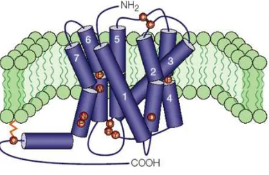
Figure 1. Characteristic structure of a GPCR in the cellular membrane. The seven transmembrane (7TM) domains are numbered. The Amino-terminus of the protein is extracellular, while the Carboxy-terminus is intracellular. The structure depicted is that of a typical rhodopsin-like GPCR. Image from Ellis (2004). 24
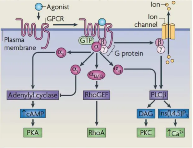
Figure 2. GPCR signaling in a classical perspective, from Ritter and Hall (2009).25
Figure 3. GPCR phylogenetic “Tree of life”, suggested by Fredriksson and Schioth (2005). The red numbers represent the time in million of years (MYA) since the split. GPCRs and their number in the different classes are represented in blue. Image from
Perez (2005). 27
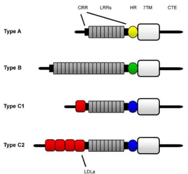
Figure 4. Representation of the major LGR types. Image from Van Hiel et al., (2011). 7TM – seven transmembrane domain; CTE – C terminal endodomain; CRR – cysteine-rich region; HR – hinge region; LDLa – low density lipoprotein receptor
domain class A; LRR – leucine-rich repeat. 28
Figure 5. General scheme of the subdivision of Ilps into insulins, insulin-like growth factors (IGFs), and relaxins based on receptor binding preference and function. 31
Figure 6. Preprohormone processing steps for Ilps. Insulin and relaxins are encoded as preprohormones and are processed twice as described in the text. IGFs do not undergo C peptide processing. “C” stands for the amino acid Cysteine. The three disulfide bonds made by the six stereotype Cysteines of the Ilp superfamily are
shown. 31
Figure 8. Evolutionary relationship between rln/insl genes in vertebrates based on Yegorov et al., (2014). While the endpoints are the seven human relaxin-like genes, not all vertebrates have these all of these genes, yet they have rln-like genes in their genome. INSL4 and INSL6 is found in placental mammals and RLN1 and RLN2 in catarrhini primates. The last common ancestor to all vertebrates is thought to have had one rln/rln3-like gene and one copy of an RXFP1/2-like receptor and one copy of an RXFP3/4 receptor (Bathgate & Wilkinson , 2007; Yegorov & Good, 2012;
Yegorov et al., 2014). 33
Figure 9. The two groups of vertebrate relaxin-like GPCR receptors. Figure adapted from Bathgate et al. (2013). RXFP1/2 are Type C1 LGRs. 7TM – seven transmembrane domain; CRR – cysteine-rich region; HR – hinge region; LDLa – low density lipoprotein receptor domain class A; LRR – leucine-rich repeat. Most of the C-terminal endodomain is not visible in this figure. The LRR is the ligand (relaxin-peptide) binding domain for RXFP1/2 receptors. We note that in both RXFP1 and RXFP2 there is an extra transmembrane domain localized N-terminally to the depicted structure (immediately upstream of the LDLa), which is nevertheless not depicted in this scheme. Whether or not this transmembrane domain serves as a processed signal peptide or is retained in the mature receptor form is not clear. 35
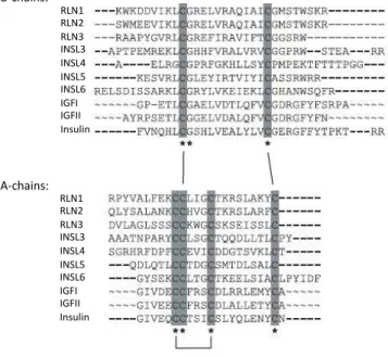
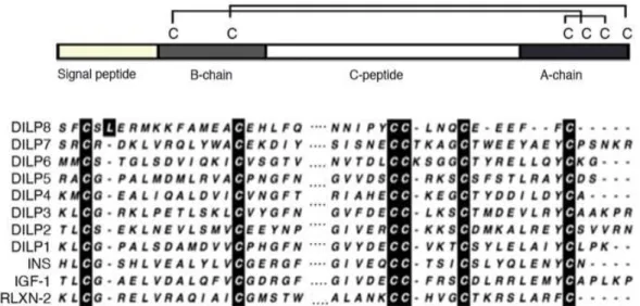
Figure 10. General Ilp scheme and protein alignment of the B and A chains of the eight Drosophila Ilps and comparison to three representatives of the human Ilp subfamilies. Image from Garelli et al. (2012). Absolutely conserved amino acids are
highlighted in dark. 37
Figure 11. Hypotheses for how Dilp8 could act as an inhibitory imaginal signal.
Image from Hariharan (2012). 41
Figure 12. Some key discoveries and methodologies used in fruit flies over the years, from “100 years of Drosophila research and its impact on vertebrate neuroscience: a history lesson for the future”, by Bellen et al., 2010. 45
Figure 14. Imaginal discs and the adult appendages that they will form, from Lewis,
2005. 48
Figure 15. Minos donor and helper constructs containing the 3xPax6/EGFP dominant marker. The helper construct expresses Minos transposase under heat-shock and is based on pCaSper. Only the transposon regions are shown.Image from
Metaxakis et al. (2005). 50
Figure 16. Flybase genome browser snapshot showing the location of the Minos element lgr3[MB06848] gene insertion (blue triangle) in the lgr3 locus on the 3rd
chromosome of the D. melanogaster genome. 51
Figure 17. Flybase genome browser snapshot showing the location of the Minos element lgr4[MB03440] gene insertion (blue triangle) in the lgr4 locus (CG34411)
on the X chromosome of the D. melanogaster genome. 51
Figure 18. Lgr3 “jump start” experiment. First virgin females of the stocks carrying the lgr3[MB06848] element, were crossed with males of a stock, carrying the MiET1 transposon insertion on a Cy balancer chromosome (Cy-Minos). The flies were transferred to new vials every day, and two days after, the old vials containing the F1 progeny were heat-shocked daily until pupariation by inserting the vials in a water-bath at 37°C for 1 h. Cy-Minos/+; lgr3[MB06848]/MKRS or TM6B male adults were selected and crossed to the balancer strain if/CyO; MKRS/TM6B. From each vial, we then selected a single eGFP-negative fly and back-crossed them to if/CyO; MKRS/TM6B and selected against the balancer chromosomes. 53 Figure 19. Lgr4 “jump start” experiment. First, virgin females of the stocks carrying the lgr4[MB03440] element, were crossed with males of a stock, carrying the MiET1 transposon insertion on a Cy balancer chromosome (Cy-Minos). The flies were transferred to new vials every day, and two days after, the old vials containing the F1 progeny were heat-shocked daily until pupariation by inserting the vials in a water-bath at 37°C for 1 h. Cy-Minos/+ males were selected and crossed to the X chromosome balancer strain Nrg[l4]/FM7C. Putative excision lines lgr4[MB03440]excision/FM7C were selected and crossed to the Nrg[l4]/FM7C
Figure 20. NZYDNA Ladder III, NZYTech. 59
Figure 21. 1kb DNA Ladder, Invitrogen. 59
Figure 22. GeneRuler 50bp DNA Ladder, ThermoScientific. 60
Figure 23. Scheme of fecundity and viability assay. 64
Figure 24. Scheme of development time assay. 65
Figure 25. lgr3 expression levels, quantified by qRT-PCR using primer pairs located in the 3’ region of the lgr3 locus. For these experiments, mRNA was isolated from male and female adult flies from different genetic backgrounds. Results showed that lgr3 levels were very low in comparison to the levels of the housekeeping gene rp49 in different backgrounds, but they were slightly higher in males than in females. Shown are the averages of three repeats. Error bars are standard deviations of the
means (SDs). 67
Figure 26. Fecundity (A) and Viability (B) analyses of lgr3 mutants. lgr3[ex1] shows no large differences in fecundity [eggs laid per female per hour (eggs/female/h)] in comparison with the control lgr3[exp2] stock. The viability (% of unfertilized embryos) of the embryos laid by the females is nevertheless strongly affected in both lgr3[ex1] and lgr3[exp2], in comparison with the other stocks. Shown are the averages +/- the standard error of the means (SEMs) of n=9 for each
genotype. 68
amplification products of the precise and imprecise excision lgr3[exp2] and lgr3[ex1], respectively, which shows that the deletion occurred downstream (to the right) of the insertion site. (C) Electrophoretic band of PCR amplification product of lgr3[ex1], from which we later obtained the gDNA sequence and identified a deletion of approximately 1.6 kb. (D) Scheme of the lgr3[ex1] deletion. The deletion removes most of the intron and completely removes exon seven and partially exon eight. The negative control used in the different experiments was dH2O, showing no
PCR amplifications, as expected. 69
Figure 28. UCSC genome browser screenshot of the D. melanogaster lgr3 locus with BLAT results (Your Sequence from Blat Search) obtained by sequencing the 1.6-kb PCR fragment obtained in Figure 29-C. This shows the breakpoints that
characterize the 3827-bp deletion in lgr3[ex1] mutant. 69
Figure 29. RT-PCR amplification pattern of lgr3 mutations and controls using mRNA isolated from males. (A) Electrophoretic pattern of PCR amplification products of lgr3[exp2] and lgr3[ex1], with the set of primers lgr3_around (set of primers in blue). The lgr3[ex1] mutant gave a smear and a major product at around 200 bp, consistent with a non-spliced transcript reading directly through the deletion breakpoint. (B) RT negative control, showing no amplification products, ensuring that any gDNA contamination, if present, was under detection levels. (C) The housekeeping gene rp49 was used as a positive control for the RT-PCR. The negative control used was dH2O. (D) Schematic representation of the lgr3 cDNA in
the lgr3[ex1] mutant and in the lgr3[exp2] control line. Blue, normal splicing and exon reading frames. Red, aberrant splicing and frame shifted exons. We were unable to sequence a specific product from the lgr3[ex1] transcript. 70
lgr3[exp2], and w1118 are less abundant. Only male cDNAs were analyzed. Shown
are the averages of three repeats +/- SDs. 71
Figure 31. Protein sequence of WT Lgr3 (Reference NP_733115.1) and the predicted Lgr3[ex1] protein. The 69 aa from I327 to T395 in the C-terminal part of the Leucine Rich Repeat (LRR) domain that were certainly removed by the lgr3[ex1] deletion are highlighted in red. The bold and underlined aa (D236) marks the end of exon 6, after which the protein most likely soon truncates due to intron readthrough of the transcript. The major domains of the Lgr3 protein are the signal peptide, LDLa domain, LRR N-terminal domain (LRRNT), LRRs, the hinge region (Hinge) and the 7TM domains. This cartoon is not to scale. It depicts the approximate region of the Lgr3[ex1] protein truncation. There could be other protein isoforms if splicing still occurs downstream of the breakpoint, but these are also predicted to encode premature stop codons and hence truncate the protein. 72
Figure 32. lgr4 expression levels, quantified by qRT-PCR. For these experiments, mRNA was isolated from male and female adult flies from different genetic backgrounds. Results showed that lgr4 levels were very low in comparison to the levels of the housekeeping gene rp49 in different backgrounds, but they were higher in males than in females. Shown are the averages of three repeats +/- SDs. 73
Figure 33. Fecundity (A) and Viability (B) analyses of lgr4 mutants and controls. (A) lgr4[ex1] lay equivalent amounts of eggs as other control lines [eggs laid per female per hour (eggs/female/h)]. (B) The viability (% of unfertilized embryos) of the embryos laid by the females is not negatively affected in lgr4[ex1] and lgr4[exp1], in comparison with the other stocks. Shown are the averages and the standard error of the means (SEMs) of n=9 for each genotype. 74
scheme, the lgr4 locus transcribes from right to the left. Exons are numbered. Colored arrows are show the approximate location of the primers used. (B) Electrophoretic pattern of PCR amplification products of the lgr4[exp1] precise excision and the lgr4[ex1] and lgr4[ex3] deletions. This shows that the lgr4[ex1] deletion occurred upstream of the insertion site while the lgr4[ex3] deletion occurred downstream. This also shows that the lgr4[exp1] is indeed a precise excision giving the expected product PCR size with the green primer pair (note that no 735-bp product is produced in the MB03440 line because of the presence of the several-kb MB03440 insertion in between the primer sites). The positive control, used to check the deletions, was the original line used in the excision protocol. As expected, the negative control used, dH2O, showed no PCR amplification. (C) Gene scheme
depicting the deletion in lgr4[ex1], showing that exon 2 was partially deleted. (D) Gene scheme depicting the deletion in lgr4[ex3], showing that the deletion affects introns two, three and four and the exons three and four. 75
Figure 35. UCSC genome browser screenshot of part of the D. melanogaster lgr4 locus containing BLAT results (Your Sequence from Blat Search) obtained by sequencing the PCR product with a set of primers upstream and downstream of the lgr4[MB03440] position (blue arrowhead). Instead of the predicted 255-bp product, we obtained a product of 96 bp, confirming the ex1 mutation. Notice that the lgr4 locus transcribes from the right to the left in this scheme. Hence, the exon to the right is exon 2, and the exon to the left is exon 3. Some of the sequences adjacent to the deletion missalign in exon 2 rather than aligning in the intron. Actually the deletion
starts immediately to the right of the MB03440 element. 76
(Hinge) and the 7TM domains. This cartoon is not to scale. It depicts the approximate region of the Lgr4[ex1] protein truncation. There could be other protein isoforms if splicing still occurs downstream of the breakpoint, but these are also predicted to encode premature stop codons and hence truncate the protein. 76 Figure 37. RT-PCR amplification pattern of lgr4 mutations and controls using mRNA isolated from males. (A) Electrophoretic pattern of RT-PCR amplification products in the depicted genotypes with the set of primers for lgr4 gene (set of primers forward in red and reverse in purple). (B) RT negative control, showing no amplification, as expected, ensuring that no gDNA contamination occurred. (C) Positive control, the housekeeping gene RP49 expression. The negative control used was dH2O, showing no PCR amplification, as expected. (D) Scheme of the lgr4
transcripts in the mutants and in the control exp1 line. Notice that in this scheme the lgr4 locus is transcribed from the right to the left. 78
Figure 38. qRT-PCR analyses of transcript levels of the lgr4[ex1] deletion mutant in comparison with the control lines, lgr4[exp1] and w[1118], and the original insertion line lgr4[MB03440]. Primer pairs in exonic sequences flanking the ex1 deletion were used and normalized to the housekeeping gene rp49. A transcript was detected in lgr4[ex1], which was nevertheless aberrant and had a distinct dissociation curve (B). One can also observe that the lgr4[MB03440] insertion induces a small reduction in lgr4 levels, as previously detected (see Figure 34), which is restored to normal in lgr4[exp1]. Only males cDNA were analyzed. Shown are the averages of three repeats, except for lgr4[exp1]. Error bars are standard deviations of the means. (B) The dissociation curves of selected qRT-PCR products shown in (A) for the three depicted genotypes. These results confirm that lgr4[ex1] produced an aberrant transcript, different from the w[1118] and lgr4[exp1] controls. 79
is depicted with a blue arrowhead. Notice that the lgr4 locus transcribes from the right to the left in this scheme. The lgr4[ex3] transcript splices perfectly from exon two to exon seven, consistent with the prediction of the deletion of all introns two, three and four, and all exons three and four. Exons 5 and 6 were also probably
deleted. 80
Figure 40. Protein sequence of WT Lgr4 (Reference NP_001096966.2) and the predicted Lgr4[ex3] protein. According to the sequence obtained, the deletion causes the formation of a truncated ~163-aa protein (153 aa of Lgr4 sequence and 10 aa of framshifted sequence from exon 7) containing the very N-terminus of Lgr4 up to the LDLa domain. In red are the aa that form due to out-of-frame splicing from exon 2 into the exon 7. The major domains of the Lgr4 protein are the signal peptide, LDLa domain, LRR N-terminal domain (LRRNT), LRRs, the hinge region (Hinge) and the 7TM domains. This cartoon is not to scale. It depicts the approximate region of the Lgr4[ex3] protein truncation. There could be other protein isoforms if splicing still occurs downstream of exon7, but most of these are also predicted to encode
premature stop codons and hence truncate the protein. 80
Figure 41. lgr3 mutation rescues the dilp8-dependent delay in the onset of pupariation. (A) Pupariation timing curves for different genotypes. Shown are averages +/- SEMs of 8-16 experiments. (B) Bar graph showing the median pupariation time of the depicted genotypes. ANOVA: p<0.001. Groups sharing the same letters are not statistically significantly different at alpha=5% according to the Tukey’s HSD post-hoc test. Shown are averages +/- SEMs of 8-16 experiments. Bar colors depict the dosage of lgr3 (blue = two wild-type alleles, red = two loss-of-function alleles; hashed bars = heterozygotes). Green arrow depicts full suppression.
Yellow arrow depicts partial suppression. 82
and one repeats, respectively. The genotype +/+; +/Df(3)BSC321 served as a negative control. (B)Bar graph showing the median pupariation time of the depicted genotypes. Shown are averages +/- SEMs of 3 experiments except for tub-dilp8/+;lgr3[ex1]/Df(3)BSC321 and tub-dilp8/+;lgr3[ex1]/lgr3[ex1], which had two and one repeats, respectively. ANOVA was not performed due to the lack of at least three repeats in the above-mentioned genotypes. Bar colors depict the dosage of lgr3 (blue = two wild-type alleles, red = two loss-of-function alleles; hashed bars = heterozygotes). Green arrows depict full suppression. Yellow arrows depict partial
suppression. 83
Figure 43. lgr4 mutation partially rescues the dilp8-dependent delay in the onset of pupariation. (A) Pupariation timing curves for different genotypes. Shown are averages +/- SEMs of 4-6 experiments. (B) Bar graph showing the median pupariation time of the depicted genotypes. ANOVA: p<0.001. Groups sharing the same letters are not statistically significantly different at alpha=5% according to the Tukey’s HSD post-hoc test. Shown are averages +/- SEMs of 4-6 experiments. Bar colors depict the dosage of lgr4 (blue = two wild-type alleles, red = two
loss-of-function alleles). Green arrows depict full suppression. 84
Figure 44. Hypothesis for the role of the Lgr3 and Lgr4 receptors, in the Dilp8
L
ist of tables
Table 1. Drosophila stocks used in this work. 49
Table 2. Mix reagents for cDNA reaction. 56
Table 3. Standard PCR reaction reagents. 58
Table 4. PCR conditions for amplification of interest genes. 58
Table 5. Lgr3 Primers used for Polymerase Chain Reaction. 58
Table 6. Lgr4 Primers used for Polymerase Chain Reaction. 58
Table 7. Primers used in qPCR assays. 62
List of abbreviations
7TM - seven transmembrane domain
20E - 20-hydroxyecdysone aa - aminoacid
AKT - serine/threonine-specific protein kinase
AMP - adenosine monophosphate
AP2 - clathrin adaptor
BLAST- basic local alignment search tool
bp - base pair(s)
Ca2+ - calcium ion
cDNA - complementary DNA
CRR - cysteine-rich region
Cys - cysteines
DAG - diacylglycerol
Df - deficiency chromossome
dH2O - distilled water
Dilp - D. melanogaster insulin-like peptides
DNA - deoxyribonucleic acid
EDTA - ethylenediaminetatracetate
FSHR - vertebrate gonadotropin receptor
Gα/Gβ/Gγ - heterotrimeric G proteins subunits
gDNA - genomic deoxyribonucleic acid
GDP - guanosine diphosphate
GFP - green fluorescent protein
GPCR – G-protein coupled receptors
GRK - G-protein coupled receptors kinases
GTP - guanosine triphosphate
H2O - water
HCl - hydrochloric acid
HR - hinge region
IGF - insulin-like growth factor
IIS - insulin/IGF Signaling
Ilp - insulin-like peptide
InR - insulin receptor
Insl - insulin-like peptide
JH - juvenile hormone
Kb - kilobase(s)
LDLa - low density lipoprotein receptor domain class A
LGR - leucine-rich repeat containing G protein-coupled receptor
LH/CGR - luteinizing hormone receptor
LRR - leucine-rich repeat
MAPK - mitogen-activated protein kinase
mRNA - messenger RNA
MYA - million years ago
NaCl - sodium chloride
NCBI - National Center for Biotechnology Information
O.N. - overnight
PCR - polymerase chain reaction
PI3K -phosphoinositide 3-kinase
PTTH - prothoracicotropic hormone
qPCR - quantitative polymerase chain reaction
RLN - relaxin
RNA - ribonucleic acid
RNAi - RNA interference
rpm - rotations per minute
RT - room temperature
RTK - tyrosine kinase receptor
RT-PCR - reverse transcription polymerase chain reaction
qRT-PCR - quantitative reverse transcription polymerase chain reaction
rp49 - housekeeping gene, also knowed as rpl32 RXFP - human relaxin receptor
SD - standard deviation
SNP - single nucleotide polymorphism
TAE - tris-acetate-ethylenediaminetatracetate
Tm - melting temperature
tRNA - transfer ribonucleic acid
TSHR - thyroid hormone receptor
Tub - tubulin promoter
1.1
G-protein Coupled receptors
G protein-coupled receptors (GPCRs) are an ancient and large family that share structural characteristics of having at least an extracellular Amino (N)-terminus, an intracellular Carboxy (C)-terminus, the characteristic seven transmembrane (7TM)-spanning helical domains, and three interhelical loops on each side of the membrane (Figure 1). GPCRs transduce a diverse array of extracellular signals, such as light, calcium, neurotransmitters, odorants, peptides, proteins, among others. Hence, GPCRs are involved in most physiological processes, from smell to vision and from taste to neurological, cardiovascular, endocrine and reproductive responses. Because of this, GPCRs are an extremely important therapeutic and pharmacological target, as well as a subject of considerable research interest (Broeck, 1996; Bargmann, 1998; Broeck, 2001; Tyndall et al., 2005; Overington et al., 2006; Bridges et al., 2008).
Figure 1. Characteristic structure of a GPCR in the cellular membrane. The seven transmembrane (7TM) domains are numbered. The Amino-terminus of the protein is extracellular, while the
Carboxy-terminus is intracellular. The structure depicted is that of a typical rhodopsin-like GPCR. Image from
Ellis (2004).
Figure 2. GPCR signaling in a classical perspective, from Ritter and Hall (2009).
G proteins are a subfamily of GTPase proteins that can bind the nucleotides guanosine triphosphate (GTP) and guanosine diphosphate (GDP) (Purves et al., 2001; Oldham & Hamm et al., 2008; Ritter & Hall, 2009). GTPases bind and hydrolyze GTP through their highly conserved G domain. Typically, when a G protein is bound to GTP it is “active”, and when it is bound to GDP, it is “inactive”. G proteins can be either monomeric (e.g., the Ras superfamily of small GTPases) or heterotrimeric (Purves et al., 2001). Heterotrimeric G proteins consist of alpha (Gα), beta (Gβ) and gamma (Gγ) subunits, with the G domain localized in the Gα subunit, the largest one (Figure 2). The Gα and Gβ subunits associate with the cellular membrane by lipid anchors.
Upon stimulation, the GPCR catalyzes the exchange of GDP to GTP in the Gα
a few subtypes (Oldham & Hamm et al., 2008; Ritter & Hall, 2009) that couples to the specific GPCR in question. The different Gα subtypes and the dissociated Gβγ
can bind to different effectors such as adenylyl cyclase, RhoGEF, phospholipase Cβ, and exercise different downstream regulation by producing second messengers (cyclic AMP, diacylglycerol – DAG, inositol-1,4,5-triphosphate) that will themselves regulate further downstream effectors like protein kinase A and C, ion channel and phospholipase Cβ (Figure 2) (Oldham & Hamm et al., 2008; Ritter & Hall, 2009).
If the stimulating ligand is still present, the activated GPCR will bind to and activate the next heterotrimeric G protein, and signaling will continue as long as ligand and inactive G proteins are available. In the absence of stimuli, the GPCR signal terminates when the Gα subunit eventually hydrolyzes the attached GTP to GDP (Oldham & Hamm et al., 2008; Ritter & Hall, 2009).
1.1.1
Role of GPCR in diseases
Many diseases are directly linked with mutations in GPCR receptors. Known diseases include, fertility disorders, congenital night blindness, familial gestational hyperthyroidism, nephrogenic diabetes insipidus, and carcinomas (Schöneberg et al., 2005; Thompson et al., 2008). Some of the mechanisms by which the mutations lead to disease comprise changes either in the GPCR gene expression levels (e.g., transcription regulatory mutations), in the abundance of the receptor in the plasma membrane (e.g., receptor recycling), in receptor signaling (e.g., G protein coupling), in receptor desensitization, or alteration of ligand binding by loss of ligand specificity or in agonist-independent receptor signaling (Sealfon, 2005; Schöneberg et al., 2005; Thompson et al., 2008).
terminating GPCR signaling, have been linked to adenoma formation. Gβ subunits mutations have been linked to hypertension (Ibegbu et al., 2012).
1.1.2
GPCR evolution
GPCRs are present in the majority of eukaryotic organisms, testifying for their extraordinary evolutionary success (Fredriksson & Schioth, 2005; Perez, 2005; Figure 3). GPCRs can be generally classified into different subfamilies the Adhesion, Secretin, Frizzled, Glutamate, and Rhodopsin families (Fredriksson, 2003). These families likely arose and expanded in bilateria about 750-430 million years ago (MYA), before the nematodes split from the chordate lineage (Fredriksson & Schioth, 2005; Perez, 2005).
Figure 3. GPCR phylogenetic “Tree of life”, suggested by Fredriksson and Schioth (2005). The red numbers represent the time in million of years (MYA) since the split. GPCRs and their number in the
The most well studied and understood GPCR family is probably the Rhodopsin GPCR family (also known as Rhodopsin-like receptor family), which were also the first to be discovered (Brody & Cravchik, 2000). This family includes Opsin receptors, receptors for biogenic Amines, Purine receptors, Peptide receptors and Orphan receptors that can be activated by diverse ligands or stimuli, such as neurotransmitters, neuropeptides, peptide hormones, light, nucleotides, prostaglandins, leukotrienes, chemotactic peptides and chemokines. These receptors vary considerably in structure, but are characterized by their conserved 7TM domain structure (Figure 1). Besides the well studied function as photoreceptors, other members of the Rhodopsin-like GPCR family, such as the Leucine-rich repeat-containing G-protein coupled receptors (LGRs) (see below) also play an important role in development (Brody & Cravchik, 2000).
The LGR subgroup of Rhodopsin-like GCPRs are structurally characterized by a large N-terminal extracellular domain flanked by a cysteine-rich region (Figure 4), in addition to the typical 7TM of the Rhodopsin-like receptor family. LGRs can be classified in three subtypes: according to the numbers of Leucine-rich repeat motifs (LRRs) in their ectodomains and the sequence of the hinge domain in between the LRRs and the 7TMs. Type A and C LGRs contain between 7-9 LRRs, while Type B LGRs have 13-18 LRRs (Figure 4; Hiel et al., 2011).
Figure 4. Representation of the major LGR types. Image from Van Hiel et al., (2011). 7TM – seven transmembrane domain; CRR – cysteine-rich region; CTE – C terminal endodomain; HR – hinge
Type A LGRs are further characterized by having the longest hinge region, with a conserved sequence containing 6 Cysteines that are expected to form disulfide bonds and stabilize the receptor structure. Examples of Type A LGRs are the Follicle Stimulating Hormone Receptor FSHR receptor (vertebrate gonadotropin receptors), the LH/CGR receptor (luteinizing hormone receptor) and the thyroid hormone receptor (TSHR). Apart from having more LRRs than Type A, Type B LGRs have a shorter hinge region with only 4 Cysteines. An example of a Type B LGR is the Drosophila Bursicon receptor, rickets, responsible for the critical wing expansion and cuticle hardening after eclosion of the fly (Baker & Truman, 2002).
Type C LGRs have the shortest hinge region among the LGRs and have the consensus sequence LxxLxHIxFDRFxYCxYAPHV at its beginning and CxPxxDGISSxEDLLSNxVLRV at its end (Hiel et al., 2011). They are further characterized by having at least one Low-density lipoprotein domain class A (LDLa) domain located N-terminally to the LRRs (Figure 4). Type C LGRs are divided in Type C1 and Type C2, depending on the number of Cysteines in the hinge region and the number of LDLa repeats in the extracellular domain. Type C2 have many LDLa repeats and four Cysteines, while type C1 has only one LDLa domain and two Cysteines in the hinge region. Examples of Type C1 LGRs are the human relaxin receptors RXFP1 and RXFP2, also known as LGR7 and LGR8, respectively. In the fruit fly genome, two LGRs type C1 genes have already been identified, Lgr3 and Lgr4 (as well known as CG31096 and CG34411 respectively) (Hiel et al., 2011). The biological functions of these two putative Drosophila Relaxin-like Type C1 LGRs are unknown and are the focus of this study.
1.2
Relaxin like-peptides
All Ilps are structurally related and are thought to originate from a common Insulin/IGF-like ancestral peptide (Blundell et al., 1980; Wilkinson & Bathgate, 2006; Bathgate et al., 2013). Ilps are now subdivided into insulins, insulin-like growth factors (IGFs), and relaxins on the basis of gene and protein structure, processing, and receptor binding preferences (Blundell et al., 1980; Wilkinson & Bathgate, 2006; Figure 5). Insulin binds to a receptor tyrosine kinase (RTK), the insulin receptor (InR), and stimulates a signaling pathway that includes phosphoinositide 3-kinase (PI3K) and serine/threonine kinase (AKT). IGFs preferentially bind related RTKs, named IGF receptors (IGFRs), which share an evolutionary history with the InR, and activate the MAPK pathway. In contrast, relaxins, as will be further described below, bind to GPCRs and regulate diverse downstream signaling pathways (Hsu et al., 2002; Hiel et al., 2011; Bathgate et al., 2013). Functionally, while the insulin/IGF pathway regulates growth, metabolism and ageing (Blundell et al., 1980; Wilkinson & Bathgate, 2007; Gronke et al., 2010), members of the vertebrate relaxin family, have antifibrotic, neuropeptide, vasodilator and cardiac stimulatory functions, affecting many processes, including - but not limited to -, female and male reproduction, such as menstruation, sperm motility, and pregnancy (reviewed in Bathgate et al., 2013). Therefore, it is important to stress, that a peptide’s appurtenance to the Ilp superfamily per se by no means implies that it will act via insulin/IGF Signaling (IIS).
Figure 5. General scheme of the subdivision of Ilps into insulins, insulin-like growth factors (IGFs), and relaxins based on receptor binding preference and function.
Figure 6. Preprohormone processing steps for Ilps. Insulin and relaxins are encoded as preprohormones and are processed twice as described in the text. IGFs do not undergo C peptide
processing. “C” stands for the amino acid Cysteine. The three disulfide bonds made by the six
stereotype Cysteines of the Ilp superfamily are shown.
apes retained the RLN1 duplication (Arroyo et al., 2014). Hence, despite the historical name, RLN2, aka as H2 relaxin in humans, is the ancestral and major form of circulating relaxin found in the great apes and is the functional equivalent to the first relaxin identified by F.L. Hisaw, which is produced by the corpus luteum and by placenta during pregnancy (Hisaw, 1926).
The evolution of vertebrate relaxin-like peptides has been focus of several studies (Maere et al., 2005; Makino et al., 2009), but there is limited consensus on how and when these peptides diverged from ancenstral Insulin/IGF-like peptides. Probably the most comprehensive analyses of the evolution of the relaxin-like peptides was carried out by Sergey Yegorov and Sara Good in 2012, who applied paleogenomic models to elucidate the role of whole genome duplications on the story of these peptides and their GPCR receptors. The most likely scenario is that a single relaxin-like peptide, probably most similar to the neuropeptide RLN3, existed in the ancestral vertebrate (Bathgate & Wilkinson , 2007; Yegorov & Good, 2012).
Figure 7. Comparison of amino-acid structures of the 10 human Ilps, adapted and modified from
Bathgate et al. (2013). Note the so called “relaxin signature” [RxxxRxxI/V] in some of the B chain of
the mature relaxin-like peptides.
Some, but not all, human relaxin-like peptides, most notably RLN1, RLN2 and RLN3, share the so called “relaxin signature” [RxxxRxxI/V] in the B chain of the mature peptide (Figure 7). These amino acids, as will be described further below,
are all important for their binding to the relaxin receptor. Despite this similarity between RLN1, RLN2 and RLN3, the human relaxin-like peptides actually have a different phylogenetic history. While RLN1 and RLN2 are intimately related, they seem more related to INLS3, 4 and 6 than to relaxin-3 and INLS5, which branch earlier in the relaxin-like peptide tree (Figure 8; Wilkinson et al., 2005).
Figure 8. Evolutionary relationship between rln/insl genes in vertebrates based on Yegorov et al., (2014). While the endpoints are the seven human relaxin-like genes, not all vertebrates have these all
of these genes, yet they have rln-like genes in their genome. INSL4 and INSL6 is found in placental
mammals and RLN1 and RLN2 in catarrhini primates. The last common ancestor to all vertebrates is
thought to have had one rln/rln3-like gene and one copy of an RXFP1/2-like receptor and one copy of
an RXFP3/4 receptor (Bathgate & Wilkinson , 2007; Yegorov & Good, 2012; Yegorov et al., 2014).
Wilkinson, 2007). INSL3 has a role in male reproductive system being involved in the mediation of testicular descent (Nef & Parada, 1999; Zimmermann et al., 1999). INSL4 plays a role during pregnancy specifically in placenta (Chassin et al., 1995; Koman et al., 1996).
While relaxin was discovered in the late 20s, as described above, the link between relaxins and GPCRs was only definitively established many decades later, in 2002 (Hsu et al., 2002; Ivell, 2002). Due to the structural similarity between relaxins and insulin/IGF-like peptides, the relaxins had been expected to also act via RTKs (Ivell, 2002; Bathgate & Wilkinson, 2007). The discovery in the late 90s, that mice lacking the relaxin family peptide INSL3 showed a similar abnormal testis descent phenotype, as mice with a disrupted GREAT gene, which encodes a GPCR, provided the first strong hint towards GPCRs as putative relaxin receptors (Nef & Parada, 1999; Zimmermann et al., 1999; Overbeek et al., 2001). The fact that relaxin stimulated cAMP in some tissues (Sherwood, 1994), but not all (Ivell, 2002), also added strength to this link. Hence, the discovery, reported in 2002, that the relaxin peptide (RLN2) was able to activate the RXFP1 and RXFP2 GPCRs (also known as LGR7 and LGR8, respectively) provided the definitive evidence that relaxins were not RTK ligands. The ligand and receptor relationship between INSL3 and GREAT was later confirmed (Kumagai et al., 2002; Bogatcheva et al., 2003), and as the mouse GREAT gene encodes the homologue of RXFP2 that is expressed in a similar distribution as INSL3 rather than RLN2, it is INSL3 that is regarded as the endogenous ligand for RXFP2 despite the fact that it can also be activated by RLN2.
et al., 2007). The receptors for INSL4 and INSL6 are currently unknown, and these peptides do not interact with any of the RXFP receptors (Bathgate et al., 2013).
Figure 9. The two groups of vertebrate relaxin-like GPCR receptors. Figure adapted from Bathgate et
al. (2013). RXFP1/2 are Type C1 LGRs. 7TM – seven transmembrane domain; CRR – cysteine-rich
region; HR – hinge region; LDLa – low density lipoprotein receptor domain class A; LRR –
leucine-rich repeat. Most of the C-terminal endodomain is not visible in this figure. The LRR is the ligand
(relaxin-peptide) binding domain for RXFP1/2 receptors. We note that in both RXFP1 and RXFP2
there is an extra transmembrane domain localized N-terminally to the depicted structure (immediately
upstream of the LDLa), which is nevertheless not depicted in this scheme. Whether or not this
transmembrane domain serves as a processed signal peptide or is retained in the mature receptor form
is not clear.
Lgr4 is encoded by 18 exons like human LGR7 and LGR8, while Lgr3 is encoded by 11 exons (Hiel et al., 2014). The function of the Drosophila relaxin-like receptors Lgr3 and Lgr4 is unknown and strikingly, there have been no loss- or gain-of-function studies on these genes to the best of our knowledge. To gain insight into the function of these receptors is the principal aim of this work.
1.2.1
The relaxin paradox
receptors (Yogorov & Good, 2012). They also suggest that the last common ancestor to all vertebrates probably had two relaxin-like peptides, one similar to RLN and the other most similar to RLN3(Yogorov & Good, 2012).
Another important fact to consider is the recent discovery of an insulin/relaxin-like peptide in the invertebrate starfish (reviewed in Mita, 2013). This peptide has a gonadotropin-like activity that is necessary for oocyte maturation and ovulation. Even though the receptor for this starfish insulin/relaxin-like peptide is still unknown, it clearly acts via G-protein and adenylyl cyclase (reviewed in Mita, 2013).
Figure 10. General Ilp scheme and protein alignment of the B and A chains of the eight Drosophila Ilps and comparison to three representatives of the human Ilp subfamilies. Image from
Garelli et al. (2012). Absolutely conserved amino acids are highlighted in dark.
et al., 2012; Garelli et al., 2012). The only IGF-like Ilp in the fly genome is Dilp6 (Okamoto et al., 2009; Slaidina et al., 2009). All other Dilps are predicted to encode a C-peptide, suggesting they are either insulin or relaxin-like (Gronke et al., 2010; Garelli et al., 2012).
The binding of insect Ilps to insect InRs has only recently been demonstrated for mosquito Ilp3/4 (Brown et al., 2008; Wen et al., 2010) and the Drosophila Dilp5 (Sajid et al., 2011). Furthermore, Dilp2 and 5 can induce tyrosine autophosphorylation of the Drosophila InR (Rulifson et al., 2002). Other Ilps are usually assumed to activate IIS based either on sequence information indicating appurtenance to the Ilp family and/or their ability to modulate InR-dependent growth in gain and loss-of-function studies (Brogiolo et al., 2001; Ikeya et al., 2002; Rulifson et al., 2002; Yang et al., 2008; Okamoto et al., 2009; Slaidina et at., 2009; Zhang et al., 2009; Gronke et al., 2010). In Drosophila, this has been assumed for Dilp1, 3, 4, 6 and 7. Indeed, the genetic evidence for Dilp1-5 acting via an InR is indeed quite strong [e.g., dilp1-5 occur in a chromosomal cluster (Brogiolo et al., 2001)], Dilp5 has been shown to bind InR (Sajid et al., 2011), and Dilp2 overexpression can rescue the growth defect of animals lacking the dilp1-5 cluster (Zhang et al., 2009). However, the evidence is weaker, contradictory or absent for Dilp6-8, respectively (see further details below for each Ilp; Brogiolo et al., 2001; Ikeya et al., 2002; Yang et al., 2008; Okamoto et al., 2009; Slaidina et al., 2009; Zhang et al., 2009; Colombani et al., 2012; Garelli et al., 2012).
Dilp6 is a fat body-derived IGF-like peptide that regulates postfeeding growth in Drosophila (Okamoto et al., 2009; Slaidina et al., 2009). It is the sole IGF-like Ilp encoded in the Drosophila genome. Genetic evidence suggests Dilp6 works via IIS (Okamoto et al., 2009; Slaidina et al., 2009), but there is no biochemical data demonstrating the direct activation of the InR by Dilp6. The biochemical identification of the Dilp6 receptor awaits further study and should bring important contributions to the understanding of Dilp6-dependent activities.
Castellanos et al., 2013), which are involved in food-based decision-making (Yang et al., 2008), and modulate tracheal growth in response to nutrition in the posterior hindgut (Linneweber et al., 2014). A previous report had found that Dilp7 overexpression induced organismal growth (Ikeya et al., 2002), and hence it was considered to be an InR agonist. Yet more recent experiments using updated sequence annotations have failed to reproduce this finding (Dr. I. Miguel-Aliaga, ICL, UK, personal communication). Accordingly, Dilp7 deletion mutations show that it is not essential for growth and development (Gronke et al., 2010). Therefore, the biochemical identification of a receptor for Dilp7 -be it the InR or not- shall certainly contribute to elucidate its molecular mechanism of action.
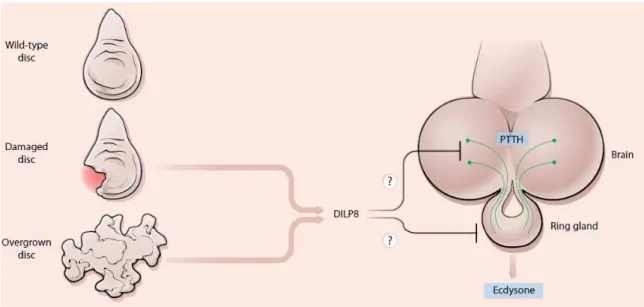
Dilp8 is an insulin/relaxin-like Ilp (Garelli et al., 2012) that couples tissue growth with developmental timing (Colombani et al., 2012; Garelli et al., 2012). Dilp8 is produced and secreted from abnormally-growing imaginal discs (the larval precursors of most adult appendages) and transiently delays the onset of metamorphosis by inhibiting the biosynthesis of the major insect molting hormone (20-hydroxyecdysone (20E)). Concomitantly, Dilp8 slows-down growth of the unaffected imaginal discs via an unknown mechanism. How Dilp8 achieves this is unknown. Interestingly, in the adult stage, Dilp8 is strongly expressed in the ovary (Garelli et al., 2012), similarly to vertebrate relaxins (Blundell & Humbel, 1980; Bathgate et al., 2013). The biology of Dilp8 is further described below.
1.3
Coordinating growth and developmental timing
In many organisms, from mammals to insects, like Drosophila, development seems to occur in a perfectly coordinated way, resulting in a rather stable adult phenotype. Formally, developmental stability is the ability of an organism to buffer given traits against environmental and intrinsic perturbations (Denver & Middlemis-Maher, 2010; Minelli & Fusco, 2010). This may involve physiological, temporal or behavioral adjustments to the developmental program (Thornhill & Moller, 1997; Moczek, 2010). The processes leading to developmental stability have been particularly well studied in insects and other arthropods (Shingleton, 2010). For instance, early in 1927, it was observed that x-ray irradiation of Drosophila larvae negatively influenced the timing of pupariation, causing a developmental delay (Hussey et al., 1927). Decades of further research suggested that the delay is mediated by a secreted signal, termed inhibitory imaginal signal, which is produced by regenerating damaged imaginal discs following irradiation (Simpson et al., 1980). Hence, the extent of the developmental delay correlates with the amount of regenerative growth (Simpson et al., 1980; Smith-Bolton et al., 2010). The inhibitory imaginal signal communicates the abnormal growth status of the discs with the neuroendocrine centers that trigger the onset of metamorphosis (Simpson et al., 1980). The major effect of the inhibitory imaginal signal is to reduce the production of 20E (Halme et al., 2010; Garelli et al., 2012; Hackney et al., 2012), the critical hormone that triggers metamorphosis (reviewed in Hill et al., 2012; Yamanaka et al., 2013). Ecdysone, the 20E precursor, is biosynthetized in the prothoracic gland, a part of the composite endocrine organ known as the “ring gland”, and is secreted directly into the hemolymph. Ecdysone is converted into 20E in the peripheral tissues. By inhibiting Ecdysone biosynthesis, the inhibitory imaginal signal induces a delay in the onset of metamorphosis in the presence of abnormal tissue growth, assuring the coordinated growth of tissues. Such abnormal growth-induced pupariation inhibition can also result from imaginal disc overgrowth (tumors) or slow growth, both of which cause a persistence of dividing mitotic cells (Menut et al., 2007; Parker et al., 2011).
neurons in the brain that directly innervate the ring gland (McBrayer et al., 2007). It is not clear what determines the synthesis of PTTH by the neurons, but a drop in the levels of the Juvenile Hormone (JH), a hormone that is produced in early larval stages and inhibits the larval to pupal transition, might be involved (Edgar, 2006).
Halme and colleagues showed that the inhibitory imaginal signal acted upstream of the synthesis of PTTH using a wing disc genetic ablation technique (Halme et al., 2010). Despite these works, the identity of the inhibitory imaginal signal remained a mystery until 2012.
Figure 11. Hypotheses for how Dilp8 could act as an inhibitory imaginal signal. Image from Hariharan (2012).
coordination of intra-organ communication in face of intrinsic errors occurring during normal development (Garelli et al., 2012).
The precise molecular mechanism by which secreted Dilp8 inhibits ecdysone biosynthesis to cause a developmental delay remains unknown, but there are many hints (Hariharan, 2012; Figure 11). Dilp8, as a member of the secreted Ilp hormone family, is expected to have a receptor. The determination of the Dilp8 receptor could allow the determination of the target tissue of Dilp8 activity. Dilp8 could act either via the sole Drosophila InR, the unexplored relaxin-like receptors or other unknown receptors. Here, as part of our objective in understanding the biological role of the Drosophila relaxin-like receptors Lgr3 and Lgr4, we plan to use genetics to test the possibility that they could act as receptors for Dilp8.
1.4 Aims
2.1
Drosophila melanogaster
as a genetic tool
Drosophila, also known as “vinegar” or “fruit” flies, are a group of small flies (measuring about 3 mm long) that are highly diverse in appearance, behavior and in reproduction habitat. The Drosophila group contains around 1500 species (Robert et al., 1999). Drosophila are dipteran (i.e., two-winged) insects of the family Drosophilidae.D. melanogaster, one of the major model organisms in genetics and development, due to, among other characteristics, its small size, relatively large brood and short generation time (see more details below), belongs to the Sophophora subgenus of Drosophila.
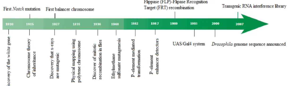
Since the discovery of the white mutation and its linkage to the X chromosome by T.H. Morgan in the early 1900s, Drosophila has been used extensively as an invertebrate model organism, rich in tools and advantages in genetic, evolutionary and developmental research. In the first 50 years of research in Drosophila, the attention of the scientific community was focused mainly in the principle of inheritance. Over the years, the development of new molecular tools, concepts and approaches (Figure 12) further revealed the enormous potential of this model, which could allow the understanding of some mechanisms in vertebrates, given the homology between vertebrates and invertebrates in critical aspects (Bellen et al., 2010).
Figure 12. Some key discoveries and methodologies used in fruit flies over the years, from “100 years of Drosophila research and its impact on vertebrate neuroscience: a history lesson for the future”, by
Bellen et al., 2010.
Drosophila has many other advantages over other invertebrate model organisms to study biological processes in vivo: flies are easy and cheap to obtain and maintain in laboratories due to their small size; their short generation time, about 10 days at room temperature (~25ºC; development may be shorter or longer in higher or lower temperatures, respectively); the small number of chromosomes: only four pairs, making genetics very simple; the relatively early availability of a very well annotated sequenced genome; most of the genetic mechanisms, such as gene transmission, linkage, sex determination, genetic interactions, chromosomal aberrations, penetrance and expressivity, are shared between the majority of organisms; it is easy to study molecular, biochemical and developmental mechanisms as well as evolutionary changes in the fruit flies, because this type of mechanisms are well conserved through the evolution; sophisticated genetic tools, such as P-elements, transgenic fly mutants carrying RNA interference (RNAi), single nucleotide polymorphism (SNP) maps, and tissue and temporal specific transgene expression through UAS-GAL4 system; the presence of balancer chromosomes that can be followed by easily observed physical characteristics; and the ability to perform large-scale forward genetic screens, to cite just a few, are some of the great advantages of Drosophila as a model organism.
of the mature oocyte is complete, it is kept in the ovaries until it is ovulated one at a time following fertilization. Fertilized females store sperm in the sperm receptaculum and spermatheca. The ovulated oocyte then passes through the oviducts and reaches the uterus where it is exposed to sperm cells. These enter the egg through a micropyle localized in its posterior dorsal side. Once the female finds a suitable place to lay the egg, the egg-laying process starts and another ovulation cycle begins.
In Drosophila, all of the embryonic development occurs out of the female. Embryogenesis lasts about one day at room temperature (around 25ºC) (Figure 13). The embryos then hatch into first instar larvae, 24 hours later, and begin feeding in the substrate they were laid upon. After one day feeding, the larvae progress to the second instar larvae, and then to the third instar larvae after another day feeding. The third instar, the last and longest larval stage, takes about two days. The third instar larva stage can be further divided into foraging and wandering stages. During wandering stage, the larva stops feeding and leaves the food towards a dry place to stop moving and start pupariation [a 12-h period that is marked by a strong pulse of Ecdysone (20E)]. During pupariation, the 3rd instar larval cuticle hardens and it becomes the puparia or pupal case. The epidermis of the larvae then detaches from the cuticle, in a process termed apolysis. When this is completed, head eversion takes place and marks the start of the true “pupal” stage of this insect, where metamorphosis occurs (pupation; see further details below). A last pupal to adult ecdysis takes place within the pupal case early in the pupal stage, producing the pharate adult. The adult fly eclodes (hatches) about 4 days after the onset of pupation.
Figure 13. The life cycle of Drosophila melanogaster, from Roote et al., 2013.
Metamorphosis consists in the process of transition from larva to adult. All significant developmental changes occur in the pupal stage and all the transformations last about 12 hours (Deepa et al., 2009). This process involves the serial destruction of larval tissues and the organ formation from the imaginal disc cells (Figure 14). Once this process is completed, adult flies are ready to emerge from the pupa. In the first hours, the newly-eclosed flies are light in color, their wings are not yet expanded and they are not sexually mature. This allows the researcher to easily distinguish recently emerged flies from the older ones in order to ensure the collection of virgin females, necessary for a controlled mating (Deepa et al., 2009).
2.2
Drosophila melanogaster
lines and breeding
Fly stocks used in this work were maintained in vials with standard cornmeal-agar medium at 25°C for experiments, or at 18°C, for stock maintenance and appropriate conditions of humidity for each experiment.
All Drosophila stocks used in this work are described in Table 1. These stocks were either obtained from the Bloomington Drosophila stock center (
or generated in the lab by genetic crosses and/or from genome excisions (as described in Metaxakis et al., 2005).
Fly virgin collections were performed by following the directions stated by Ashburner for all the crosses along the work (Ashburner et al., 2007).
Figure 14. Imaginal discs and the adult appendages that they will form, from Lewis, 2005.
2.3 Excision protocol
the genome. These stocks are publically available and provide a valuable tool for many different experimental approaches.
Table 1. Drosophila stocks used in this work. Stock
number Name Origin Genotype
1 w[1118] M. Dominguez’s lab w[1118]
2 iso-1 Bloomington #2057
so-1 : y[1]; Gr22b[1] Gr22d[1] cn[1] CG33964[R4.2] bw[1] sp[1]; LysC[1] MstProx[1] GstD5[1] Rh6[1]
3 Oregon R Bloomington #5 Oregon-R-C
4 if/CyO;MKRS/TM6B A Jacinto’s lab w[1118]; if/CyO; MKRS/TM6B
5 FM7 Bloomington #5708 Nrg[l4]/FM7C
6 MKRS/TM6B Generated in the lab MKRS/TM6B
7 Minus Transposase - if/Cy-Minus Bloomington
#24613
w[1118]; if/P{w[+mC]=hsILMiT}2.4 (from 24613); Mkrs/Tm6
8 Minus Transposase Bloomington #24613 w[1118]; sna[Sco]/SM6a,
P{w[+mC]=hsILMiT}2.4
9 tub-dilp8 M. Dominguez’s lab tub-dilp8 (1)
10 lgr3[MB06848] Bloomington #25253 w[1118];; Mi{ET1}lgr3[MB06848]
11 lgr3[ex1] Generated in the lab w[1118];; lgr3[ex1]
12 lgr3[exp1] Generated in the lab w[1118];; lgr3[exp1]/ TM6B
13 lgr3[exp2] Generated in the lab w[1118];; lgr3[exp2]/ TM6B
14 lgr3[exp3] Generated in the lab w[1118];; lgr3[exp3]/ TM6B
15 tub-dilp8; lgr3[ex1] Generated in the lab w[1118]; tub-dilp8(1); lgr3[ex1]
16 lgr4[MB03440] Bloomington #23615 w[1118] lgr4[MB03440]
17 lgr4[ex1] Generated in the lab w[1118] lgr4[ex1]
18 lgr4[ex3] Generated in the lab w[1118] lgr4[ex3]
19 lgr4[exp1] Generated in the lab w[1118] lgr4[exp1]
20 lgr4[exp2] Generated in the lab w[1118] lgr4[exp2]
21 lgr4[ex1]; tub-dilp8(1) Generated in the lab w[1118] lgr4[ex1]; tub-dilp8(1)
22 lgr4[ex3]; tub-dilp8(1) Generated in the lab w[1118] lgr4[ex3]; tub-dilp8(1)
23 lgr4[ex1];; lgr3[ex1]/TM6B Generated in the lab w[1118] lgr4[ex1];; lgr3[ex1]/TM6B
24 lgr4[exp1];; lgr3[ex1]/TM6B Generated in the lab w[1118] lgr4[exp1];; lgr3[ex1]/TM6B
25 lgr4[ex1];; lgr3[exp2]/TM6B Generated in the lab w[1118] lgr4[ex1];; lgr3[exp2]/TM6B
If crossed to transgenic stocks that express the Transposase enzyme, the remobilization of these transposable elements can be induced in these lines. Usually, the remobilization of the transposable element will not leave any molecular scar in the locus where it was inserted, so that the event is called a “precise” excision. However, in some cases, the remobilization event is not performed and/or repaired correctly by the cellular machinery, producing a deletion mutation in the locus where the transposable element was once inserted. These rarer events are called imprecise excisions. Imprecise excisions can generate deletions of several kilobases, making them an interesting way of producing loss-of-function alleles in the genes that have been targeted with these genetically modificed transposable elements by the fly community over the years. It must be considered that in some cases the presence of a transposable element insertion per se within a gene locus can alter the activity of the gene, typically in a negative fashion. This is more common when the transposable elements fall into regulatory regions (e.g., promoters) or coding regions (exons). However, some insertions, like intronic insertions, are silent and/or cause weak effects on gene transcription and/or splicing of the gene.
Figure 15. Minos donor and helper constructs containing the 3xPax6/EGFP dominant marker. The
helper construct expresses Minos transposase under heat-shock and is based on pCaSper. Only the
transposon regions are shown.Image from Metaxakis et al. (2005).