Artigo Original
Luana Cristina Berwig1 Ana Maria Toniolo da Silva1 Eliane Castilhos Rodrigues Côrrea2 Anaelena Bragança de Moraes3 Márlon Munhoz Montenegro4 Rodrigo Agne Ritzel1
Descritores
Palato duro/anatomia & histologia Medidas Respiração bucal Estudo comparativo
Keywords
Palate, hard/anatomy & histology Measures Mouth breathing Comparative study
Correspondence address: Luana Cristina Berwig
R. Araújo Viana, 173/101, Nossa Senhora de Fátima, Santa Maria (RS), Brasil, CEP: 97015-040.
E-mail: [email protected]
Received: 4/7/2011
Accepted: 7/25/2011
Study carried out at the Undergraduate Program in Speech-Language Pathology and Audiology, Universidade Federal de Santa Maria – UFSM – Santa Maria (RS), Brazil, with grants from Coordenação de Aperfeiçoamento de Pessoal de Nível Superior –CAPES.
(1) Department of Speech-Language Pathology and Audiology, Universidade Federal de Santa Maria – UFSM – Santa Maria (RS), Brazil.
(2) Department of Physical Therapy, Universidade Federal de Santa Maria – UFSM – Santa Maria (RS), Brazil. (3) Department of Statistics, Universidade Federal de Santa Maria – UFSM – Santa Maria (RS), Brazil. (4) School of Dentistry, Universidade Federal do Rio Grande do Sul – UFRGS – Porto Alegre (RS), Brazil.
from different etiologies
Dimensões do palato duro de respiradores nasais e orais
por diferentes etiologias
ABSTRACT
Purpose: To compare the hard palate dimensions of nasal-breathing children, mouth breathers from obstructive etiology, and habitual mouth breathers. Methods: The sample comprised 76 children, 37 boys and 39 girls, with mean age of 9.32±1.16 years, distributed according to the diagnosis of breathing mode and to the etiology of mouth breathing. Plaster cast models of the subjects’ superior dental arch were obtained in order to measure the hard palate with a digital caliper. Measurements of transverse, vertical and anteroposterior palatal length were taken. The hard palate measures were compared among the groups through statistical analysis. Results: The comparison of hard palate dimensions observed in nasal and mouth breathers showed differences regarding the distance and depth of second premolars, and the distance of irst molars. Differences were also found between the groups of mouth breathers regarding the hard palate depth at the level of canines. Conclusion: Mouth breathers showed narrower hard palate at the level of second premolars and irst molars, and deeper palate in the level of second premolars, when compared to nasal breathers. It is evidenced that habitual mouth breathers pre-sented deeper hard palate at the level of canines, when compared to mouth breathers from obstructive etiology.
RESUMO
INTRODUCTION
Nasal breathing enables the physiological position of oro-facial structures, favoring the appropriate performance of the other functions of the oral sensorimotor system. Under these conditions, the muscles act in balance over the facial’s hard tissue, becoming a stimulus for the harmonious craniofacial growth and development(1,2).
The naso-respiratory function can be replaced by a compen-satory oral pattern, due to obstructive or habitual causes(3). The
obstructive mouth breathing occurs when there is a mechanical hindrance to the airlow passage through the upper airways, because of enlarged adenotonsilar tissues, among other causes. On the other hand, in the habitual mouth breathing there is no upper airways obstruction, and it occurs as a result of laccidity or bad positioning of orofacial muscles, transitory swelling of the nasal mucosa, and repaired airway obstruction(4-6). Being a
multifactorial pathology, studies have been carried out in order to verify the effects of the different etiological factors of mouth breathing in the orofacial complex(7-10).
In general, the establishment of mouth breathing mode may alterations in miofunctional aspects, body posture, craniofacial morphology, and dental occlusion, as well as in the behavior and the quality of life of the subjects(11). Among the morphological
alterations mentioned, there are the hard palate modiications, which are expressed by the following classiication: deep and atretic(6,12); deep and narrow(13); high and narrow(11); ogival and
narrow(14); ogival(15); deep(5,16).
If the hard palate is morphologically altered, the functions and resting position depending on this structure may have been adapted. In view of this, the careful anatomical examination is indispensable. Hence, professionals should use quantitative assessments of the hard palate, which allows more accuracy in the diagnosis and assists the clinician in the assessment of this structure.
Based on the hypothesis that the mouth breathing mode, as well as the mixed breathing mode, may produce alterations in the hard palate morphology, and that different manifestations can be observed according to the etiology of mouth breathing, the current study had the aim to compare the hard palate di-mensions of nasal-breathing children, mouth-breathing chil-dren from obstructive etiology, and habitual mouth-breathing children.
METHODS
The present study was approved by the Research Ethics Committee of Universidade Federal de Santa Maria, under protocol number 220.0.243.000-8. Children agreed to take part in the study, and their parents/guardians signed a written consent form.
Caucasian children from both genders, with ages ranging from 7 to 11 years, who presented mixed dentition were included in the study. The exclusion criteria were: history of speech-language pathology and/or orthodontic and/or or-thopedic treatment; evident signs of neurological impairment and/or syndromes; cognitive limitations; and craniofacial
malformation.
In order to verify the inclusion and exclusion criteria of the study, 273 children from four public schools underwent speech-language pathology screening composed by anamnesis with their parents and orofacial inspection. The anamnesis focused on questions related to identiication data, general development, general health, feeding habits, oral habits, sle-eping habits, previous and current treatments. The orofacial inspection veriied the resting position of lips, tongue and mandible, and the usual respiratory mode. The respiratory mode classiication was based on data obtained in the oro-facial inspection and the answers obtained in the anamnesis (snoring, nocturnal slaver, mouth dryness). Breathing was classiied as nasal when it was carried out predominantly through the nasal cavity and there was any sealing point in the oral cavity; mixed mode when breathing was carried out through the nose and the mouth; and mouth breathing when it was carried out predominantly through the oral cavity(6).
After screening, 76 children were selected, 37 boys and 39 girls, with mean age of 9.32±1.16 years. The selected children underwent otorhinolaryngologic evaluation to diagnose the respiratory mode and the mouth-breathing etiology. The same otorhinolaryngologist conducted the anamnesis and the clinical exam, which included oroscopy, anterior rhynoscopy, otoscopy, and nasopharyngoscopy. The endoscopic exam was used to classify the degree of pharyngeal tonsils during evaluation, and was carried out using a lexible nasoibroscopy Machida®, with 3.2 mm, microcamera Asap®.
The hyperthophy degree of the pharyngeal tonsils was classiied as(17): Degree 1 – pharyngeal tonsils without contact
with adjacent pharyngeal structures; Degree 2 – pharyngeal tonsils in contact with torus tubarius; Degree 3 – pharyngeal tonsils in contact with torus tubarius and vomer; Degree 4 – pharyngeal tonsils in contact with torus tubarius, vomer and soft palate during rest.
The hyperthophy degree of the palatine tonsils was clas-siied during the oroscopy in(18): Degree 1 – palatine tonsils
occupying up to 25% of the space between anterior pillars and oropharynx; Degree 2 – palatine tonsils occupying from 25% to 50% of the space between anterior pillars and oropha-rynx; Degree 3 – palatine tonsils occupying from 50 to 75% of the space between anterior pillars and oropharynx; Degree 4 – palatine tonsils occupying from 75% to 100% of the space between anterior pillars and oropharynx.
Children were distributed into nasal-breathing (NB) and mouth-breathing (MB) groups, according to the speech- language pathology screening and the otorhinolaryngologic diagnosis. In addition, mouth-breathing children were classiied according to the etiology of the breathing mode (obstructive – MBO, and habitual – MBH).
besides signs and symptoms of daytime and/or nocturnal mouth breathing during the othorhinolaryngologic assessment. The MBO group (n=24) was composed by children with diagnosis of mechanical upper airway obstruction, i.e., pharyngeal and/or palatine tonsils hypertrophy degrees 3 and 4, associated or not to rhinitis. The MBH group (n=28) was constituted by children with diagnosis of absence of upper airway obstruction, who presented transitory swelling of nasal mucosa (intermittent rhinitis), treated or not, and who maintained habitual mouth breathing even without obstruction.
All children in the sample were clinically examined by a dentist, who made alginate impressions and obtained cast mo-dels from the upper arch. Transversal (width), vertical (depth) and antero-posterior length measurements of the hard palate were obtained through these models.
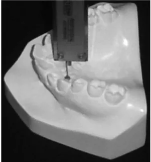
To measure the dimensions of the hard palate, reference points were marked in the most apical palatal points of the maxillary canines, irst and second premolars at the junction of the tooth and gingival margin(19). In the irst molars, the marked
point corresponded to the union of the gingival margin with the palate groove(4). The most anterior point of the hard palate
was marked at the saggital line between the superior central incisives(Figure 1).
These measurements were taken using a digital caliper (Western®) with 0.01 mm of resolution and ±0.02 mm of
preci-sion. For the transversal and the anteroposterior length measures of hard palate, internal measuring faces of the instrument were utilized. For the vertical measures, a 0.05 mm stainless wire was cut with orthodontics pliers in the corresponding length to the transversal measurement obtained, and ixed with dental wax between the points previously set in the level of each of the considered tooth. Having ixed the wire, the depth was measured with the caliper extremity.
The measurements were carried out from zero in the caliper digital scale, and the value obtained in this scale was compared to the analogical scale. In case of discrepancy, the measurement was repeated, not considering the irst value obtained.
Transversal and vertical measures of the hard palate were taken, obeying the order as follows:
a) Canine distance: transversal distance in millimeters between the points of the gingival margin of the maxillary canine region.
b) Canine depth: Vertical measures in millimeters from the midpalatal line to the stainless wire linking the gingival margin of the maxillary canine region (Figure 2).
c) First premolar distance: transversal distance in millimeters between the points in the gingival margin of the maxillary irst premolars.
d) First premolar depth: vertical measure in millimeters from the midpalatal palatine line to the stainless wire linking the gingival margin of the maxillary irst premolars.
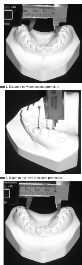
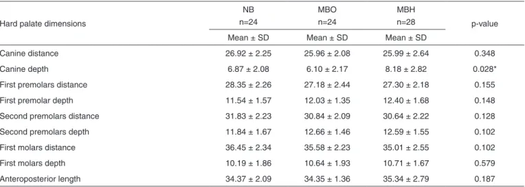
e) Second premolar distance: transversal distance in milli-meters of the gingival margin of the maxillary second premolars region points (Figure 3).
f) Second premolar depth: vertical measure in millimeters from the midpalatal line to the stainless wire linking the gingival margin of the maxillary second premolars region (Figure 4).
g) Molar distance: transversal distance in millimeters between the points of the gingival margin of the maxillary irst molars (Figure 5).
h) Molar depth: vertical measure in millimeters from the midpalatal line to the stainless wire linking the gingival margin of the maxillary irst molars region.
i) Anteroposterior length: distance between the most anterior point of the dental arch to the stainless wire that linked the gingival margin of the maxillary irst molars region. Considering that those children were in the mixed dentition period, for describing these measurements the anatomical region of the permanent teeth (gingival margin) was adopted as nomenclature. Therefore, the children’s measurements with irst and second deciduous molars were described by means of irst and second permanent premolars, once the localization in the gingival margin is the same.
In case of one tooth or both teeth were absent or had not erupted yet, these points were not marked and the measurements were not made in the respective level of the teeth.
After tabulation of the aforementioned measurements, the value of 0.05 mm, corresponding to the diameter of the stainless
Figure 1. Reference points for measuring the hard palate
wire was subtracted from the four depth and anteroposterior length measurements.
After 30 days, 30% of the models were randomly selected and reexamined by the same examiner to conirm the reprodu-cibility of the palate measurements and verify the agreement between the irst and second measurements by Intraclass correlation coeficient. It was veriied signiicant agreement between all measurements.
The Lilliefors test veriied the normality of the variables studied. For the comparison of the hard palate dimensions be-tween NB and MB, Student’s t and Mann Whitney tests were used. The variance analysis (ANOVA) and Kruskall-Wallis tests were used for the comparison among NB, MBO and MBH. When the statistical signiicance was veriied in these tests, Tukey’s multiple comparison was carried out. A signiicance level of 5% was set for all analyses (p<0.05).
RESULTS
It was veriied a signiicant difference (p<0.05) in the second premolars depth and distance and in the irst molars distance between the MB and NB groups (Figures 3, 4 and 5). These indings indicate that MB children presented hard palate narrower than NB children (Table 1).
Some difference between groups regarding the palate dep-th in dep-the canine region was found (Figure 2). From multiple comparisons, it was ascertained that this difference occurred between the MBO and MBH groups. Therefore, MBH children presented greater depth in the hard palate compared to those in the MBO group. (Tables 2 and 3)
DISCUSSION
Among the different instruments for quantitative evaluation of the hard palate, the tridimentional compass Korkahaus has been the most used(7,9,20,21), followed by the caliper(4,22). The
choice of the digital caliper is justiied by its accuracy as an instrument, with low cost, and for being frequently used in the orofacial motricity ield.
Research regarding the hard palate dimension in different respiratory modes have carried out transversal and/or vertical measurements in the canine region(7,9,21-23), second molars
deci-duous(7,9,21,22) and permanent premolars(4). The present research
was based on the method of another study developed with Turner syndrome patients(19), in which the same transversal and
vertical measurements of the hard palate were obtained. The use of such method(19) justiies itself, once the aim of this study
was to measure the width and depth of the hard palate from the canine teeth to the irst molar regions. It was taken into account that morphological changes in different parts of the hard palate may determine different adaptations in the habitual positioning of the tongue apex and back, as well as the orofacial functions.
By analyzing the hard palate dimensions, according to the respiratory mode, it was observed that MB children presented all the mean value of the stance in the second premolars and irst molars region. Such results agree with those in a similar study(21), with 6 and 10-year-old children, which difference Figure 3. Distance between second premolars
Figure 4. Depth at the level of second premolars
Table 1. Comparison of the hard palate dimensions between nasal breathing and mouth breathing groups
Hard palate dimensions
NB n=24
MB
n=52 p-value Mean ± SD Mean ± SD
Canine distance 26.92 ± 2.25 25.98 ± 2.38 0.145
Canine depth 6.87 ± 2.08 7.25 ± 2.73 0.681
First premolars distance 28.35 ± 2.26 27.25 ± 2.28 0.054
First premolar depth 11.54 ± 1.57 12.23 ± 1.53 0.078
Second premolars distance 31.83 ± 2.23 30.73 ± 2.14 0.045*
Second premolars depth 11.84 ± 1.67 12.62 ± 1.50 0.033*
First molars distance 36.45 ± 2.34 35.26 ± 2.41 0.049*
First molars depth 10.19 ± 1.86 10.68 ± 1.78 0.296
Anteroposterior length 34.37 ± 2.09 34.89 ± 2.29 0.364
* Significant values (p≤0.05) – Student’s t test
Note: NB = nasal breathing; MB = mouth breathing; SD = standard deviation
Table 2. Comparison of the hard palate dimensions among nasal-breathing, and mouth-breathing from obstructive and habitual etiology groups
Hard palate dimensions
NB n=24
MBO n=24
MBH
n=28 p-value
Mean ± SD Mean ± SD Mean ± SD
Canine distance 26.92 ± 2.25 25.96 ± 2.08 25.99 ± 2.64 0.348
Canine depth 6.87 ± 2.08 6.10 ± 2.17 8.18 ± 2.82 0.028*
First premolars distance 28.35 ± 2.26 27.18 ± 2.44 27.30 ± 2.18 0.155
First premolar depth 11.54 ± 1.57 12.03 ± 1.35 12.40 ± 1.68 0.148
Second premolars distance 31.83 ± 2.23 30.84 ± 2.09 30.64 ± 2.22 0.128
Second premolars depth 11.84 ± 1.67 12.66 ± 1.46 12.59 ± 1.55 0.102
First molars distance 36.45 ± 2.34 35.58 ± 2.23 35.01 ± 2.55 0.102
First molars depth 10.19 ± 1.86 10.64 ± 1.93 10.71 ± 1.67 0.579
Anteroposterior length 34.37 ± 2.09 34.35 ± 1.36 35.34 ± 2.79 0.187 *Significant values (p≤0.05) – Kruskal-Wallis test
Note: NB = nasal breathing; MBO = mouth breathing of obstructive etiology; MBH = habitual mouth breathingy; SD = standard deviation
Table 3. Difference of the mean values in the depth measurement of the hard palate in the canine region in the multiple comparison among nasal-breathing, and mouth-breathing from obstructive and habitual etiology groups
Comparison Depth at canine level (—XA – —XB) p-value
NB x MBO 0.77 0.954
NB x MBH 1.30 0.389
MBH x MBO 2.08 0.025*
* Significant level (p≤0.05) – Kruskal-Wallis multiple comparison test
Note: NB = nasal breathing; MBO = mouth breathing of obstructive etiology; MBH = habitual mouth breathing;
—
X = mean
between NB and MB was also observed in the distance of the second molars deciduous, i.e., permanent second premolars in the current study.
From the results obtained, it is believed that there is a tendency in the MB group to the hard palate narrowing in the
posterior area. In general, the literature reports that MB children present narrower hard palate due to the reduced airway low passage through the nasal cavity, which impairs the lateral maxilla growth(24).
On the other hand, a difference between NB and MB groups in the canine and irst premolar distance was not veriied, sug-gesting the MB group is not related to the anterior narrowing of the hard palate. Such indings conirm data of other studies, which did not ind any difference in the hard palate width in the canine region between NB and MB children as well(7,9,21).
The MB children presented all mean values of the vertical hard palate dimensions greater than NB children, with statistical signiicance of the second premolars depth. Such a inding, indicates not only an increase of the hard palate depth in the posterior region, but also that the deepest point of the hard palate lays in the region of the second premolars, once the greatest mean value of the palatine depth in these teeth was found.
pos-sible explanation for the increased vertical dimension of the hard palate in these patients is the enlarged air pressure in the oral cavity related to the nasal one(7,9).
The hard palate depth increase and width decrease also may occur due to the alteration in the lips and tongue habitual posi-tion, frequently observed in these patients(14). In such conditions,
the external restraint provided by the lips is absent. Moreover, by being lowered in the oral loor, the tongue does not perform the enlargement and modeling functions of the hard palate(4,22).
In the analysis considering the different etiologies of MB, MBO and MBH, children presented smaller transversal dimen-sions and greater vertical dimendimen-sions than the NB group. There was a signiicant difference in the depth in the canine region. This result could not be compared to the literature, once neither studies comparing different etiologies of MB nor studies having measured the depth palate in the canine teeth were found.
The multiple comparisons showed that MBH presented greater mean value of the hard palate depth in the canine region than MBO. The remaining measures were similar between both groups. From this result, it can be inferred that the MBH, when the patient breathes through the mouth, even having permeated airways, can be as harmful as or more than MB caused by upper airway mechanical obstruction.
The MBO seems to be more harmful, once many pa-tients sometimes have to undergo surgical intervention for re-establishing the nasal respiratory mode. It is believed that the persistent obstruction to the airlow passage through the nose may predispose to craniofacial morphology changes. However, not always these changes will be greater than the ones found in the MBH.
The severity degree of changes in the orofacial complex, not only in the MB from obstructive ethiology but also in the MBH, will also depend on the age of the establishment and the length of time of the mouth breathing, respiratory mode (mixed or oral), genetic and concomitant oral habits presented by the patient. Other environment factors involved that may favor the mouth breathing should be considered.
Based on the results of the current study, it became evi-dent that mouth breathing had inluence over the hard palate morphology at some measurements in vertical and transversal levels. Moreover, a standardized terminology must be sought to characterize the hard palate or decide for the use of terms referring to the vertical and transversal plane.
The quantitative measurements studied enable greater ac-curacy in the diagnoses of morphological changes in the hard palate, minimizing the doubts during clinical evaluation. Never-theless, additional research is needed aiming the establishment of normative parameters related to the hard palate dimensions, contributing thus for an effective use in the clinical practice.
CONCLUSION
Through the analysis of the results of this study, it was possi-ble to conclude that mouth breathing children present narrower hard palate in the second premolars and irst molars and deeper in the second premolars compared to nasal breathing children. It was also evident an increase in the hard palate depth in the
canine teeth in habitual mouth-breathing children compared to obstructive mouth-breathing children.
REFERENCES
1. Faria PT, de Oliveira Ruellas AC, Matsumoto MA, Anselmo-Lima WT, Pereira FC. Dentofacial morphology of mouth breathing children. Braz Dent J. 2002;13(2):129-32.
2. Ambrosio AR, Trevilatto PC, Martins LP, Santos-Pinto AD, Shimizu RH. Electromyographic evaluation of the upper lip according to the breathing mode: a longitudinal study. Braz Oral Res. 2009;23(4):415-23. 3. Vianna-Lara MS, Caria PH. Electromyographic analysis of the upper lip
in nose and mouth breathers. Braz J of Oral Sci. 2006;5(19):1203-8. 4. Oliveira MO, Vieira MM. Influência da respiraçäo bucal sobre a
profundidade do palato. Pró-Fono. 1999;11(1):13-20.
5. Frasson JM, Magnani MB, Nouer DF, Siqueira VC, Lunardi N. Comparative cephalometric study between nasal and predominantly mouth breathers. Braz J Otorhinolaryngol. 2006;72(1):72-82.
6. Berwig LC, Silva AM, Busanello AR, Almeida FL, Bolzan GP, Hennig TR, et al. Alterações no modo respiratório, na oclusão e na fala em escolares: ocorrências e relações. Rev CEFAC. 2010;12(5):795-802. 7. de Freitas FCN, Bastos EP, Primo LS, de Freitas VL. Evaluation of
the palate dimensions of patients with perennial allergic rhinitis. Int J Paediatr Dent. 2001;11(5):365-71.
8. Di Francesco RC, Passerotii G, Paulucci B, Miniti A. Respiração oral na criança: repercussões diferentes de acordo com o diagnóstico. Rev Bras Otorrinolaringol. 2004;70(5):665-70.
9. Ghasempour M, Mohammadzadeh I, Garakani S. Palatal arch diameters of patients with allergic rhinitis. Iran J Allergy Asthma Immunol. 2009;8(1):63-4.
10. Souki BQ, Pimenta GB, Souki MQ, Franco LP, Becker HM, Pinto JA. Prevalence of malocclusion among mouth breathing children: do expectations meet reality? Int J Pediatr Otorhinolaryngol. 2009;73(5):767-73.
11. Marchesan IQ, Krakauer LR. The importance of respiratory activity in myofunctional therapy. Int J Orofacial Myology. 1996;22:23-7. 12. Castelluci e Barbosa M, Knop LA, Lessa MM, de Araujo TM.
Avaliação da radiograia cefalométrica lateral como meio de diagnóstico da hipertrofia de adenóide. R Dental Press Ortodon Ortop Facial. 2009;14(4):83-91.
13. Bianchini AP, Guedes ZC, Vieira MM. A study on the relationship between mouth breathing and facial morphological pattern. Braz J Otorhinolaryngol. 2007;73(4):500-5.
14. Cattoni DM, Fernandes FD, Di Francesco RC, Latorre MR. Characteristics of the stomatognathic system of mouth breathing children: anthroposcopic approach. Pró-Fono. 2007;19(4):347-51. 15. Gouveia SA, Nahás AC, Cotrim-Ferreira FA. Estudo cefalométrico
das alterações dos terços médio e inferior da face em pacientes com diferentes padrões respiratórios e tipos faciais. R Dental Press Ortodon Ortop Facial. 2009;14(4):92-100.
16. Coelho AR, Tanaka O, Ribeiro JS, Machado MA, Camargo ES. Transverse craniofacial dimensions in Angle Class II, Division 1 malocclusion according to breathing mode. Braz Oral Res. 2010;24(1):70-5.
17. Parikh SR, Coronel M, Lee JJ, Brown SM. Validation of a new grading system for endoscopic examination of adenoid hypertrophy. Otolaryngol Head Neck Surg. 2006;135(5):684-7.
18. Brodsky L, Koch RJ. Anatomic correlates of normal and diseased adenoids in children. Laryngoscope. 1992;102(11):1268-74.
19. Laine T, Alvesalo L, Lammi S. Palatal dimensions in 45,X-females. J Craniofac Genet Dev Biol. 1985;5(3):239-46.
20. Drevensek M, Papi JS. The inluence of the respiration disturbances on the growth and development of the orofacial complex. Coll Antropol. 2005;29(1):221-5.
22. Moreira M, de Paiva Lino A. Evaluation of palatal depth and width in mouth breathers with primary dentition. Int J Orofacial Myology. 1989;15(1):19-24.
23. Nieto Perea P, Acosta Quiñones JM, Meneses López A. Determinación de la profundidad del paladar en niños con respiración bucal de 6-8 años de edad. Rev Estomatol Hered. 2005;15(1):50-3.