0103 - 5053 $6.00+0.00
Article
* e-mail: [email protected]
Phytochemistry of
Trattinnickia burserifolia
,
T. rhoifolia
, and
Dacryodes hopkinsii
:
Chemosystematic Implications
M. da Paz Limaa, Patrícia A. de Campos Bragab, Mario Lopes Macedob, M. Fátima das G. F. da Silva*,b, A. Gilberto Ferreirab, João B. Fernandesb and Paulo C. Vieirab
a
Instituto Nacional de Pesquisa da Amazônia, Coordenação de Pesquisas em Produtos Naturais, CP 478, 69011-970 Manaus - AM, Brazil
b
Departamento de Química, Universidade Federal de São Carlos, CP 676, 13565-905 São Carlos - SP, Brazil
O estudo de Trattinnickia burserifolia levou ao isolamento dos triterpenos conhecidos ursanos α-amirenona, α-amirina, 3-epi-α-amirina, 3α,16β-diidroxiurs-12-eno; oleananos β-amirenona, β -amirina, 3-epi-β-amirina, 3α,16β-diidroxiolean-12-eno; tirucalanos ácidos 3α -hidroxitirucal-8,24-dien-21-óico, 3α-hidroxitirucal-7,24-dien-21-óico, e 3-oxotirucal-8,24-dien-21-óico; damaranos dammarenediol-II e 3α,20(S)-diidroxidamar-24-eno. Além desses foram ainda isolados o monoterpeno novo 2(S*)-fenilacetoxi-4(R*)-p-menta-1(7),5-dieno, e os triterpenos novos 3β -fenilacetoxiurs-12-eno, 3β-fenilacetoxiolean-12-eno e 3β,16β,11α-triidroxiurs-12-eno. Os triterpenos de T. burserifolia,T. rhoifolia e Dacryodes foram analisados em mistura. Os espectros de RMN 13C
mostraram que os principais triterpenos eram α-amirina e β-amirina em T. burserifolia; α-amirina, β -amirina, 3-epi-α-amirina, 3-epi-β-amirina, lupenona, ácidos 3α-hidroxitirucal-8,24-dien-21-óico e 3α-hidroxitirucal-7,24-dien-21-óico em T. rhoifolia; α-amirina, β-amirina, lupeol, tirucalol, sitosterol e estigmasterol em D. hopkinsii. A quimiossistemática da tribo Protieae é discutida.
Trattinnickia burserifolia has yielded the known ursanes, α-amyrenone, α-amyrin, 3-epi-α -amyrin, 3α,16β-dihydroxyurs-12-ene, the oleananes β-amyrenone, β-amyrin, 3-epi-β-amyrin, 3α,16β-dihydroxyolean-12-ene, the tirucallane acids 3α-hydroxytirucall-8,24-dien-21-oic, 3α -hydroxytirucall-7,24-dien-21-oic and 3-oxotirucall-8,24-dien-21-oic, the dammaranes dammarenediol-II and 3α,20(S)-dihydroxydammar-24-ene. Besides it was isolated the new monoterpene 2(S*)-phenylacetoxy-4(R*)-p-mentha-1(7),5-diene and, the new triterpenes 3β -phenylacetoxyurs-12-ene, 3β-phenylacetoxyolean-12-ene and 3β,16β,11α-trihydroxyurs-12-ene. The triterpenes from T. burserifolia,T. rhoifolia and Dacryodes were analyzed in mixture. Their 13C
NMR spectra showed that the major triterpenes were in T. burserifoliaα-amyrin and β-amyrin; in T. rhoifoliaα-amyrin, β-amyrin, 3-epi-α-amyrin, 3-epi-β-amyrin, and lupenone; in T. rhoifoliaα -amyrin, β-amyrin, 3-epi-α-amyrin, 3-epi-β-amyrin, 3α-hydroxytirucall-8,24-dien-21-oic acid and 3α-hydroxytirucall-7,24-dien-21-oic acid; in D. hopkinsiiα-amyrin, β-amyrin, lupeol, tirucallol, sitosterol and stigmasterol. Aspects of chemosystematic of the tribe Protieae are discussed.
Keywords: Burseraceae, monoterpene, triterpenes, chemosystematic
Introduction
The Burseraceae has usually been considered to contain 21 genera and nearly 600 species. Engler (1931) classified these genera into three tribes.1 The Protieae
consist of four genera exhibiting many morphological characters regarded as primitives. Three Protieae genera occur in tropical America and one in Asia. The following group the Boswellieae contain eight genera centred in
Africa and Asia. In contrast, the Canarieae, represented by nine genera, appear more advanced in their morphology. This tribe is predominantly Paleotropical, therefore, two genera occur in South America. Later Lam (1932) recognised these tribes but replaced the name Boswellieae by Bursereae.2
Crepidospermum Hook. is a member of the Protieae
and consists of five species distributed in the tropical South America. Swart in 1942 on morphological grounds described the genus Hemicrepidospermum to
their morphology have led Daly (1989) to consider
Hemicrepidospermum a section of Crepidospermum; the
two sections have three and two species, respectively.4 The
following tropical S. American genera of the Protieae,
Tetragastris and Protium, have long been considered
closely related, in fact, many specimens of each genus have been mistakenly referred to the other.4Garuga is the
only representative of Asian Protieae and its morphology is easily recognisable.4
Trattinnickia was also a member of the Protieae,
however, morphological and anatomical evidence have led Daly (1989) to transfer it into the Canarieae and to propose a taxonomic position close to Dacryodes.4
Within tribe Protieae phytochemical data were not available for Crepidospermam, Tetragastris, Trattinnickia
and Dacryodes. As part of our chemosystematic interest in
the Brazilian Burseraceae, we recently reported the phytochemical investigation of Crepidospermam rhoifolium Benth. and Tetragastris altissima (Aublet)
Swart.5 Thus, we have now examined the resin, stem bark
and branches of Trattinnickia burserifolia Engl., T. rhoifolia var. willdenowii Engl. and Dacryodes hopkinsii
Daly.4
Resulsts and Discussion
Chemical composition of the extracts
A chloroform-soluble fraction of the resin of T. burserifolia afforded one new monoterpene (1), three new
triterpenes (2-4) and the known ursanes, α-amyrenone,6α
-amyrin (5), 3-epi-α-amyrin, 3α,16β-dihydroxyurs-12-ene
(6),7 the oleananes β-amyrenone,6 β-amyrin (7), 3-epi-β
-amyrin, 3α,16β-dihydroxyolean-12-ene (8),7 the
tirucallane acids 3α-hydroxytirucall-8,24-dien-21-oic,8
3α-hydroxytirucall-7,24-dien-21-oic,6
3-oxotirucall-8,24-dien-21-oic,8 and the dammaranes dammarenediol-II and
3α,20(S)-dihydroxydammar-24-ene.5
The 1H NMR spectrum (Table 1) of compound 1 showed
signals for a terminal methylene (δ 5.08, br s, and 5.00, br s), two olefinic protons which were coupled to each other (δ 5.80, dd, J 10.1 and 1.0 Hz; 6.14, dd, J 10.1 and 2.5 Hz),
two methyl doublet (δ 0.88, d, J 6.8 Hz; 0.86, d, J 6.8 Hz),
an oxymethine (δ 5.57, dd, J 5.0 and 2.8 Hz), an aryl
substituted methylene (δ 3.61, 2H, s) and five protons as a multiplet between δ 7.30 and 7.25, clearly indicating the presence of a phenyl group. From HMBC experiments the observed correlations between the methylene protons at δ
5.08 and 5.00 and the 13C signals at δ 127.2 and 71.8,
requiring the presence of a conjugated double bond and an allylic proton attached to a carbon adjacent to an oxygen atom. HSQC experiments showed correlations of terminal methylene and conjugated double bond protons (δ 5.80 and 6.14) with the 13C signals at δ 115.4, 133.7 and 127.2,
respectively. HSQC also permitted the assignment of the signal at δ 41.8 to aryl substituted methylene at δ 3.61, which showed cross peaks with the 13C signals of the
aromatic ring (C-1’, C-2’ and C-6’) and carboxyl at δ 171.1, indicating a phenylacetoxyl substituent. This group must be connected allylic to terminal methylene, due to the observed downfield shifted proton signal at δ 5.57, which showed one-bond correlation with the 13C signal at δ 71.8.
These correlations resulted in the construction of a CH2=C[CH(R)OCOCH2Ph]CH=CHR system.
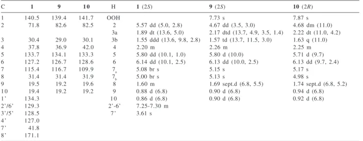
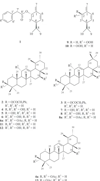
Table 1.13C and 1H NMR spectral data for compounds 1 and the model compounds 9 and 10
C 1 9 1 0 H 1 (2S) 9 (2S) 10 (2R)
1 140.5 139.4 141.7 OOH 7.73 s 7.87 s
2 71.8 82.6 82.5 2 5.57 dd (5.0, 2.8) 4.67 dd (3.5, 3.0) 4.68 dm (11.0)
3a 1.89 dt (13.6, 5.0) 2.17 dtd(13.7, 4.9, 3.5, 1.4) 2.22 dt (11.0, 4.2) 3 30.4 29.0 30.1 3b 1.55 ddd (13.6, 9.8, 2.8) 1.57 td (13.7, 11.5, 3.0) 1.63 q (11.0)
4 37.8 36.9 42.0 4 2.20 m 2.26 m 2.25 m
5 133.7 134.1 133.3 5 5.80 dd (10.1, 1.0) 5.80 d (10.0) 5.71 d (9.7)
6 127.2 126.7 128.6 6 6.14 dd (10.1, 2.5) 6.13 dd(10.0, 2.5) 6.13 dd(9.7, 2.4)
7 115.4 116.7 109.9 7a 5.08 br s 5.15 s 5.17 s
8 31.4 31.4 31.9 7b 5.00 br s 5.13 s 4.98 s
9 19.5 19.2 19.6 8 1.60 m 1.69 sept.d(6.8, 5.5) 1.74 sept.d (6.8, 5.2)
1 0 19.4 19.2 19.2 9 0.88 d (6.8) 0.90 d (6.8) 0.94 d (6.8)
1’ 134.3 1 0 0.86 d (6.8) 0.90 d (6.8) 0.92 d (6.8)
2’/6’ 129.3 2’-6’ 7.25-7.30 m
3’/5’ 128.5 7’ 3.61 s
4’ 127.0
7’ 41.8
8’ 171.1
The remaining unassigned 13C signals accounted for 2
CH3, CH2 and 2 CH, of which two methyls were coupled to a methine, suggesting the presence of a monoterpene p
-menthane skeleton to be substituted with phenylacetoxyl. The identification of the nucleus as a p-menthane was
supported by comparison of the 13C NMR spectrum (Table
1) with those of 2(S)-hydroperoxy-4(R)-p
-mentha-1(7),5-diene (9) and 2(R)-hydroperoxy-4(R)-p
-mentha-1(7),5-diene (10) obtained from photooxygenation of (-)-(R)-α -phellandrene.9 Based on the 1H and 13C NMR data for 9
and 10, in compound 1 the resonances for H-2 (δ 5.57, dd,
J 5.0 and 2.8 Hz) and C-4 (δ 37.8) were characteristic of 2(S)-4(R)-p-menthane derivative, but this has not been
confirmed. Compound 1 is thus
2(S*)-phenylacetoxy-4(R*)-p-mentha-1(7),5-diene.
The two new triterpenes 2 and 3 showed a single spot
on TLC in various solvent systems and attempts to separate this mixture into its constituents were not successful. They
also showed the spectral characteristics of a phenylacetoxyl substituent. The 1H and 13C NMR spectra of this mixture in
addition to signals described above for phenylacetoxyl, revealed resonances for C-1 and H-1 to C-30 and H-30 in close agreement with those for α-amyrin (5) and β-amyrin (7), respectively7, 10 (Table 2). The downfield shift of the
signals for C-3 (δ 81.4) and H-3 (δ 4.48) in the 1H and 13C
NMR spectra, when compared with 5 and 7, determined
the position of the phenylacetoxyl at C-3 in both the compounds of the mixture. The phenylacetoxyl present at C-3β was evident by resonance at δ 4.48 with a large coupling constant (J 11.0 and 5.1 Hz). The structure of the
new natural products were thus established as 3β -phenylacetoxyurs-12-ene (2) and 3β -phenylacetoxyolean-12-ene (3).
The new triterpene 4 was identified on the basis of the
following data. The 1H NMR spectrum indicated the
presence of three signals characteristics of protons attached to a carbon adjacent to an oxygen atom (δ 3.23, dd, J 10.4
and 5.8 Hz; 4.23, dd, J 11.1 and 5.2 Hz; 4.27, dd, J 8.7 and
3.2 Hz), one olefinic proton (δ 5.24, d, J 3.2 Hz), and eight Table 2.13C NMR spectrum data for compounds 2, 3 and the model
compounds 5 and 7
C 2 3 5 7 C 2 3
1 38.5 38.4 38.7 38.7 1’ 134.5 134.5 2 27.9 27.0 27.2 27.3 2’ 129.3 129.3 3 81.4 81.4 78.3 79.0 3’ 128.5 128.5 4 37.8 37.8 38.7 38.8 4’ 126.9 126.9 5 55.3 55.3 55.2 55.3 5’ 128.5 128.5 6 18.2 18.2 18.3 18.5 6’ 129.3 129.3 7 32.5 32.5 32.9 32.8 7’ 42.1 42.1 8 40.1 40.1 40.0 38.8 8’ 171.3 171.3 9 47.7 47.6 47.7 47.7
methyl groups, six of them on quaternary carbons and two of them on a methine group, suggesting a urs-12-ene skeleton. From the HMBC experiments (Table 3) the observed correlations between the two methyl protons at
δ 0.77 and 0.97 and the 13C signals at δ 78.7 (3J; CH by
DEPT), 55.4 (3J; CH), 39.3 (2J; quaternary carbon), 28.4 (3J
for methyl at δ 0.97; CH3) and 15.7 (3J for methyl at δ 0.77;
CH3) led to their assignments as C-3, C-5, C-4, C-23 and C-24, respectively. Based on the HSQC experiments the signal at δ 0.77, 0.97 and 3.23 were then assigned to Me-24, Me-23 and H-3, respectively. The methyl proton at δ
1.06 (δC 17.0)showed long-range correlation with the 13C
signal for C-5 (δ 55.4), permitting the assignment of these signals to H3-25 and C-25, respectively. The signal for H3 -25 also showed correlations with the 13C signals at 55.3
(CH), 40.9 (CH2) and 38.1 (quaternary carbon), showing that these signals correspond to C-9, C-1 and C-10, respectively. The olefinic proton at δ 5.24 showed cross peaks with the C-9 signal (δ 55.3), and was coupled to the
1H signal at δ 4.27, thus indicating a hydroxyl group to be
located at C-11and a double bond at C-12. The late oxymethine proton showed one-bond correlation with the
13C signal at δ 68.2 and long-range correlation with the 13C signals at δ 141.3 and 129.2, allowing the assignment
of these to C-11, C-13 and C-12, respectively. Moreover, the existence of correlations between H-12 and the 13C
signals at δ 59.9 (CH) and 44.3 (quaternary carbon) led to their assignments as C-18 and C-14, respectively. A fourth methyl proton at δ 1.20 (δC 24.3) was attributed to H3-27 by its correlations with the C-13 (δ 141.3) and C-14 (δ
44.3) signals. The H3-27 signal also showed a cross-peak with the signal at δ 43.6, confirming a methyl group at C-8. In the same way, the unsubstituted C-15 emerged from the correlation between the H3-27 signal and the 13C signal
at δ 36.0 (3J; CH
2), which showed one-bond correlation
with the 1H signal at δ 1.36 (m). This signal was coupled to
the 1H signal at δ 4.23 (dd, J 11.1 and 5.2 Hz), requiring
the presence of a hydroxyl function at C-16. The coupling constants indicated that the hydroxyl group was attached
β (equatorial) to C-16 and was coupled only to H2-15, indicating C-17 fully substituted. This was supported by the relationship of the H-16 (δ 4.23) signal to the 13C signal
at δ 21.9, which showed one-bond correlation with the methyl proton at δ 0.73, and long-range correlation with the 13C signals for C-18, C-16 and at δ 38.5 (quaternary
carbon) and 35.2 (CH2). The signals at δH0.73, δC21.9,
38.5 and 35.2 were then assigned to H3-28, C-28, C-17 and C-22, respectively. A sixth methyl proton at δ 1.05 (δ 18.0) was attributed to H3-26 by its correlation with the C-9 and C-14. H3-26 signal also showed cross peaks with the 13C
signal at δ 43.6 (quaternary carbon) and 33.7 (CH2), which were attributed to C-8 and C-7, respectively. A seventh methyl proton at δH 0.82 (d, J 6.3 Hz; δC 17.8) was attributed to H3-29 by its correlation with the C-18 signal. H3-29 signal also showed cross peaks with the 13C signal at
δ 39.1 (CH) and (or) 39.5 (CH), suggesting a methine for C-20, indicating a methyl group to be located at C-20 and confirming a urs-12-ene skeleton. Thus, the eighth methyl proton at δH0.91 (d, J 5.9 Hz;δC21.5) was attributed to H3-30. The signal for C-21 was established as δ 30.4 (CH2;
δH 1.42 m, by HSQC) by the existence of a correlation between the H3-30 signal and this 13C signal.
The stereochemistry suggested for 4 was based on the
biosynthesis of urs-12-enes. However, for C-3, C-11 and C-16 the stereochemistry were assigned by coupling constants and NOESY experiments. A model shows that, in compound 4, ring A is nearer to a chair conformation, in
which H-3 and H-5 are on the α-side of the molecule. This was supported by NOESY experiments (Table 3), which showed correlation of the signal of H-3α (δ 3.23; OH-3β) with the signal of H-5α (δ 0.72 m, by HSQC). Moreover, the existence of a correlation from H-3 to H3-23 (δ 0.97) confirmed that Me-23 is in the α-configuration. In addition, the signal of H-11 (δ 4.27) showed cross-peaks with the signals of H3-25 (δ 1.06) and H3-26 (δ 1.05), suggesting a spatial proximity of H-11 to Me-25 and Me-26, which requires 11-OH to be in the α-configuration. The
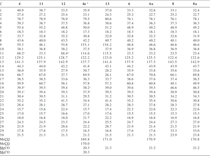
Table 3. HMBC assignments for 4 and 4a, and G-NOESY for 4
4 HMBC 4a HMBC
H C H C
3 2 4 3 Ac (170.0), 23, 24
9 5, 8, 10, 11, 26, 25
1 1 12, 13 1 1 8, 10, 13
1 2 9, 14, 18 1 2 9, 14, 18
1 5 13, 16, 17
1 6 2 8 1 6 Ac (169.8), 28
1 8 12, 13, 14, 16, 17, 19 (or 20) 1 8 12, 13, 14, 17
2 3 3, 4, 5, 24 2 3 3, 4, 5, 24
2 4 3, 4, 5, 23 2 4 3, 5, 23
2 5 1, 5, 9, 10 2 5 1, 5, 9, 10
2 6 7, 8, 9, 14 2 6 7, 8, 14
2 7 8, 13, 14, 15 2 7 8, 13, 14, 15
2 8 16, 17, 18, 22 2 8 16, 17, 18, 22
2 9 18, 19 (or 20) 2 9 18, 19, 20
3 0 19 (or 20), 21 3 0 19, 20, 21
4 G-NOESY
H H
relationship of H-16 signal (δ 4.23) to the H3-27 (α, δ 1.20) indicated that 16-OH is in the β-configuration.
The ESI-MSMS showed ions at m/z 457 [M - H]-, 439
[M - H - H2O]- and 421 [M –H - H
2O - H2O]-, confirming the
presence of hydroxyl groups and thus the molecular formula (C30H50O3). Based in the above evidence the structure of this compound was thus established as 3β,16β,11α-trihydroxyurs-12-ene (4). The structural
assignment was also supported by comparison of the 13C
NMR spectrum (Table 4) with those of 3β,16β -dihydroxyurs-12-ene (11)7 and 3α,11α
-dihydroxyurs-12-ene (12).10 In order to confirm of the assignments for 6a
and 8a discussedbelow, 4 was acetylated. This reaction
involved dehydration of C-11 alcohol and acetylation of the C-3 and C-16 hydroxyl groups to give 3β,16β -diacetoxyurs-9(11),12-diene (4a). The 1H NMR spectrum
of 4a revealed the downfield shift of the signals for H-3 (δ
4.51, dd, J 11.4 and 4.9 Hz) and H-16 (δ 5.46, dd, J 11.4
and 5.5 Hz). From the HMBC experiments (Table 3) the observed correlations between the two methyl protons at
δ 0.88 and 0.90 and the 13C signals at δ 79.5, 50.0 (3J; CH),
36.8 (2J; quaternary carbon), 27.1 (3J, CH
3) and 15.7 (3J,
CH3) led to their assignments as C-3, C-5, C-4, C-23 and C-24, respectively. The oxymethine proton at δ 4.51 showed long-range correlation with the 13C signal at δ
170.0 and with the C-24 (δ 15.7) and C-23 (δ 27.1) signals, confirming this proton signal to H-3 and allowing the assignment of the signal at δ 170.0 to C-3 acetoxyl group. Moreover, the existence of correlations between H3-28 (δ
0.91) and the 13C signals at δ 69.9 (CH), 57.8 (CH), 36.3
(quaternary carbon) and 34.4 (CH2) led to their assignments as C-16, C-18, C-17 and C-22, respectively. Thus, the
Table 4.13C NMR spectrum data for compounds 4, 4a, 6a, 8a,and the model compounds 6, 8, 11, 12 and 1310, 26-29
C 4 1 1 1 2 4a b 1 3 6 6 a 8 8 a
1 40.9 38.7 33.5 35.9 37.0 33.3 32.6 33.1 32.4
2 27.4 27.2 25.4 23.1 24.3 25.2 22.7 25.2 22.7
3 78.7 78.9 76.0 79.5 80.6 76.1 78.1 76.1 78.1
4 39.3 38.7 37.5 36.8 38.6 37.4 36.3 37.3 36.3
5 55.4 55.2 48.8 50.0 51.2 48.9 49.2 48.9 49.2
6 18.3 18.3 18.2 17.3 18.2 18.3 18.1 18.3 18.1
7 33.7 32.8 35.2 30.8 32.0 32.8 32.3 32.6 31.9
8 43.6 40.0 43.5 42.2 40.7 40.2 40.2 40.1 40.0
9 55.3 46.1 55.8 153.1 154.2 46.8 46.6 46.6 46.6
1 0 38.1 36.8 38.2 37.5 37.9 36.9 36.8 36.9 36.8
1 1 68.2a 23.3 68.4a 114.6 115.5 23.3 23.2 23.5 23.4
1 2 129.2 125.1 128.7 122.2 123.0 125.2 125.3 122.4 122.6
1 3 141.3 137.9 142.9 137.7 141.4 137.9 137.5 143.5 142.9
1 4 44.3 44.0 42.2 41.8 43.1 44.2 43.9 43.9 43.7
1 5 36.0 35.9 27.9 30.7 28.2 35.9 33.8 35.6 33.6
1 6 66.7 67.0 27.7 69.9 26.1 67.0 70.8 66.1 69.8
1 7 38.5 38.5 33.6 36.3 33.7 38.6 37.6 37.4 36.5
1 8 59.9 60.7 58.1 57.8 57.3 60.8 60.8 49.1 49.9
1 9 39.5c 39.5 39.4 38.2c 39.0 39.6 39.5 46.6 46.5
2 0 39.1c 39.4 39.3 37.5c 39.5 39.5 39.4 30.9 30.8
2 1 30.4 30.5 31.1 29.5 31.2 30.5 30.5 34.2 34.3
2 2 35.2 35.2 41.3 34.4 41.4 35.2 35.4 30.6 30.8
2 3 28.4 28.1 28.7 27.1 28.2 28.3 27.8 28.3 27.8
2 4 15.7 15.6 22.4 15.7 17.4 22.3 22.6 22.3 22.6
2 5 17.0 15.7 16.6 17.0 17.6 15.5 15.5 15.3 15.3
2 6 18.0 16.8 18.0 21.7 22.2 16.9 16.8 16.9 16.8
2 7 24.3 24.5 23.3 24.4 25.5 24.7 24.4 27.3 27.0
2 8 21.9 21.9 28.6 22.2 28.7 21.9 21.4 21.5 21.9
2 9 17.8 17.6 17.5 16.5 16.8 17.6 17.6 33.3 33.0
3 0 21.5 21.3 21.3 21.2 21.5 21.3 21.3 23.9 23.8
MeCO 169.8 171.0 170.9 170.8
MeCO 170.0
MeCO 20.3 21.3 21.2 21.2
MeCO 20.3
Assignments based on HSQC, HMBC and G-NOESY for 4, HMBC for 4a and DEPT for 6a and 8a. In 4a: C-3 acetoxyl group δ 170.0; C-16 acetoxyl group δ169.8. The use of compound 13 as a model permitted to find out that Mahato and Kundu10 published the 13C NMR data
for 3β-hydroxyurs-9(11),12-diene, however, they were for 3β-acetoxyurs-9(11),12-diene (see reference 26); a The structural assignment was
also supported by comparison of the 13C NMR spectrum with those of olean derivatives, 11α-methoxyolean-12-ene and 11β
proton signal at δ 5.46 (δC 69.9) was confirmed to H-16 and the 13C signals at δ 169.8 and 22.2 were attributed to
C-16 acetoxyl group and C-28, respectively, due their correlations with the H-16 signal. The 1H NMR also showed
an olefinic proton to be coupled to H-12 (δ 5.49, d, J 5.6
Hz; 5.61 d, J 5.6 Hz,), thus indicating the second double
bond between C-9 and C-11. The olefinic proton signal at
δ 5.49 was attributed to H-12 by its correlation with the C-18 (δ 57.8) signal. H-12 signal also showed cross peaks with the 13C signal at δ 41.8 (quaternary carbon), which
was attributed to C-14. In the same way, the olefinic proton signal at δ 5.61 to H-11 emerged from the correlations between the H3-25 and H-11 signals with the 13C signals at
δ 37.5, assigned to C-10. H-11 signal also showed cross peaks with the 13C signal at δ 42.2, which was attributed to
C-8. Moreover, H3-25 showed cross peaks with the C-5 signal (δ 50.0) and the 13C signals at δ 153.1 and 35.9,
leading to their assignments as C-9 and C-1, respectively. The signal for C-13 was established as 137.7 by the existence of a correlation between the H3-27 signal (δ 0.99) and this 13C signal. H
3-27 signal also showed cross peaks
with the 13C signals at δ 30.7, which was attributed to
C-15. The relationship of 1H signal at δ 1.69 (d, J 11.1 Hz)
to the C-13, C-14, C-17 signals and the 13C signal at δ
122.2 led to their assignments as H-18 and C-12. Thus, the second olefinic carbon at δ 114.6 was attributed to C-11. The ursa-9(11),12-diene system was also supported by the
13C NMR spectrum which agreed closely with published
data for 3β-acetoxyurs-9(11),12-diene (13).10 In the HMBC
experiments several other long-range correlations were observed, which also confirmed the attribution of all the
13C signals of the molecule (Table 3 and 4).
Compounds 3α,16β-dihydroxyurs-12-ene (6) and
3α,16β-dihydroxyolean-12-ene (8) have previously been
isolated from Canarium album.7 In the present
inves-tigation the physical separation of these compounds was not achieved, even after acetylation with anhydride in pyridine, but the 13C NMR data (Table 4) left no doubt that
they were 3α,16β-diacetoxy derivatives 6a and 8a. The
presence of urs- and olean-12-ene systems were indicated by the characteristic olefinic 13C resonances at δ 125.3
and 137.5, 122.6 and 142.9, respectively. The latter 13C
signals were more intense than the former, indicating the olean-12-ene derivative as the major compound. In the same way, the intense olefinic 1H signal at δ 5.26 (t, J 3.6
Hz) was then assigned to H-12 of oelan-12-ene, whereas δ
5.20 (t, J 3.6 Hz) to H-12 of urs-12-ene derivative. The 1H
and 13C NMR spectra revealed the presence of two acetoxyl
groups. The identification of the A-ring containing only 3α-acetoxyl group as substituent was supported by the coupling constants for H-3 (δ 4.63, t, J 2.7 Hz, 8a; 4.50, t,
J 2.7 Hz, 6a) and 13C NMR spectrum, which showed that
the acetylation accentuated the α-effect (C-3, δ 78.1) and diminished the β-effect (C-2, δ 22.7; C-4, δ 36.3) when compared with published data for C-2 to C-4 in 6 and 8.
Inspection of the 13C NMR data of various hydroxy
urs-and olean-12-enes, revealed that introduction of a hydroxyl group only on D- and E-ring causes a significant alteration of the chemical shift of C-18 (δ 58.9, 5; 60.8, 6; 47.4, 7;
49.1, 8).10 Although, in urs- and olean-12-enes containing
21β-, 21α-, 22α- and 22β-hydroxyl groups, the C-21 and C-22 resonate at δ 70.0 to 76.7.10 In olen-12-enes
containing 15α- or 16β-hydroxyl groups, these carbinyl carbons resonate at δ 65.9 to 68.2.10 The coupling constants
of 1H signals of an oxymethine for 6a and 8a (δ 5.46, dd, J
11.9 and 5.0 Hz, 8a; 5.48, dd, J 11.9 and 5.0 Hz, 6a)
indicated that the acetoxyl group would be attached equatorial to C-15 (α) or C-16 (β), thus the above compounds are excellent models. The presence of a hydroxyl group at C-15α has a pronounced effect on the olefinic carbon resonances. The chemical shift of C-12 and C-13 in 3β,15α-dihydroxyolean-12-ene appear at δ
123.0 and 146.1, respectively. The corresponding resonances for 3β,16β-dihydroxyolean-12-ene appear at
δ 122.2 and 143.4, i.e. C-13 is shielded by ∆δ 2.7.10 The
shielded resonance observed for 8a (δ 122.6 and 142.9) is typical of 16β-hydroxyolean-12-ene, determining the position of the second acetoxyl group at C-16β. Although, no urs-12-ene containing a 15α-hydroxyl group appears to have been isolated so far. However, for the analogous structural situation in 3β-hydroxyoelan-12-en-27,28-dioic (C-13, δ 138.1) and 3β-hydroxyurs-en-27,28-dioic (C-13,
δ 134.2)acids a similar shield is observed for C-13 in both systems [C-13, δ 145.1 (7) – 138.1 = ∆δ 7.0; δ 139.3 (5)
– 134.2 = ∆δ 5.1].10 Placement of the acetoxyl group at
C-16 received further support from acetylation of 4, which
yielded 4a. In the 13C NMR spectrum of 4a (Table 4) the
signal for C-16 was observed at δ 69.9, in close agreement with the resonance for the corresponding carbon in 6a (δ
70.8)and 8a (δ 69.8). All the above data suggested that 6a
was 3α,16β-diacetoxyurs-12-ene and 8a was 3α,16β -diacetoxyolean-12-ene.
The 1H NMR spectra of concentrated MeOH extracts
of stem bark of T. burserifolia, branch and resin of T. rhoifolia, branch and resin of Dacryodes hopkinsii showed
signals corresponding to triterpenes isolated from resin of
T. burserifolia. Thus, only CH2Cl2-soluble fractions of
these extracts were further examined by their 13C NMR
spectra which showed that the major triterpenes were in stem bark of T. burserifoliaα-amyrin (5) and β-amyrin (7);
resin of T. rhoifoliaα-amyrin (5), β-amyrin (7), 3-epi-α -amyrin, 3-epi-β-amyrin, 3α -hydroxytirucall-8,24-dien-21-oic acid8 and 3α-hydroxytirucall-7,24-dien-21-oic acid;6
in branch of D. hopkinsii α-amyrin (5), β-amyrin (7),
lupeol,10 tirucallol,12 sitosterol and stigmasterol; and in
resin of D. hopkinsiiα-amyrin (5) and β-amyrin (7).
Chemosystematic implications
Burseraceous genera are characterised by the production of tetracyclic tirucallane, dammarane, cycloartane, lanostane, and pentacyclic lupane, ursane and oleanane triterpenes.13 Ursanes and/or oleananes are
omnipresent. All genera produce volatile oils which are often represented by many different sesquiterpenes, aromadendranes, humulanes, germacranes, eudesmanes, elemanes, guaianes, pseudo-guaianes, bourbonanes, caryophyllanes, cubebanes, cadinanes, copaanes and bisabolanes.13 Furosesquiterpenes have been isolated from
Commiphora species.13 Lignans are less common and were
for long known only from Bursera, but have now been
recorded in Commiphora and Protium.14, 15
Until recently the phytochemical knowledge of Protieae were the records of three dammaranes (dammaradienol in
Garuga pinnata,16 3α,20(S)-dihydroxydammar-24-ene in
Crepidospermumrhoifolium,5 and cabraleadiol in Protium apiculatum5), one tirucallane (butirospermol in G.
pinnata16), two ursanes (α-amirin and 3-epi-α-amirin in G. pinnata,16 and only the former in P. paniculatum Engl17and
P. icicariba5), one oleanane (β-amirin in P. paniculatum17
and P. icicariba5), one cycloartane (3β
,24-dihydroxy-cycloart-25-ene in C. rhoifolium5), one multiflorane
(secoisobryononic acid in Tetragastris altissima5), one
friedelane (friedelin in T. altissima5), one taraxerane (taraxerol
in T. altissima5), one lupane (lupeol in P. icicariba5 and P.
apiculatum5), four sterols (sitosterol, stigmasterol,
campesterol and 3β-O-β-D-glucopyranosylsitosterol in C. rhoifolium5,only the two first in P. paniculatum;17 and only
the former in P. opacum Swart,18 P. apiculatum5 and T.
altissima5), four lignans
[(+)-(2S,3S)-2-(3’,4’-methylenedioxy-acetophenone)-butyrolactone, (-)-cubebin epimers in P. tenuifolium Engl,15 parabenzolactone in C. rhoifolium5 and (-)-savinin in T. altissima5], one coumarin
(propacin in P. opacum18), one biflavonoid (amentoflavone
in G. pinnata19) and two macrocyclic biphenyl ether
(garuganin I and III in G. pinnata16). Protieae genera have
been shown to have a chemical profile that is comparable to those of all other burseraceous tribes.5, 13
All the burseraceous tribes yielded tirucallanes, ursanes, oleananes and lupanes, while dammaranes appear to have been recorded only from Protieae and Boswellieae
genera.5Trattinnickia has in common tirucallane, ursane
and oleanane types with Dacryodes. Lupanes have been
found only in Dacryodes. Furthermore, Dacryodes contains
peculiar 3,4-secolupanes13 which could be taken as
indicative of an affinity to the Boswellieae where similar lupanes occur.13 It is also chemical evidence favouring its
classification in the Canarieae, notably by the co-occurrence of 3,4-secolupanes in Canarium muelleri and C. zeylanicum.13
The co-occurrence of dammaranes in Trattinnickia, Garuga,17Crepidospermum,5Protium5 and Commiphora20, 21
suggests some affinity between Protieae and Boswellieae (Bursereae). The simplest dammaranes, in which the side chain is undegraded (as in dammarenediol-II and 3α ,20(S)-dihydroxydammar-24-ene from T. burserifolia), are typical
of the Protieae and that increasing ability to lose the entire C-17 side chain (as in mansumbinanes21) occurs through
the Boswellieae (in Commiphora). This can be seen as a
further advance in oxidative mechanisms and appears to agree closely with the suggested phylogenetic sequence within the Burseraceae; Protieae considered the most primitive, Boswellieae intermediate. Thus, the isolation of two undegraded dammaranes from T. burserifolia
suggest that Trattinnickia appears to have a less
pronounced relationship to the Canarieae than to the Protieae, since they do not occur, at present, in the former. Thus, the presence of dammaranes in T. burserifolia does
not support Daly’s taxonomic conclusions.5
The most common tetracyclic triterpenes in the Rutales families are tirucall-7-en derivatives which are the precursors of the limonoids and quassinoids that are major chemotaxonomic characters of the order.22, 23 The Burseraceae
is an exception, no quassinoids or limonoids have so far been isolated. Burseraceous genera have heretofore yielded relatively few variety of compounds. This fact can be rationalised by the presence in their species of massive quantities of tannins (possibly also essential oils), general defences which make the presence of specific alleochemics superfluous.24 Essential oils represented by many different
sesquiterpenes, appear to inhibit the formation of squalene, potential precursor of limonoids.25
Experimental
General
0.25 mm ID, 0.25 µm film thickness) capillary column with helium as the carrier gas at a flow rate of 1.6 mL min-1.
The temperature was programmed initially at 60 °C for 2 min, then increased with a rate of 3 °C min-1 to 240 °C. The
injection was split and its temperature was 225 °C. The interface temperature was 250 °C. The chromatograph was coupled to a Shimadzu QP5000 mass selective detector at 70 eV; IR (BOMEN - Ft/IR). [α]D: Perkin Elmer 241 instrument; IR (KBr, BOMEN - Ft/IR); R-HPLC: Recycling High-Performance Liquid Chromatography on a model Shimadzu LC-6AD; the column used was a Shim-pack Prep-Sil (H), 250 mm X 20 mm, 5 mm particle size, 100 Å pore diameter; eluant: CHCl3; flow rate: 8.0 mL min-1 and
5.0 mL min-1; detection (Shimadzu SPD-6AV): UV
λ 254 nm.
Plant material
Trattinnickia burserifolia, T. rhoifolia and Dacryodes hopkinsii were collected from Forest Reserve Adolpho
Ducke, Amazonas, Brazil; vouchers (184.962, 178.219, 178.240, respectively) were deposited in the Herbarium of Instituto Nacional de Pesquisa da Amazônia (INPA), Manaus, AM.
Extraction and isolation from resin of T. burserifolia
The resin was dissolved in CHCl3,filtered and concentrated under vacuum. The concentrated (60 g) was partitioned into CHCl3, MeOH and H2O soluble fractions. The concentrated CHCl3-soluble fraction was subjected to column chromatography over silica gel. Elution with a hexane-CH2Cl2-MeOH gradient afforded 6 fractions (3 hexane-fractions, 1 hexane/CH2Cl2 fraction, 1 CH2Cl2 fraction and 1 MeOH fraction). The hexane fractions were combined in 2 groups on the basis of analytical TLC. The hexane-fraction 2-3 gave a mixture (12.6 g) of α-amyrin (5) and β-amyrin (7). The hexane-fraction 1 was subjected
to column chromatography over silica gel eluting with a hexane-EtOAc gradient to afford a mixture of 5 and 7 (3 g)
and fractions A, B and C. Fraction A was twice flash chromatographed on silica gel, eluting with hexane-EtOAc gradient and finally with hexane-EtOAc (98:2) affording impure 1. Compound 1 was purified by R-HPLC (CHCl3;
detection UV λ 254 nm, flow rate: 8.0 mL min-1; see above)
affording pure 1 at 1 first peak (2.4 mg). Fraction B was
flash chromatographed on silica gel, eluting with hexane-EtOAc gradient yielding 32 fractions. These fractions were combined in 5 groups on the basis of analytical TLC. The 5 groups were monitored by 1H NMR (200 MHz) and were
examined only those which showed features of taxonomic
interest. Group 2 (fractions B17-24) was twice flash chromatographed on silica gel, eluting with benzene-CH2Cl2 (9:1) and finally with hexane-EtOAc 95:5 affording a mixture (5 mg) of 2 and 3. Group 3 (fractions B25-27)
was flash rechromatographed on silica gel eluting with hexane-EtOAc 95:5 yielding an amorphous solid which was purified by preparative TLC (silica gel; hexane-CH2Cl2-THF, 10:1.0:0.25) to yield a mixture (7.9 mg) of α -and β-amyrenone. Group 4 (fraction B28) was purified three times by preparative TLC (silica gel; benzene) to yield
3-epi-α-amyrin (8.4 mg). Group 5 (fractions B29-32) was flash rechromatographed on silica gel eluting with hexane-EtOAc 9:1 yielding 3-epi-β-amyrin (13 mg) after crystallization in hexane. Fraction C yielded a precipitate (458 mg) from which 80 mg were methylated with CH2N2 yielding the corresponding methyl ester of 3α -hydroxytirucall-8,24-dien-21-oic acid.
Hexane/CH2Cl2 fraction from the concentrated CHCl3 -soluble fraction of resin was subjected to column chromatography over silica gel eluting with a hexane-EtOAc gradient to afford 42 fractions. These fractions were combined in 7 groups on the basis of analytical TLC. The 7 groups were monitored by 1H NMR (200 MHz) and were
examined only those which showed features of taxonomic interest. Group 3 (fractions G3D17-19) yielded a precipitate which was dissolved in Me2CO and kept in the refrigerator overnight. The residue was flash rechromatographed on silica gel eluting with hexane-CH2Cl2-MeOH 20:5:1 yielding a mixture of 3α-hydroxytirucall-8,24-dien-21-oic acid and 3α-hydroxytirucall-7,24-dien-21-oic acid. The filtrate was evaporated and the residue was rechromatographed as above affording a mixture (160 mg) of α-amyrin (5) and β-amyrin (7) and 3α-hydroxytirucall-8,24-dien-21-oic acid (6.7 mg). Group 5 (fractions G5E28-37) was twice flash chroma-tographed on silica gel, eluting with hexane-CH2Cl2-MeOH 10:1:1 and finally with hexane-EtOAc (8:2) affording fraction G5E-X and 3-oxotirucall-8,24-dien-21-oic acid (16 mg) after crystallisation in hexane-Me2CO. Fraction G5E-X was acetylated with Ac2O-pyridine to give acetate derivatives which were subsequently purified by flash chromatography eluting with hexane-Me2CO 8:2 affording fraction G5E-Xa and fraction G5E-Xb. Fraction G5E-Xa was flash rechromatographed on silica gel eluting with benzene-EtOAc 95:5 to yield a mixture (102 mg) of 6a and 8a.
Fraction G5E-Xb was rechromatographed as above to afford a mixture (4.4 mg) of C3-epimers 3-acetoxyldammarenediol-II and 3α-acetoxy-20(S)-hydroxydammar-24-ene.
fractions were combined in 7 groups on the basis of analytical TLC. The 7 groups were monitored by 1H NMR
(200 MHz) and were examined only those which showed features of taxonomic interest. Group 4 (fractions G4F5-8) was three times flash rechromatographed on silica gel eluting with CH2Cl2-EtOAc 7:3, then CH2Cl2-EtOAc 7:3 and finally CH2Cl2-MeOH 95:5, affording 4 (2.6 mg).
Compound 4 was allowed to react overnight with an excess
of Ac2O in pyridine. Work-up as usual yielded 3β,16β -diacetoxyurs-9(11),12-diene (4a).
Extractions from stem bark of Trattinnickia burserifolia, branch of T. rhoifolia, resin of T. rhoifolia, branch of Dacryodes hopkinsii and resin of D. hopkinsii
These organs ground were extracted with MeOH. The
1H NMR spectra of concentrated MeOH extracts showed
signals corresponding to triterpenes isolated from resin above. They were partitioned into hexane, CH2Cl2 and MeOH soluble fractions. The 1H NMR spectra of these
fractions showed that the major triterpenes were in concentrated CH2Cl2-soluble fraction. The 13C NMR spectra
of these fractions showed that the major triterpenes were in: a) stem bark of T. burserifolia, α-amyrin (5) and β-amyrin (7); b) branch of T. rhoifolia, α-amyrin (5), β-amyrin (7), 3-epi-α-amyrin, 3-epi-β-amyrin, lupenone and sitosterol; c) resin of T. rhoifolia, α-amyrin (5), β-amyrin (7), 3-epi-α -amyrin, 3-epi-β-amyrin, 3α -hydroxytirucall-8,24-dien-21-oic acid and 3α-hydroxytirucall-7,24-dien-21-oic acid; d) branch of Dacryodes hopkinsii, α-amyrin (5), β-amyrin (7),
lupeol, tirucallol, sitosterol and stigmasterol; e) resin of D. hopkinsii, α-amyrin (5) and β-amyrin (7).
2(S*)-Phenylacetoxy-4(R*)-p-mentha-1(7),5-dien (1)
Amorphous solid; [α]D26 + 2.5°(CHCl
3; c 0.0024); IR
νmax/cm-1: 2956, 2923, 2851 (aliphatic CH), 1733 (ester),
1457 (aromatic C=C), (liq. film); 1H NMR (400 MHz,
CDCl3): see Table 1; 13C NMR (100 MHz, CDCl 3,
multiplicities assigned from DEPT 135 experiment): see Table 1; HMBC and HSQC (400/100 MHz, CDCl3): see discussion. GC-MS: Rt 22.27,EIMS: m/z (rel. int.): 1 failed
to give an [M]+•, the main fragments observed being 150
for [M - C6H5CH2CO - H] +• (10), 149 for [150 – H]+ (100),
119 for [C6H5CH2CO]+ (10), 91 for [C
6H5CH2]+ (20).
Mixture of 3β-phenylacetoxyurs-12-en (2) and 3β -phenylacetoxyolean-12-en (3)
Amorphous solid; IR νmax/cm-1: 2937, 2857 (aliphatic
CH), 1733 (ester), 1455 (aromatic C=C), (liq. film); 1H NMR
(400 MHz, CDCl3): δ 5.16 (1H, t, J 3.6 Hz, H-12, 3); 5.11
(1H, t, J 3.6 Hz, H-12, 2), 4.48 (1H, dd, J 11.0, 5.1 Hz, H-3);
3.60 (2H, s, H-7’); 1.11-0.76 (16 Me, s). 13C NMR (100
MHz, CDCl3, multiplicities assigned from DEPT 135 experiment): see Table 2. GC-MS: Rt: 54.6 min. and 54.13 min. EIMS: m/z (rel. int.): 544 [M]+• (5), 218 (100):
associated with retro-Diels-Alder cleavage of C-ring, 119 for [C6H5CH2CO]+ (20), 91 for [C
6H5CH2]+ (60).
3β,16β,11α-Trihydroxyurs-12-en (4)
Amorphous solid; [α]D24 + 13.5°(CHCl
3; c 0.002); IR
νmax/cm-1 3424 (OH), 2928, 2867 (aliphatic CH), 1710
(ester), (liq. film); 1H NMR (400 MHz, CDCl
3; resonances
were confirmed by HSQC and HMBC experiments): δ 5.24 (1H, d, J 3.2 Hz, H-12), 4.27 (1H, dd, J 8.7, 3.2 Hz, H-11),
4.23 (1H, dd, J 11.1, 5.2 Hz, H-16), 3.23 (1H, dd, J 10.4, 5.8
Hz, H-3), 2.20 (2H, dt, J 13.5, 3.4 Hz, H2-1), 2.03 (2H, dt, J
13.9, 3.1 Hz, H2-7), 1.65 (2H, m, H2-2), 1.62 (2H, m, H2-22), 1.59 (1H, m, H-6a), 1.57 (1H, d, J 11.3, H-18), 1.48 (1H, d, J 8.7, H-9), 1.42 (2H, m, H2-21), 1.38 (1H, m, H-6b), 1.36
(2H, m, H2-15), 1.20 (3H, s, Me-27), 1.06 (3H, s, Me-25), 1.05 (3H, s, Me-26), 0.97 (3H, s, Me-23), 0.91 (3H, d, J 5.9
Hz, Me-30), 0.82 (3H, d, J 6.3 Hz, 29), 0.77 (3H, s,
Me-24), 0.73 (3H, s, Me-28), 0.72 (1H, m, H-5). 13C NMR (100
MHz, CDCl3, multiplicities assigned from DEPT 135 experiment): see Table 4. ESI-MSMS (probe) 25 eV, m/z
(rel. int.): 457 [M – H]- (100), 439 [M – H – H
2O]- (40), 421
[M – H – H2O - H2O]- (10), 247 (30).
3β,16β-diacetoxyurs-9(11),12-diene (4a)
Amorphous solid; 1H NMR (400 MHz, CDCl 3;
resonances were confirmed by HMBC experiments): δ 5.61 (1H, d, J 5.6 Hz, H-11), 5.49 (1H, d, J 5.6 Hz, H-12), 5.46
(1H, dd, J 11.4 and 5.5 Hz, H-16), 4.51 (1H, dd, J 11.4 and
4.9 Hz, H-3), 2.06 (3H, s, MeCO), 2.05 (3H, s, MeCO), 1.69 (1H, d, J 11.1 Hz, H-18), 1.23 (3H, s, Me-25), 1.20 (3H, s,
Me-26), 0.99 (3H, s, Me-27), 0.95 (3H, d, J 4.6 Hz, Me-30),
0.91 (3H, s, 28), 0.90 (3H, s, 23), 0.88 (3H, s, Me-24), 0.81 (3H, d, J 6.5 Hz, Me-29). 13C NMR (100 MHz,
CDCl3): see Table 4.
Mixture of compounds 3α,16β-diacetoxyurs-12-ene (6a) and 3α,16β-diacetoxyolean-12-ene (8a)
1H NMR (400 MHz, CDCl
3): δ 5.20 (1H, t, J 3.6 Hz,
H-12, 6a), 5.26 (1H, t, J 3.6 Hz, H-12, 8a), 5.46 (1H, dd, J 11.9
and 5.0, H-16, 8a), 5.48 (1H, dd, J 11.9 and 5.0, H-16, 6a),
4.63 (1H, t, J 2.7 Hz, H-3, 8a), 4.50 (1H, t, J 2.7 Hz, H-3, 6a),
1.30-0.83 (16 Me). 13C NMR (100 MHz, CDCl
Acknowledgements
The authors thank Conselho Nacional de Desenvolvimento Científico e Tecnológico (CNPq), Fundação de Amparo à Pesquisa do Estado de São Paulo (FAPESP), Coordenação de Aperfeiçoamento de Pessoal de Ensino Superior (CAPES), Financiadora de Estudos e Projetos (FINEP) for financial support.
References
1. Engler, A. von In Die Natürlichen Pflanzenfamilien; Engler, A.; von ; Prantl, K., eds., 2nd ed., Engelmann: Leipzig, 1931,
vol. 10, p.187.
2. Lam, H. J.; Bull. Jard. Bot. BuitenzorgIII1932, 12, 281.
3. Swart, J. J.; A Monograph of the Genus Protium and some Allied Genera (Burseraceae); Dukkerij Koch en Knuttel:
Gouda, 1942.
4. Daly, D. C.; Britonia1989, 41, 17.
5. Lima, M. da P.; Castro, F. B. G. de; Ferreira, A. G.; Rodrigues Fo, E.; Silva, M. F. das G. F. da; Fernandes, J. B.; Vieira, P. C.;
Rev. Latinoamer. Quim. 2001, 29, 135.
6. Guang, L.; Gray, A. I.; Waterman, P. G.; Phytochemistry1988, 27, 2283.
7. Tamai, M.; Watanabe, N.; Someya, M.; Kondoh, H.; Omura, S.; Ling, Z. P.; Chang, R.; Ming, C. W.; Planta Med. 1989, 55, 44.
8. Sawadogo, M.; Tessier, A. M. V.; Delaveu, P.; Ann. Pharm. Fr. 1985, 43, 89.
9. Matusch, R.; Schmidt, G.; Helv. Chim. Acta 1989, 72, 51. 10. Mahato, S. B.; Kundu, A. P.; Phytochemistry1994, 37, 1517.
11. Wenkert, E.; Baddeley, G. V.; Burfitt, I. R.; Moreno L. N.; Org. Magn. Reson. 1978, 11, 337.
12. Polonsky, J.; Varon, Z.; Rabanal, R. M.; Jacquemin, H.; Isr. J. Chem.1977, 16, 16.
13. Khalid, S. A. In Chemistry and Chemical Taxonomy of the Rutales;Waterman, P. G.; Grundon, M. F.; eds., Academic Press: New York, 1983, p. 281.
14. O’Sullivan, J. In Chemistry and Chemical Taxonomy of the Rutales; Waterman, P. G.; Grundon, M. F.; eds., Academic
Press: New York, 1983, p. 267.
15. Siqueira, J. B. G.; Zoghbi, M. das G. B.; Cabral, J. A.; Wolter-Filho, W.; J. Nat. Prod.1995,58, 730.
16. Mishra, A. K.; Haribal, M. M.; Sabata, B. K.; Phytochemistry 1985,24, 2463.
17. Zoghbi, M. das G. B.; Siqueira, J. B. G.; Wolter, E. L. A.; Júnior, O. L. P.; Acta Amazonica 1994, 24, 59.
18. Zoghbi, M. das G. B.; Roque, N. F.; Gottlieb, O. R.;
Phytochemistry1981,20, 180.
19. Ansari, F. R.; Ansari, W. H.; Rahman, W.; Indian J. Chem., Sect. B 1978,16B, 846.
20. Ampofo, S.; Waterman, P. G.; Phytochemistry 1985, 24, 2925.
21. Provan, G. J.; Waterman, P. G.; Phytochemistry 1986, 25, 917. 22. Silva, M.F. das G.F. da; Gottlieb, O.R.; Dreyer, D.L.; Biochem.
Syst. Ecol. 1984,12, 299.
23. Silva, M.F. das G.F. da; Gottlieb, O.R.; Biochem. Syst. Ecol. 1987,15, 85.
24. Gottlieb. O. R.; J. Ethnopharmacol.1982, 6, 227.
25. Kaplan, M. A. C.; Gottlieb. O. R.; Interciencia 1990,15, 26. 26. Matsunaga, S.; Tanaka, R.; Akagi, M.; Phytochemistry 1988,
27, 535.
27. Mathiasa, L.; Vieira, I. J. C.; Braz-Filho, R.; Rodrigues-Filho, E., J. Braz. Chem. Soc.2000, 11, 195.
28. Barreiros, M. L.; David, J. M.; Pereira, P. A. P.; Guedes, M. L. S.; David, J. P.; J. Braz. Chem. Soc.2002, 13, 669.
29. Barnes, R. A.; Pereira, A. L.; Scofield, C. V.; Braz-Filho, R.; Pinto, A. C. Chem. Pharm. Bull. 1984, 32, 3674.
Received: July 17, 2003
Published on the web: May 10, 2004