J. Braz. Chem. Soc., Vol. 18, No. 7, 1405-1409, 2007. Printed in Brazil - ©2007 Sociedade Brasileira de Química 0103 - 5053 $6.00+0.00
ArticleArticleArticleArticleArticle
*e-mail: [email protected]
A New Antifungal Phenolic Glycoside Derivative, Iridoids and Lignans
from
Alibertia sessilis
(Vell.) K. Schum. (Rubiaceae)
Viviane C. da Silva,a Vanderlan da S. Bolzani,a Maria C. M. Young b and Márcia N. Lopes*,a
a
Departamento de Química Orgânica, Instituto de Química, Universidade Estadual Paulista, CP 355, 14801-970 Araraquara-SP, Brazil
b
Seção de Fisiologia e Bioquímica de Plantas, Instituto de Botânica, CP 4005, 01061-970 São Paulo-SP, Brazil
Um novo derivado fenólico glucosilado antifúngico, 3,4,5-trimetoxifenil-1-O-β-D-(5-O
-siringoila)-apiofuranosil-(1→6)-β-D-glicopiranosideo (1), juntamente com quatro iridóides
conhecidos, ácido geniposídico (2), geniposídeo (3), 6α-hidroxigeniposideo (4) e 6β
-hidroxigeniposideo (5); duas lignanas, (+)-lioniresinol-3α-O-β-D-glicopiranosideo (6),
(-)-lioniresinol-3α-O-β-D-glicopiranosideo (7); e dois ácidos fenólicos, ácidos clorogênico (8) e
salicílico (9) e D-manitol (10), foram isolados do extrato etanólico dos galhos de Alibertia
sessilis. As estruturas de 1 e dos compostos conhecidos foram determinadas por análise espectroscópica. Todos os compostos isolados foram avaliados quanto à atividade antifúngica
frente aos dois fungos fitopatogênicos Cladosporium cladosporioides e C. sphaerospermum
por bioautografia direta.
A new antifungal phenolic glycoside, 3,4,5-trimethoxyphenyl-1-O-β-D-(5-O
-syringoyl)-apiofuranosyl-(1→6)-β-D-glucopyranoside (1), together with four known iridoids, geniposidic
acid (2), geniposide (3), 6α-hydroxygeniposide (4) and 6β-hydroxygeniposide (5); two lignans,
(+)-lyoniresinol-3α-O-β-D-glucopyranoside (6), (-)-lyoniresinol-3α-O-β-D-glucopyranoside (7);
and two phenolic acids, chlorogenic (8) and salicylic acids (9) and D-manitol (10), were isolated
from the ethanolic extract of the stems of Alibertia sessilis. Structures of 1 and of the known
compounds were determined by spectroscopic analysis. All compounds isolated were evaluated
for their antifungal activities against two phytopathogenic fungi strains Cladosporium
cladosporioides and C. sphaerospermum by direct bioautography.
Keywords: Rubiaceae, Alibertia sessilis, phenolics, iridoids, antifungal
Introduction
In our continuing search for antifungal agents from plants from the Cerrado and Atlantic Forest in the State of São Paulo, Brazil, we screened several species of the Rubiaceae family, which is recognized as a rich source of bioactive metabolites.1-5 We particularly studied plants of the Alibertia
genus, since relatively few chemical studies have been reported. Ourprevious investigation with the leaves of A. macrophylla resulted in the isolation of antifungal iridoid aglycones, and caffeic acid ester derivatives.6,7 Additionally,
flavones and coumarin have been reported for A. myrciifolia.8
This paper reports the bioassay-guided isolation and structure elucidation of the new
3,4,5-trimethoxyphenyl-1-O-β-D-(5-O-syringoyl)-apiofuranosyl-(1→6)-β -D-gluco-pyranoside (1) (Figure 1), and the known lignans (+)-lyoniresinol-3α-O-β-D-glucopyranoside (6), (-)-lyoniresinol-3α-O-β-D-glucopyranoside (7), which are being described for the first time for Rubiaceae. This paper also reports the isolation of iridoids geniposidic acid (2), geniposide (3), 6α -hydroxygeniposide (4) and 6β-hydroxygeniposide (5), phenolic acids,chlorogenic (8) and salicylic acid (9) and D-manitol (10). The antifungal activity of compound 1 against Cladosporium sphaerospermum and C. cladosporioides was detected by using direct bioautography.
Results and Discussion
-1406 A New Antifungal Phenolic Glycoside Derivative, Iridoids and Lignans J. Braz. Chem. Soc.
butanol. These sub-extracts were tested against Cladosporium sphaerospermum and C. cladosporioides using direct bioautography showing that only the ethyl acetate fraction was active. This sub-extract was bioassay-guide fractioned to afford a new phenolic derivative 1
along with several known compounds.
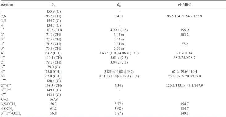
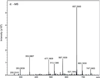
Compound 1 was isolated as a brown amorphous powder. The molecular formula was determined as being C29H38O17 from the HRESIMS at m/z 657.2065 [M-H]–. The UV spectrum showed an absorption
maximum at 279 nm, which is compatible with a phenolic structure. The IR spectrum showed a characteristic absorption band attributable to the following groups: carbonyl (1708 cm-1), hydroxyl
(3418 cm-1), and aromatic (1605 and 1509 cm-1), as
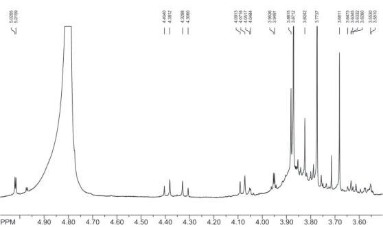
well as a glycosidic linkage (1063 cm-1). The 1H and 13C NMR spectra (Table 1) revealed signals for two
1,3,4,5-tetrasubstituted symmetrical aromatic rings. The 1H NMR exhibited singlet signals corresponding
to five aromatic methoxyls at δH 3.77 (6H, OCH3-3,5), 3.68 (3H, OCH3-4), and 3.87 (6H, OCH3-3′′′,5′′′) and two sets of aromatic proton signals at δH 6.41 (2H, H-2,6) and 7.34 (2H, H-2′′′,6′′′) due to a 3,4,5-trimethoxy phenyl derivative and symmetrical 3,5-dimethoxy-4-hydroxy-benzoyl moieties, respectively.9 This was
supported by the gHMBC spectrum, which showed cross-peaks corresponding to a long-range coupling of hydrogen at δH 6.41 and carbon signals at δC 155.9 (C-1), 154.7 (C-3,5), 134.7 (C-4) and 96.5 (C-2,6) and also between hydrogen at δH 7.34 and carbon signals at δC 167.9 (C=O), 149.1 (C-3′′′,5′′′), 143.1 (C-4′′′),
120.6 (C-1′′′) and 108.5 (C-2′′′,6′′′). Others signals observed in 1H and 13C NMR spectra are consistent
with glycosyl moieties. According to literature,10 the
comparison of the 13C NMR data of the glycosidic
moiety of various other analogous compounds, the structure of 1 seems to be composed of a glucosyl [δC
103.2 (C-1′), 74.9 (C-2′), 77.9 (C-3′), 71.5 (C-4′), 76.9 (C-5′) and 68.2 (C-6′)] and an apiosyl [δC 110.4
(C-1′′), 78.7 (C-2′′), 79.0 (C-3′′), 75.0 (C-4′′) and 67.9 (C-6′′)] moieties. The 1H, 13C NMR and TOCSY spectra
confirmed the presence of these two sugar moieties due to the presence of two anomeric signals at δH 4.79
(d, J 7.5 Hz)/δC 103.2 (CH) for glucose and δH 5.01
(d, J 2.3 Hz)/δC 110.4 (CH) for apiose. Furthermore, the coupling constants J 7.5 Hz of the anomeric proton signal of the D-glucosyl moiety as well as the chemical shift (δC 110.4) of the anomeric carbon of the D-apiosyl
moiety, demonstrated that both sugar moieties have
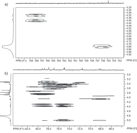
β-anomeric configurations. The gHMBC spectrum of
1 revealed cross-peaks corresponding to long-range couplings of hydrogens H-6′ of β-glucose at δH 3.63
(d, J 10.0 Hz) and 4.06 (d, J 10.0 Hz) and carbon C-1′′ of apiose at δC 110.4; in this way, the β-D-apiosyl
moiety was attached to C-6′ of the glucose. The chemical shift of the oxymethylene carbon C-6′ (δC
68.2) was used to confirm the disaccharide linkage as apiofuranosyl-(1→6)-glucopyranoside.10 In addition,
long-range correlations between H-5′′ signals of β -D-apiose [δH 4.31 (d, J 11.4 Hz) and 4.39 (d, J 11.4 Hz)]
and the syringoyl carbonyl group (δC 167.9) indicated that the 3,5-dimethoxy-4-hydroxy-benzoyl group was attached to C-5′′ of the β-D-apiose. Finally, the proton at δH 4.79 (H-1′ of glucose) was correlated with C-1
of the 3,4,5-trimethoxy phenyl group (δC 155.9), indicating that the β-D-glucosyl moiety was located at C-1. Therefore, the structure of compound 1 was established as being 3,4,5-trimethoxyphenyl-1-O-β -D-(5-O-syringoyl)-apiofuranosyl-(1→6)-β -D-gluco-pyranoside.
The known compounds 2-10 were identified by comparing their 1H and 13C NMR data with those
previously published in literature.11-16
The antifungal activity of compounds 1, 2, 6-10 against Cladosporium cladosporioides and C. sphaerospermum was evaluated by direct bioautography on a TLC plate.17,18 Only
compound 1 exhibited moderate activity (MIC of 100 µg), when compared with the standard nystatin (1.0 µg).
Experimental
General experimental procedures
1D- (1H, 13C, TOCSY) and 2D- (gHMQC and gHMBC)
NMR experiments were recorded on a Varian INOVA 500 spectrometer (11.7 T) at 500 MHz (1H) and 126 MHz
(13C), using adequate solvent with TMS as internal
1407 Silva et al.
Vol. 18, No. 7, 2007
out in a Varian HPLC, using a Phenomenex Luna RP18 (2) column (250 × 21.20 mm i.d. × 10 µm).
Plant material
Alibertia sessilis was collected in the Estação Ecológica e Experimental de Mogi-Guaçu (Ecological and Experimental Reserve of Mogi-Guaçu), São Paulo, Brazil in November 2003 by Dr M.C.M. Young and identified by Dr I. Cordeiro. The voucher specimen (SP 370.914) was deposited at the Botanical Institute Herbarium, São Paulo, Brazil.
Extraction and isolation of constituents
The dried and powdered stems (50.0 g) of A. sessilis were extracted exhaustively with ethanol, at room temperature, to give a crude material (4.15 g), which was partitioned in hexane, EtOAc and n-BuOH. After evaporation, the EtOAc extract (1.30 g) was submitted to column chromatography on Sephadex LH-20 using MeOH as eluent to give 15 fractions after TLC analysis. Fraction 1 (116.2 mg) was composed of a precipitate, which was recrystalized with H2O to give D-manitol (10) (63.7 mg). Fraction 2 (146.4 mg) was subjected to column chromatography on RP-18 using a gradient mixture of H2O/MeOH and MeOH/EtOAc as eluent to give 11 subfractions. Subfraction 2.1 (80.0 mg) was
re-chromatographed on RP-18, eluted with H2O with increasing amounts of MeOH to afford geniposidic acid (2) (11.8 mg). Subfraction 2.7 (29.6 mg) was also submitted to column chromatography on RP-18, using a gradient solvent system of H2O/MeOH and MeOH/ EtOAc as eluent to yield 7 subfractions after TLC analysis. Subfraction 2.7.6 (18.8 mg) was chromato-graphed by preparative HPLC (C18 column; mobile phase: MeOH/H2O (3:7 v/v); flow rate: 10 mL min-1; UV
detector: 238 nm) to separate the mixture of the diastereoisomeric lignans (+)-lyoniresinol 3α-O-β -D-glucopyranoside (6) (6.5 mg) and (-)-lyoniresinol 3α -O-β-D-glucopyranoside (7) (5.8 mg).19 Subfraction 2.7.7
(6.7 mg) was identified as 3,4,5-trimethoxyphenyl-1-O
-β-D-(5-O-syringoyl)-apiofuranosyl-(1→6)-β -D-gluco-pyranoside (1). Fraction 4 (274.2 mg) was fractioned on RP-18 using H2O with increasing amounts of MeOH as mobile phase to give salicylic acid (9) (6.4 mg). Fractions 5 and 6 (104.9 mg) were combined and submitted to column chromatography on silica gel eluting with hexane/EtOAc and EtOAc/MeOH to give 6.9 mg of chlorogenic acid (8).
The n-butanol extract (0.55 g) was submitted to column chromatography on Sephadex LH-20 using MeOH as eluent to give 7 fractions after TLC analysis. Fraction 3 (180.0 mg) was subjected to column chromatography on RP-18 using a gradient mixture of H2O/MeOH to give 9 subfractions. Subfraction 3.4 (10.2 mg) was identified as Table 1. 1H and 13C NMR (500 and 126 MHz, respectively; J in Hz).and gHMBC data 1 (in CD
3OD)
position δC δH gHMBC
1 155.9 (C)
-2,6 96.5 (CH) 6.41 s 96.5/134.7/154.7/155.9
3,5 154.7 (C)
-4 134.7 (C)
-1′ 103.2 (CH) 4.79 d (7.5) 155.9
2′ 74.9 (CH) 3.43 m 103.2
3′ 77.9 (CH) 3.52 m
4′ 71.5 (CH) 3.34 m 77.9
5′ 76.9 (CH) 3.60 m
6′ 68.2 (CH2) 3.63 d (10.0)/4.06 d (10.0) 71.5/110.4
1′′ 110.4 (CH) 5.01 d (2.3) 68.2/75.0/78.7
2′′ 78.7 (CH) 3.94 d (2.3)
3′′ 79.0 (C)
-4′′ 75.0 (CH2) 3.85 m/ 4.08 d (9.7) 67.9/ 79.0/ 110.4
5′′ 67.9 (CH2) 4.31 d (11.4)/ 4.39 d (11.4) 75.0/ 78.7/ 79.0/167.9
1′′′ 120.6 (C)
-2′′′,6′′′ 108.5 (CH) 7.34 s 120.6/143.1/149.1/167.9
3′′′,5′′′ 149.1 (C)
-4′′′ 143.1 (C)
-C=O 167.9
-3,5-OCH3 56.7 3.77 s 154.7
4-OCH3 61.2 3.68 s 134.7
1408 A New Antifungal Phenolic Glycoside Derivative, Iridoids and Lignans J. Braz. Chem. Soc. an iridoid mixture 3-5 and subfraction 3.6 (8.6 mg) as a
lignan mixture 6-7.
3,4,5-trimethoxyphenyl-1-O-β-D -(5-O-syringoyl)-apiofuranosyl-(1→6)-β-D-glucopyranoside (1)
Brown amorphous powder (6.7 mg, 0.16%). [α]2 D 5
–2.36 (c 0.021, MeOH). UV λmax /nm (MeOH) 279. IR (KBr) νmax/cm-1: 3418, 2931, 1708, 1605, 1509, 1461,
1427, 1338, 1228, 1125, 1063, 816, 764. HRESIMS m/z 657.2065 [M - H]– (calcd for C
29H37O17, m/z 657.2031 [M
- H]–).1H and 13C NMR: see Table 1.
Known compounds isolated
The structures of known compounds were established by 1H and 13C NMR and gHMBC data and by comparing
their spectroscopy data with those reported in literature.
Geniposidic acid (2) (0.28%)
The 1H and 13C NMR data, in CD
3OD, were in
accordance with those reported in reference 11.
Geniposide (3); 6α-hydroxygeniposide (4); 6β -hydroxygeniposide (5) (0.24%)
The 1H and 13C NMR data of the mixture, in CD 3OD,
were in accordance with those reported in references 12 and 13. The compounds were identified in mixture mainly on the basis on their TOCSY spectra.
(+)-Lyoniresinol-3α-O-β-D-glucopyranoside (6) (0.15%) and (-)-lyoniresinol-3α-O-β-D-glucopyranoside (7) (0.14%)
The 1H and 13C NMR data, in CD
3OD, were in
accordance with those reported in reference 14.
Chlorogenic acid (8) (0.16%)
The 1H and 13C NMR data, in DMSO-d
6, were in
accordance with those reported in reference 15.
Salicylic acid (9) (0.15%)
The 1H and 13C NMR data, in CD
3OD, were in
accordance with those reported in reference 16.
D-manitol (10) (1.53%)
The 1H and 13C NMR data, in D
2O, were in accordance
with those reported in reference 16.
Antifungal Assay
The microorganisms used in the antifungal assays Cladosporium sphaerospermum (Penzig) SPC 491 and C.
cladosporioides (Fresen) SPC 140 had been kept at the Botanical Institute of São Paulo, Brazil. For the antifungal assay, solutions containing 100, 50, 25, 10, 5 and 1 µg of the test compound were prepared. 10.0 µL of each solution was applied to pre-coated TLC plates eluted with EtOAc/ MeOH (7:3 v/v) and dried to completely remove all solvents. The chromatograms were sprayed with a spore suspension of Cladosporium sphaerospermum or C. cladosporioides in glucose and salt solution and incubated for 72 h in darkness in a moistened chamber at 25 °C. A clear inhibition zone appeared against a dark background indicating the minimal amount of the compound required to eliminate the fungus (detection limits - minimum amount required for the inhibition of fungal growth on TLC plates). Nystatin was used as the positive control (1 µg).17,18
Conclusions
Lignans 6 and 7 are here described for the first time as being found in Rubiaceae and the antifungal phenolic glucoside 1 is being reported for the first time in literature. Compound 10 was previously isolated from Alibertia myrciifolia. Phenolics 8 and 9 are here reported for the first time as being found in genus Alibertia. Iridoids 2
and 3-5 are in agreement with chemosystematic correlations and botanical occurrence in the Rubiaceae family. This study will contribute significantly to improve knowledge about secondary metabolites and biological activity for one more species from the Brazilian Cerrado.
Acknowledgments
The authors are grateful to the Fundação de Amparo à Pesquisa do Estado de São Paulo (FAPESP) for financial support and also to the Conselho Nacional de Desenvolvimento Científico e Tecnológico (CNPq) for a fellowship granted to V.C.S.
Supplementary Information
Supplementary data are available free of charge at http://jbcs.sbq.org.br, as PDF file.
References
1. Bolzani, V. da S.; Young, M. C. M.; Furlan, M.; Cavalheiro, A. J.; Araújo, A. R.; Silva, D. H. S.; Lopes, M. N.; An. Acad. Bras. Cienc. 1999, 71, 181.
1409 Silva et al.
Vol. 18, No. 7, 2007
3. Young, M. C. M.; Araújo, A. R.; da Silva, C. A.; Lopes, M. N.; Trevisan, L. M. V.; Bolzani, V. da S.; J. Nat. Prod. 1998, 61, 936.
4. Bolzani, V. da S.; Trevisan, L. M. V.; Izumisawa, C. M.; Gunatilaka, A. A. L.; Kingston D. G. I.; Young, M. C. M.; Phytochemistry 1997, 46, 308.
5. Bolzani, V. da S.; Trevisan, L. M. V.; Izumisawa, C. M.; Young, M. C. M.; J. Braz. Chem. Soc. 1997, 7, 157.
6. Young, M. C. M.; Braga, M. R.; Dietrich, S. M. C.; Gottlieb, H. E.; Trevisan, L. M. V.; Bolzani, V. da S.; Phytochemistry 1992, 31, 3433.
7. Bolzani, V. da S.; Trevisan, L. M. V.; Young, M. C. M.; Phytochemistry 1991, 30, 2089.
8. Luciano, J. H. S.; Lima, M. A. S.; Souza, E. B.; Silveira, E. R.; Biochem. Syst. Ecol. 2004, 32, 1227.
9. Jung, M. J.; Kang, S. S.; Jung, Y. J.; Choi, J. S.; Chem. Pharm. Bull. 2004, 52, 1501.
10. Warashina, T.; Nagatani, Y.; Noro, T. Phytochemistry2004, 65, 2003.
11. Guarnaccia, R.; Madyastha, K. M.; Tegtmeyer, E.; Coscia, C. J.; Tetrahedron Lett. 1972, 50, 5125.
12. Damtoft, S.; Jensen, R. S.; Nielsen, B. J.; Phytochemistry 1981, 20, 2717.
13. Bianco, A. In Studies in Natural Products Chemistry, Atta-ur-Rahman, ed., Elsevier: Amsterdam, 1990, p. 439.
14. Achenbach, H.; Löwel, M.; Waibel, R.; Gupta, M.; Solis, P.; Planta Med. 1992, 58, 270.
15. Maruta, Y.; Kawabata, J.; Niki, R.; J. Agric. Food Chem. 1995, 43, 2592.
16. Pouchert, C. J.; Behnke, J.; The Aldrich Library of 13C and 1H FT NMR Spectra, Aldrich Chemical Company: Milwakee, 1993. 17. Homans, A. L.; Fuchs, A.; J. Chromatogr. 1970, 51, 327. 18. Rahalison, L.; Hamburger, M.; Monod, M.; Frenk, E.;
Hostettmann, K.; Planta Med. 1994, 60, 41.
19. da Silva, V. C.; Silva, G. H.; Bolzani, V. da S.; Lopes, M. N. Ecl. Quim. 2006, 31, 55.
Received: May 23, 2007 Web Release Date: November 14, 2007
J. Braz. Chem. Soc., Vol. 18, No. 7, S1-S7, 2007. Printed in Brazil - ©2007 Sociedade Brasileira de Química 0103 - 5053 $6.00+0.00
Supplementary InformationSupplementary InformationSupplementary InformationSupplementary InformationSupplementary Information
*e-mail: [email protected]
A New Antifungal Phenolic Glycoside Derivative and Iridoids and Lignans
from
Alibertia sessilis
(Vell.) K. Schum. (Rubiaceae)
Viviane C. da Silva,a Vanderlan da S. Bolzani,a Maria C. M. Young b and Márcia N. Lopes*,a
a
Departamento de Química Orgânica, Instituto de Química, Universidade Estadual Paulista, CP 355, 14801-970 Araraquara-SP, Brazil
b
Seção de Fisiologia e Bioquímica de Plantas, Instituto de Botânica, CP 4005, 01061-970 São Paulo-SP, Brazil
S2 A New Antifungal Phenolic Glycoside Derivative and Iridoids and Lignans J. Braz. Chem. Soc.
Figure S3. HRESIMS spectrum of compound 1 (negative mode).
Figure S4. 1H NMR spectrum of compound 1 (CD
S3 Silva et al.
Vol. 18, No. 7, 2007
Figure S5. Expansion of 1H NMR spectrum of compound 1 (CD
3OD, 500 MHz).
S4 A New Antifungal Phenolic Glycoside Derivative and Iridoids and Lignans J. Braz. Chem. Soc.
Figure S7. 13C NMR spectrum of compound 1 (CD
3OD, 126 MHz).
S5 Silva et al.
Vol. 18, No. 7, 2007
S6 A New Antifungal Phenolic Glycoside Derivative and Iridoids and Lignans J. Braz. Chem. Soc.
S7 Silva et al.
Vol. 18, No. 7, 2007
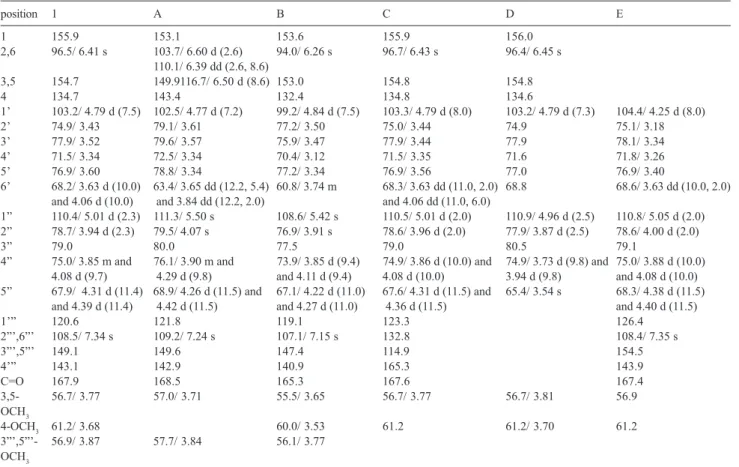
Table S1. 1H and 13C NMR data of analogous compounds of 1 in literature
position 1 A B C D E
1 155.9 153.1 153.6 155.9 156.0
2,6 96.5/ 6.41 s 103.7/ 6.60 d (2.6) 94.0/ 6.26 s 96.7/ 6.43 s 96.4/ 6.45 s 110.1/ 6.39 dd (2.6, 8.6)
3,5 154.7 149.9116.7/ 6.50 d (8.6) 153.0 154.8 154.8
4 134.7 143.4 132.4 134.8 134.6
1’ 103.2/ 4.79 d (7.5) 102.5/ 4.77 d (7.2) 99.2/ 4.84 d (7.5) 103.3/ 4.79 d (8.0) 103.2/ 4.79 d (7.3) 104.4/ 4.25 d (8.0)
2’ 74.9/ 3.43 79.1/ 3.61 77.2/ 3.50 75.0/ 3.44 74.9 75.1/ 3.18
3’ 77.9/ 3.52 79.6/ 3.57 75.9/ 3.47 77.9/ 3.44 77.9 78.1/ 3.34
4’ 71.5/ 3.34 72.5/ 3.34 70.4/ 3.12 71.5/ 3.35 71.6 71.8/ 3.26
5’ 76.9/ 3.60 78.8/ 3.34 77.2/ 3.34 76.9/ 3.56 77.0 76.9/ 3.40
6’ 68.2/ 3.63 d (10.0) 63.4/ 3.65 dd (12.2, 5.4) 60.8/ 3.74 m 68.3/ 3.63 dd (11.0, 2.0) 68.8 68.6/ 3.63 dd (10.0, 2.0) and 4.06 d (10.0) and 3.84 dd (12.2, 2.0) and 4.06 dd (11.0, 6.0)
1” 110.4/ 5.01 d (2.3) 111.3/ 5.50 s 108.6/ 5.42 s 110.5/ 5.01 d (2.0) 110.9/ 4.96 d (2.5) 110.8/ 5.05 d (2.0) 2” 78.7/ 3.94 d (2.3) 79.5/ 4.07 s 76.9/ 3.91 s 78.6/ 3.96 d (2.0) 77.9/ 3.87 d (2.5) 78.6/ 4.00 d (2.0)
3” 79.0 80.0 77.5 79.0 80.5 79.1
4” 75.0/ 3.85 m and 76.1/ 3.90 m and 73.9/ 3.85 d (9.4) 74.9/ 3.86 d (10.0) and 74.9/ 3.73 d (9.8) and 75.0/ 3.88 d (10.0) 4.08 d (9.7) 4.29 d (9.8) and 4.11 d (9.4) 4.08 d (10.0) 3.94 d (9.8) and 4.08 d (10.0) 5” 67.9/ 4.31 d (11.4) 68.9/ 4.26 d (11.5) and 67.1/ 4.22 d (11.0) 67.6/ 4.31 d (11.5) and 65.4/ 3.54 s 68.3/ 4.38 d (11.5)
and 4.39 d (11.4) 4.42 d (11.5) and 4.27 d (11.0) 4.36 d (11.5) and 4.40 d (11.5)
1’” 120.6 121.8 119.1 123.3 126.4
2”’,6”’ 108.5/ 7.34 s 109.2/ 7.24 s 107.1/ 7.15 s 132.8 108.4/ 7.35 s
3”’,5”’ 149.1 149.6 147.4 114.9 154.5
4’” 143.1 142.9 140.9 165.3 143.9
C=O 167.9 168.5 165.3 167.6 167.4
3,5- 56.7/ 3.77 57.0/ 3.71 55.5/ 3.65 56.7/ 3.77 56.7/ 3.81 56.9 OCH3
4-OCH3 61.2/ 3.68 60.0/ 3.53 61.2 61.2/ 3.70 61.2
3”’,5”’- 56.9/ 3.87 57.7/ 3.84 56.1/ 3.77 OCH3