Article
J. Braz. Chem. Soc., Vol. 27, No. 1, 91-98, 2016. Printed in Brazil - ©2016 Sociedade Brasileira de Química 0103 - 5053 $6.00+0.00
A
*e-mail: [email protected]
Antimicrobial Potential of Bio-Oil for Use in Diesel Oil B10
Sabrina A. Beker,*,a Maria Elisabete Machado,b Gabriela P. S. Maciel,b Rosângela Silva,c
Renato Cataluña,b Elina B. Caramãob and Fatima M. Bentoa
aLaboratório de Biodeterioração de Combustíveis, Biocombustíveis e Biocidas (LAB-BIO),
Departamento de Microbiologia, Imunologia e Parasitologia and bDepartamento de Química
Inorgânica, Universidade Federal do Rio Grande do Sul, 90050-170 Porto Alegre-RS, Brazil
cPontifícia Universidade Católica do Rio Grande do Sul, 90619-900 Porto Alegre-RS, Brazil
The aim of this study was to evaluate the antimicrobial activity of bio-oil (obtained by pyrolysis of biomass: soybean oil, eucalyptus sawdust and coffee grounds) added to the blend B10 (diesel and biodiesel). The bio-oil is compatible to diesel oil and it contains promising compounds that can exhibit antimicrobial activity during fuel storage. We evaluated the antimicrobial activity of bio-oil added to diesel oil B10 by determining the minimum inhibitory concentration at different concentrations (0-10%) using oil-deteriorating microorganisms for 10 days at 30 °C. Bio-oil was separated from the aqueous phase by solid phase extraction using ultrapure water and sodium hydroxide. The aqueous phase containing the solubilized compounds of bio-oil was characterized by comprehensive two-dimensional gas chromatography with time-of-flight mass spectrometric detection. Minimum inhibitory concentration in the range of 0.25% to 4% was observed for the tested inocula. The chromatographic analysis of both extracts allowed the identification of several oxygenated compounds, which the majority of the analytes consisted of phenolic compounds, followed by ketones.
Keywords: antimicrobial, bio-oil, phenolic compounds, diesel, biodiesel, blend
Introduction
Oil and coal are responsible for most power generated in the world. However, in view of the limitation of these fossil energy sources and the environmental issues involved, the search for new renewable energy sources is becoming more intense. For this reason, there is great interest in the exploitation of biomass energy for producing liquid fuels for transportation sector.1 The biomass consists of a natural resource comprising all organic material capable of being transformed into energy. One process used for this transformation is through pyrolysis, the heat-mediated decomposition of organic matter in the absence of oxygen, generating solid, gaseous and fluid products, such as the widely explored bio-oil. This product is particularly promising for being a biofuel that can be used as a component in the formulation of diesel oil.2,3
Another form of biomass energy, however, resulting from transesterification, is biodiesel. In Brazil, law 11.097 made the addition of biodiesel to diesel oil mandatory, and since
then biofuel is effectively part of the national energy matrix. The addition of biodiesel to diesel, at variable proportions, is called diesel oil B, currently used at 7% (B7).
control and eliminate microbial contamination.5 However, this practice is not legal in Brazil, since there are many doubts in oil and petroleum sector concerning their use in fuels.
It is a great challenge for research and industry to find a compound with the ability to inhibit microbial growth without affecting the fuel quality and characteristics. Bio-oil meets these demands, since tests with diesel engines were successful.8 In addition, some studies indicate the presence of antimicrobial activity for being phenolic compounds-rich, thus representing an alternative preservative.9-12
The aim of this study was to evaluate the antimicrobial potential of bio-oil when added to B10 blend (diesel and biodiesel) with oil-deteriorating microorganisms and to characterize bio-oil soluble compounds in the aqueous phase by two-dimensional gas chromatography with time-of-flight mass spectrometry (GC×GC/TOF MS) detection, to identify the compounds responsible for the antimicrobial action.
Experimental
Bio-oil
Bio-oil used was produced as described by Cataluña et al.8 using a fixed bed reactor, immobilized with a 1:1:1 (v/m/m) mixture of soybean oil, coffee grounds and eucalyptus sawdust, using the binders calcium hydroxide [Ca(OH)2] and sodium hydroxide (NaOH) in an inert argon atmosphere at a heating rate of 10 °C min−1 from room temperature to 700 °C, maintaining this temperature for 15 min.8
Fuels
The following fuels were used: low sulfur content (50 ppm) diesel oil (diesel A) and biodiesel, 80% soy and 20% tallow (v/v), provided by Ipiranga Produtos de Petróleo S/A (Porto Alegre, RS, Brazil), and Granol Indústria Comércio e Exportação S/A (Porto Alegre, RS, Brazil), respectively. In the laboratory, biodiesel (B100) was blended with diesel oil to prepare a 10% (v/v) biodiesel blend (B10). The blend was sterilized by vacuum filtration through membrane pores (0.22 µm, Millipore, Merck), using a sterilized Büchner flask, autoclaved for 15 min at 121 °C and 1 atm. Thereafter, the fuel was stored in vials sterilized in the same way and then hermetically sealed. To avoid photo-oxidation, the vials were protected from light with aluminum foil and stored at room temperature.
Microorganisms
In this study we used oil-deteriorating microorganisms isolated from diesel and biodiesel storage tanks in
Brazil. These species are included in the bacteria and mycology collection of the Laboratory of Fuel and Biofuel Biodeterioration (Universidade Federal do Rio Grande do Sul, RS, Brazil).13-15 The studied microorganisms were a filamentous fungus (Paecilomyces variotii) isolated from biological sludge obtained from fuel storage tanks;15 a yeast (Candida silvicola) isolated from a diesel oil tank;13 and a bacterium (Bacillus pumilus) previously isolated from sediment resulting from biodiesel centrifugation.
Preparation of inoculum from the isolated microorganisms
The inoculum of the filamentous fungus Paecilomyces variotii was prepared from 7 days old cultures on agar-malt agar in inclined tubes in an incubator at 29 ± 1 °C, by adding 2 mL sterile saline (0.85%) and 2 mL of surfactant (Tween 80) at 0.01%. The inoculum for the yeast Candida silvicola was obtained from yeast grown in agar-malt in slanted tubes by adding 2 mL of sterile saline solution (0.85%). Inoculum of the bacterium Bacillus pumilus was prepared from Petri culture containing Luria-Bertani agar after 24 h of incubation at 30 °C. Spores and cells were counted in a Neubauer chamber, and the suspension in each flask had a final concentration of 105 spores or cells mL−1.15
Preparation of uncharacterized inoculum
Uncharacterized inoculum was prepared according to American Society for Testing and Materials (standard
practice ASTM E1259).16 Briefly, 2% of blend B10
(previously filtered and sterilized) was added to 100 mL Bushnell-Haas mineral medium in an Erlenmeyer flask, which was then inoculated with 5 mL of microbiological sludge from a contaminated tank. The flask was incubated in an orbital shaker (CIENTEC CT-712, Belo Horizonte, MG, Brazil) at 30 °C and 200 rpm for 7 days. The final cell concentration in the bottles was 105 colony-forming unit (CFU) mL−1.
Evaluation of antimicrobial activity
different concentrations of blends of bio-oil and B10 were added to each glass flask. The experiment was carried out in quadruplicate. Finally, the inoculum was added at a concentration of 105 spores or cells mL−1 prepared as described above and the flasks were sealed with sterile cotton and placed in an incubator at a temperature of 30 °C for 10 days.
Analyses
Evaluation of the minimum inhibitory concentration
The MIC was determined by the broth dilution method (single dilution series), where the biofilm formation and the turbidity of the media in the flask were observed by naked eye.16
Solid phase extraction of bio-oil
For the characterization of the soluble bio-oil compounds in the aqueous phase, which possibly contribute to its antimicrobial effect, solid phase extraction (SPE) was performed. Samples were prepared by two procedures. First, a test tube was filled with 0.5 mL of bio-oil and 5 mL of ultrapure water. The solution was stirred and the separation of phases awaited. Then, the aqueous phase was collected, and was passed through a solid-phase extraction cartridge of octadecyl-bonded silica (C18; Agilent Technologies; 500 mg per 3 mL), which was previously prepared by adding 4 mL methanol under vacuum in a Büchner flask and washed with 20 mL of ultrapure water. After preparing the cartridge, the samples were percolated in sequential fractions of 1 mL (total of 5 mL). After drying the cartridge, the analytes were eluted with 5 mL of dichloromethane (DCM) and 5 mL of chloroform. In parallel, the same procedure was performed with 1 mol L−1 NaOH solution instead of ultrapure water. The extracts were transferred to a beaker containing DCM for solubilization. After evaporation of the solvents, the samples were chromatographically analyzed. The procedures were performed in duplicate.
Analysis of chromatographic extracts
Analyses were performed with two-dimensional gas chromatography with time-of-flight mass spectrometry
(GC×GC/TOF MS), using a Pegasus-IV system (LECO Corporation, MO, USA) equipped with a liquid nitrogen quad-jet modulator and a CTC CombiPAL Autosampler. The columns used in the first and second dimensions were: a DB-5 column (5% phenyl, 95% dimethylpolysiloxane, length 60 m, inner diameter 250 µm, phase thickness 0.25 µm), and a DB-17 column (50% phenyl, 50% dimethylpolysiloxane, length 2.15 m, inner diameter 180 µm, phase thickness 0.18 µm; Agilent Technologies, J & W Scientific, Agilent, CA, USA). The carrier gas was helium under a constant flow rate of 1 mL min−1 and the sample injection volume was 1 µL. The injection temperature was 280 °C and the samples were injected in splitless mode. The temperature program of the first column was set to begin at 40 °C for 1 min at a heating rate of 3 °C min−1 until reaching the final temperature of 300 °C. The compounds were identified using software ChromaTOF version 3.32 (LECO Corporation, MO, USA), as described by Cataluña et al.8
Results and Discussion
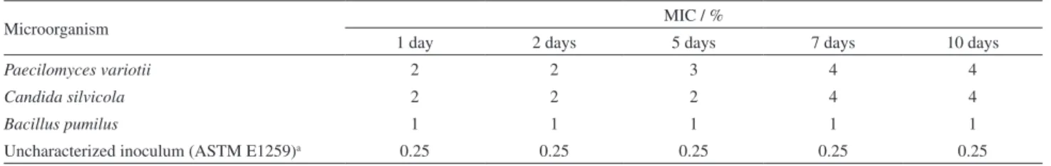
The different concentrations of bio-oil in blend B10 were evaluated for antimicrobial activity for 10 days and the results of the MIC are listed in Table 1.
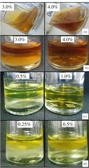
After 10 days of incubation with the filamentous fungus Paecilomyces variotii, the MIC rose from 2% to 4%, which was also clearly inhibitory for the yeast Candida silvicola (Figures 1a and 1b, respectively).
Figure 1a shows the formation of biomass at the water-oil interface, which is characteristic of the development of filamentous fungus, due to the set of hyphae (vegetative fungal structures). The growth of fungus Paecilomyces sp. was evaluated in other diesel blends and biodiesel B5, B10 and B20, and in pure biodiesel by Bücker et al.15 Cazarolli et al.18 also evaluated the oil-deteriorating capacity of this microorganism, but in the presence of pure soybean biodiesel (B100), biomass formation was clearly observed at the water-oil interface.18 No growth was observed in the reagent flask at a concentration of 4% bio-oil (Figure 1a). Probably, in the flask containing the mixture at a concentration of 3% bio-oil, the fungus
Table 1. Percentage values of the minimum inhibitory concentration (MIC) of the blend (bio-oil + B10) for the tested inocula
Microorganism MIC / %
1 day 2 days 5 days 7 days 10 days
Paecilomyces variotii 2 2 3 4 4
Candida silvicola 2 2 2 4 4
Bacillus pumilus 1 1 1 1 1
Uncharacterized inoculum (ASTM E1259)a 0.25 0.25 0.25 0.25 0.25
tolerated the presence of bio-oil compounds. However, at a concentration of 4%, growth was inhibited and also eradicated. It is possible to indicate that for Paecilomyces variotii the minimum inhibitory concentration was 4% after 10 days of incubation.
Candida sp. has often been cited for its ability to degrade products derived from diesel oil, kerosene, lubricating oil, and biodiesel.13,15,19,20 The species C. silvicola was therefore tested in this experiment. The MIC was measured by the turbidity of the aqueous medium (Figure 1b). As for P. variotii, MIC for C. silvicola was 4%.
In our assessment, after 10 days of incubation the MIC for the oil-deteriorating bacterium Bacillus pumilus was 1% (Figure 1c and Table 1). Species of the genus Bacillus are characterized as Gram-positive, forming endospores
and are strict or facultative aerobes.21 Spore-forming are known to be more resistant than non-spore-forming species. For the uncharacterized inoculum, MIC was 0.25% (Figure 1d and Table 1). These results were lower than for groups of microorganisms evaluated separately. The use of an uncharacterized inoculum has been recommended
by ASTM E125916 for antimicrobial assessments of
fuels. Uncharacterized inoculum was collected from tanks contaminated with microorganisms, which was assumed to be an oil-deteriorating microbial population. No survey investigated which species were actually present, but the results indicated that bio-oil components at a concentration of 0.25% were effective to inhibit the microbial population. These results suggested that the microbial community used in this evaluation was highly sensitive to bio-oil components.6,15 It must be remembered that the uncharacterized inoculum has different microorganisms with different capabilities of growing under limited conditions, so it might be suggested that the microorganisms present in this consortium could compete among them for energy source, leading to the decrease of viable microorganisms.
The bio-oil used in this study resulted from pyrolysis of the biomass of soybean oil, eucalyptus sawdust and coffee grounds.8 The use of GC×GC/TOF MS characterization of the compounds found in crude bio-oil allowed the identification of the following chemical classes: ketones, alcohols, ethers, phenols, aliphatic and aromatic hydrocarbons, and nitrogen compounds. The most-represented compounds were ketones and nitrogen. The compounds from the aqueous phase were SPE-extracted to analyze the compounds of bio-oil that were solubilized in aqueous media and had antimicrobial activity, since the microorganisms developed in the oil-water interface or in the aqueous phase.
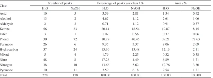
Table 2 shows the results for the SPE extract obtained with ultrapure water and NaOH by GC×GC/TOF MS (Figure S1 and S2 of Supplementary Information section, respectively). The compounds were tentatively identified when the similarity between the sample and library spectra was greater than 750 and after a detailed analysis of the spectra. In total, 278 peaks were tentatively identified with ultrapure water, and the largest class contained ketones (56 compounds), followed by nitro compounds (48 compounds). The results for the use of 1 mol L−1 NaOH solution to extract the compounds from the aqueous phase of bio-oil are also shown in Table 2.
The results showed a total number of tentatively identified compounds of 178, i.e., 100 compounds less than when ultrapure water was used for extraction. The major class contained phenols, most represented both Figure 1. Experimental flasks containing inocula after 10 days of
with regard to the number of compounds (72) and the area (78.6%). Comparing these results with those of extracted with ultrapure water, it was found that the latter was more effective than NaOH solution with regard to the largest number of compounds extracted for all classes (Table 2).
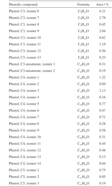
Specific isomer structural assignment is not possible in the absence of authentic standards. It is only possible to determine the number of carbons in the side chain but not the exact position of it. Tables 3 and 4 shows the tentatively identified phenolic compounds, their molecular formula and area %.
However, with regard to the percentage of extracted phenolic compounds, the extraction efficiency of SPE with NaOH was higher than with water. The results suggested that the presence of phenol may be related to fungal growth inhibition, since the oxygenated compounds, including phenolic compounds, are concentrated in the aqueous phase by affinity. Similar results were obtained by Mohan et al.11 in an assessment of the antifungal capacity of bio-oil resulting from pyrolysis of sawdust and its lignin-rich fractions obtained by extraction with ethyl-acetate.These authors observed that the extract, which contained more phenolic compounds, had a greater fungicidal effect on the fungi tested than pure bio-oil.
Several studies suggested the efficacy of phenolic compounds derived from lignin as antimicrobial agents.11,22,23 In many cases, bio-oil was tested as an alternative wood preservative, in view of its growth-inhibiting capacity of oil-deteriorating microorganisms.9,11,24 Also the insecticidal activity of pyrolysis bio-oils from coffee grounds has previously been studied.12 Probably the different components of most biomass materials, lignin rich sawdust and coffee grounds, can synergistically produce a potent
Table 2. Distribution of compounds, number of peaks and their areas in the extract with ultrapure water and 1 mol L−1 NaOH
Class Number of peaks Percentage of peaks per class / % Area / %
H2O NaOH H2O NaOH H2O NaOH
Acid 10 5 3.59 2.81 1.34 0.92
Alcohol 13 2 4.67 1.12 2.61 1.06
Aldehyde 2 2 0.71 1.12 0.91 0.37
Ketone 56 33 20.14 18.54 12.87 8.15
Ester 3 1 1.07 0.56 0.37 0.06
Phenol 30 72 10.79 40.45 39.21 78.63
Furanone 26 6 9.35 3.37 8.06 2.09
Hydrocarbon 37 24 13.30 13.48 12.13 2.11
Mixed 5 4 1.79 2.25 0.32 0.38
Nitro 48 8 17.26 4.49 6.89 1.71
Nitrogen 38 10 13.66 5.62 12.76 3.30
Pyranone 10 11 3.59 6.18 2.54 1.21
Total 278 178 100.00 100.00 100.00 100.00
Table 3. Identification of the phenolic compounds obtained by the extract with ultrapure water
Phenolic compound Formula Area / %
Phenol C6H6O 37.58
Phenol, C1, isomer 1 C7H8O 14.83
Phenol, C1, isomer 2 C7H8O 20.72
Phenol, C1, isomer 3 C7H8O2 4.20
Benzenediol, methoxy- C7H8O3 0.12
Phenol, C2, isomer 1 C8H10O 0.99
Phenol, C2, isomer 2 C8H10O 1.96
Phenol, C2, isomer 3 C8H10O 7.24
Phenol, C2, isomer 4 C8H10O 4.73
Phenol, C2, isomer 5 C8H10O 1.13
Phenol, C3, isomer 2 C9H12O 0.32
Phenol, C3, isomer 3 C8H10O 1.83
Phenol, C3, isomer 4 C9H12O 0.18
Phenol, C3, isomer 5 C9H12O 0.65
Phenol, C3, isomer 6 C9H12O 0.53
Phenol, C3, isomer 7 C9H12O 0.16
Phenol, C3, isomer 8 C9H12O 0.44
Phenol, C3, isomer 9 C9H12O 0.38
Phenol, C3, isomer 1 C9H12O 0.34
Phenol, C3, isomer 2 C9H12O 0.17
Phenol, C3, isomer 3 C9H12O 0.29
Phenol, methoxy, C2 C9H12O2 0.63
Phenol, dimethoxy- C8H10O3 0.41
Benzenetriol C6H6O3 0.07
bio-oil antimicrobial. Further tests are required to identify which biomass material produce the bio-oil with the strongest antimicrobial activity.
The hydroxyl (−OH) groups of phenolic compounds were described for their inhibitory action, since these groups can interact with the cell membrane and break the structures thereof, causing leakage of intracellular components.25,26 Active groups such as hydroxyls (−OH) promote electron movement in the membrane, acting as electron exchangers and causing a reduction in the electron gradient across the membrane. This process causes the collapse of the proton-driving force and the decrease of adenosine triphosphate (ATP) and ultimately leads to cell death.27 Similarly, Farag et al.28 reported that these hydroxyl groups can easily connect to the active site of enzymes altering the cellular metabolism of microorganisms. The above modes of action illustrate
the importance of phenolic compounds and their hydroxyl groups in the antimicrobial activity.28
Oliveira et al.29 studied phenolic extracts and their antifungal effect on the Aspergillus flavus and observed that the development of the filamentous fungus was inhibited by the action of the extracts.29 Aside from the filamentous fungi, yeasts were also inhibited in studies conducted by Kim et al.30 by thymol, a phenolic essential oil found in oregano.30 The same compound was tested on Saccharomyces cerevisiae, in which a minimum inhibitory concentration of 128 µg mL−1 of thymol was found.31
Compared with the MIC values for fungi, the bacterium Bacillus pumilus responded to a lower concentration of bio-oil, being more susceptible to the presence of phenols. Possibly this was due to the cellular makeup. The cell wall of Gram-positive bacteria consists of a thick peptidoglycan Table 4. Identification of the phenolic compounds obtained by the extract with 1 mol L−1 NaOH
Phenolic compound Formula Area / %
Indanol C9H10O 0.60
Indanol, C1, isomer 1 C10H12O 0.40
Indanol, C1 C10H12O 0.67
Indanol, C1 C10H12O 0.47
Indanol, C1 C10H12O 0.50
Indanol, C1 C10H12O 0.17
Phenol C6H6O 3.20
Phenol, methoxy, isomer 1 C7H8O2 0.11
Phenol, methoxy, isomer 2 C7H8O2 0.02
Phenol, methoxy, C2 C9H12O2 1.16
Phenol, methoxy, C3 C10H14O2 0.04
Phenol, allyl C9H10O 0.03
Phenol, C1, isomer 1 C7H8O 5.26
Phenol, C1, isomer 2 C7H8O 25.07
Phenol, C1, isomer 3 C7H8O 0.92
Phenol, C2, isomer 1 C8H10O 0.51
Phenol, C2, isomer 2 C8H10O 8.40
Phenol, C2, isomer 3 C8H10O 0.06 Phenol, C2, isomer 4 C8H10O 10.89
Phenol, C2, isomer 5 C8H10O 0.03
Phenol, C2, isomer 6 C8H10O 2.70
Phenol, C2, isomer 7 C8H10O 3.77
Phenol, C3, isomer 1 C9H12O 0.69
Phenol, C3, isomer 2 C8H10O 4.00
Phenol, C3, isomer 3 C9H12O 0.24 Phenol, C3, isomer 4 C9H12O 0.77
Phenol, C3, isomer 5 C9H12O 5.94
Phenolic compound Formula Area / %
Phenol, C3, isomer 6 C9H12O 6.21
Phenol, C3, isomer 7 C9H12O 2.78
Phenol, C3, isomer 8 C9H12O 0.45
Phenol, C3, isomer 9 C9H12O 2.04
Phenol, C3, isomer 10 C9H12O 0.62
Phenol, C3, isomer 11 C9H12O 1.19
Phenol, C3, isomer 12 C9H12O 0.56
Phenol, C3, isomer 13 C9H12O 0.23
Phenol, C3-unsaturate, isomer 1 C10H12O 0.31
Phenol, C3-unsaturate, isomer 2 C10H12O 0.19
Phenol, C4, isomer 1 C10H14O 1.22
Phenol, C4, isomer 2 C10H14O 0.09
Phenol, C4, isomer 3 C10H14O 2.13
Phenol, C4, isomer 4 C10H14O 0.16
Phenol, C4, isomer 5 C10H14O 0.77
Phenol, C4, isomer 6 C10H14O 0.47
Phenol, C4, isomer 7 C10H14O 0.71
Phenol, C4, isomer 8 C10H14O 0.28 Phenol, C4, isomer 9 C10H14O 0.58
Phenol, C4, isomer 10 C10H14O 0.31
Phenol, C4, isomer 11 C10H14O 0.44
Phenol, C4, isomer 12 C10H14O 0.46
Phenol, C4, isomer 13 C10H14O 0.13
Phenol, C4, isomer 14 C10H14O 0.04
Phenol, C5, isomer 1 C10H14O 0.75 Phenol, C5, isomer 2 C11H16O 0.05
layer containing teichoic and lipoteichoic acids and proteins associated with this cytoplasmic membrane and layer, while the fungi have a fundamentally different cell wall and cell membrane from that of bacteria and other eukaryotes. Both structures (cell membrane and cell wall) are vital for the cell and their disruption can lead to cell death.21
The differences between the cell envelopes of the different microorganisms may have influenced the values of MIC. Phenolic compounds, known to destabilize the cytoplasmic membrane of microorganisms, may have affected the bacterial membrane more effectively than that of the fungus evaluated in this test, with a consequently lower value of MIC of bio-oil.32
The endospores in Gram-positive bacteria are characterized by different coating layers that are rigid and highly resistant to several physical and chemical elimination methods, including phenolic compounds. The endospores are mostly eliminated by exposure to high temperature for a long time. In this study, it was observed that bacterium B. pumilus was not resistant to the phenols, with relatively low MIC (0.5%), compared to that of fungus. Most likely, the bacteria were probably unable to form spores in this environment under these conditions.
Walsh et al.33 investigated the antibacterial activity and the action mechanisms of two phenolic compounds and found leakage of components through the cell membrane of the bacteria Escherichia coli and Staphylococcus aureus, i.e., both Gram-negative as well as Gram-positive bacteria were affected by the action of phenolic products.33 Dong et al.34 observed inhibition of gram-positive bacteria (S. aureus and Listeria monocytogenes) when using phenolic extract from bio-oil of corn stover residue (lignin-rich). However, in the tests using E. coli O157:H7
and Salmonella enteritidis, both Gram-negative, no
antimicrobial action of the phenolic extract was observed.34
Conclusions
Depending on the challenge organisms used, inhibition ranges of 0.25 to 4% were observed for bio-oil added to mixture B10. The major class of compounds from both extracts (with ultrapure water and NaOH solution) was that of phenols followed by ketones. However, ultrapure water extracted a greater diversity of compounds than NaOH (278 vs. 178 compounds, respectively).
In Brazil, the diesel and biodiesel (in its pure form) must comply with physical and chemical specifications established by the National Agency Petroleum, Natural Gas and Biofuels (ANP) and the mandatory percentage is the blend B7 (7% of biodiesel in petrol diesel). The use of biocides as agents to control microbial contamination in fuel storage tanks is still
a relatively unknown option for the oil sector in Brazil, with many questions to be clarified. Which biocide is better in a particular case? What concentration should be used? How can a particular phase (oil, interface or aqueous) be treated? How long is the preservation time? How should the aqueous phase from biocide treated diesel and biodiesel tanks be safely discarded?6 In case of the bio-oil from pyrolysis be recognized with the antimicrobial properties, it is important and desirable that its addition to the fuel does not cause any reaction that compromises characteristics as ignition, lubricity and flammability. Effects on emissions, filterability and corrosion potential to other fuel distribution components have to be evaluated, as well.
Supplementary Information
Supplementary data (bidimentional and tridimentional color diagram of the extract with H2O GC×GC/TOF MS and with NaOH GC×GC/TOF MS) are available free of charge at http://jbcs.sbq.org.br as PDF file.
Acknowledgements
The authors are indebted to the Brazilian Council for Scientific and Technological Development (CNPq) and the Brazilian Federal Agency for Support and Evaluation of Graduate Education (CAPES) for funding this project.
References
1. Santos, F. A.; Queiróz, J. H.; Colodette, J. L.; Fernandes, S. A.; Guimarães, V. M.; Rezende, S. T.; Quim. Nova2012, 35, 1004. 2. Minkova, V.; Marinov, S. P.; Zanzi, R.; Bjornbom, E.;
Budinova, T.; Stefanova, M.; Lakov, L.; Fuel Process. Technol.
2000,62, 45.
3. Chum, H. L.; Overend, R. P.; Fuel Process. Technol.2001, 71, 187.
4. Bento, F. M.; Beech, I.; Gaylarde, C.; Englert, G.; Muller, I.; World J. Microbiol. Biotechnol.2005a, 21, 135.
5. Passman, F. J.; Int. Biodeterior. Biodegrad.2013, 81, 88. 6. Zimmer, A.; Cazarolli, J.; Teixeira, R. M.; Viscardi, S. L. C.;
Cavalcanti, E. S. H.; Gerbase, A. E.; Ferrão, M. F.; Piatnicki, C. M. S.; Bento, F. M.; Fuel2013, 112, 153.
7. http://www.abntcatalogo.com.br/norma.aspx?ID=322742, accessed in September 2015.
8. Cataluña, R.; Kuamoto, P.; Petzhold, C.; Caramao, E.; Machado, M. E.; Silva, R.; Energy Fuels2013, 27, 6831.
9. Meier, D.; Anderson, B.; Irbe, I.; Chirkova, J.; Faix, O.; Prog. Thermochem. Biomass Convers. 2001, 2, 1550.
10. Mourant, D.; Yang, D.-Q.; Lu, X.; Roy, C.; Wood Fiber Sci.
11. Mohan, D.; Shi, J.; Nicholas, D. D.; Pittman Jr., C. U.; Steele, P. H.; Cooper, J. E.; Chemosphere2008, 71, 456.
12. Bedmutha, R.; Bookera, C. J.; Ferrante, L.; Briens, C.; Berruti, F.; Yeunga, K. K. C.; Scott, I.; Conn, K.; J. Anal. Appl. Pyrolysis2011, 90, 224.
13. Bento, F. M.; Gaylarde, C. C.; Int. Biodeterior. Biodegrad.2001, 47, 107.
14. Meyer, D. D.; Beker, S. A.; Bücker, F.; Peralba, M. C. R.; Frazzon, A. P. G.; Osti, J. F.; Andreazza, R.; Camargo, F. A. O.; Bento, F. M.; Int. Biodeterior. Biodegrad. 2014, 95, 356. 15. Bücker, F.; Santestevan, N. A.; Roesch, L. F.; Jacques, R. J. S.;
Peralba, M. C. R.; Camargo, F. A. O.; Bento, F. M.; Int. Biodeterior. Biodegrad.2011, 65, 172.
16. http://www.astm.org/Standards/E1259.htm accessed in September 2015.
17. Bushnell, C. D.; Haas, H. F.; J. Bacteriol. 1941, 41, 654. 18. Cazarolli, J. C.; Guzatto, R.; Samios, D.; Peralba, M. C. R.;
Cavalcanti, E. H. S.; Bento, F. M.; Int. Biodeterior. Biodegrad.
2013, 95, 364.
19. Bento, F. M.; Englert, G. E.; Gaylarde, C. C.; Muller, I. L.; Mater. Corros.2004, 55, 577.
20. Miranda, R. C.; de Souza, C. S.; Gomes, E. B.; Lovaglio, R. B.; Lopes, C. E.; Sousa, M. F. V. Q.; Braz. Arch. Biol. Technol.
2007, 50, 147.
21. Harvey, R. A.; Champe, P. C.; Fisher, B. D.; Microbiologia Ilustrada, 2ª ed.; Artmed: Porto Alegre, 2008.
22. Mazela, B.; Waste Manage.2006, 27, 461.
23. Kim, K. H.; Jeong, H. S.; Kim, Y. J.; Han, G. S.; Choi, I. G.; Choi, J. W.; Chemosphere2012,89, 688.
24. Suzuki, T.; Doi, S.; Yamakawa, M.; Yamamoto, K.; Watanabe, T.; Funaki, M.; Holzforschung1997, 5, 214.
25. Lai, P. K.; Roy, J.; Curr. Med. Chem. 2004, 11, 1451. 26. Xu, J.; Davidson, P. M.; Zhong, Q.; J. Agric. Food Chem. 2013,
61, 12720.
27. Ultee, A.; Bennik, M. H. J.; Moezelaar, R.; Appl. Environ. Microbiol.2002, 68, 1561.
28. Farag, R. S.; Daw, Z. Y.; Hewedi, F. M.; El-Baroty, G. S. A.; J. Food Protect.1989,52, 665.
29. Oliveira, I.; Sousa, A.; Valentão, P.; Andrade, P. B.; Ferreira, I. C. F. R.; Ferreres, F.; Bento, A.; Seabra, R.; Estevinho, L.; Pereira, J. A.; Food Chem. 2007, 105, 1018.
30. Kim, J.; Campbell, B.; Mahoney, N.; Chan, K.; Molyneux, R.; May, G.; Biochem. Biophys. Res. Commun.2008, 372, 266. 31. Bi, X.; Guo, N.; Jin, J.; Liu, J.; Feng, H.; Shi, J.; Xiang, H.;
Wu, X.; Dong, J.; Hu, H.; Yan, S.; Yu, C.; Wang, X.; Deng, X.; Yu, L.; J. Appl. Microbiol. 2010,108, 712.
32. Chapman, J. S.; Int. Biodeterior. Biodegrad.2003, 51, 133. 33. Walsh, S. E.; Maillard, J. Y.; Russel, A. D.; Catrenich, C. E.;
Charbonneau, D. L.; Bartolo, R. G.; J. Hosp. Infect.2003, 55, 98.
34. Dong, X.; Dong, M.; Lu, Y.; Turley, A.; Jin, T.; Wua, C.; Ind. Crop. Prod.2011, 34, 1629.
Submitted: July 1, 2015
Published online: September 29, 2015