ABSTRACT
I n vit ro
effects of
Melaleuca alt ernifolia
essential
oil on growth and production of volatile sulphur
compounds by oral bacteria
Talita Signoreti GRAZIANO1, Caroline Morini CALIL2, Adilson SARTORATTO3, Gilson César Nobre FRANCO4, Francisco Carlos GROPPO5, Karina COGO-MÜLLER6
1- Universidade Estadual de Campinas, Faculdade de Odontologia de Piracicaba, Área de Microbiologia e Imunologia, Departamento de Diagnóstico Oral, Piracicaba, SP, Brasil.
2- Centro de Diagnóstico de Halitose – Halicenter, São Paulo, SP, Brasil.
3- Universidade Estadual de Campinas, Centro Pluridisciplinar de Pesquisas Químicas, Biológicas e Agrícolas, Campinas, SP, Brasil.
4- Universidade Estadual de Campinas, Faculdade de Odontologia de Piracicaba, Área de Farmacologia, Anestesiologia e Terapêutica, Departamento de Ciências Fisiológicas, Piracicaba, SP, Brasil.
6- Universidade Estadual de Campinas, Faculdade de Ciências Farmacêuticas, Campinas, SP, Brasil.
Corresponding address:
6XEPLWWHG)HEUXDU\0RGL¿FDWLRQ0D\$FFHSWHG-XQH
O
bjective: Halitosis can be caused by microorganisms that produce volatile sulphur compounds (VSCs), which colonize the surface of the tongue and subgingival sites. Studies have reported that the use of natural products can reduce the bacterial load and, consequently, the development of halitosis. The aim of this study was to evaluate the antimicrobial activity of the essential oil of Melaleuca alt ernifolia on the growth and volatile sulphur compound (VSC) production of oral bacteria compared with chlorhexidine. Material and Methods: The effects of these substances were evaluated by the Minimum Inhibitory Concentration (MIC) and Minimum Bactericidal Concentration (MBC) in planktonic cultures of Porphyrom onas gingivalis and Porphyrom onas endodont alis. In addition, gas chromatography analyses were performed to measure the concentration of VSCs from bacterial cultures and to characterize M. alt ernifolia oil components. Results: The MIC and MBC values were as follows: M. alt ernifolia - P. gingivalis (MIC and MBC=0.007%),P. endodont alis (MIC and MBC=0.007%=0.5%); chlorhexidine - P. gingiv alis and P. endodont alis (MIC and MBC=1.5 Pg/mL). M. alt ernifolia
2S) by P. gingivalis (p<0.05, ANOVA-Dunnet) and the H2S and methyl mercaptan (CH3SH) levels of P. endodont alis (p<0.05, ANOVA-Dunnet). Chlorhexidine reduced the growth of both microorganisms without altering the production of VSC in P. endodont alis. For P. gingivalis, the production of H2S and CH3SH decreased (p<0.05, ANOVA-Dunnet). Conclusion: M. alt ernifolia can reduce bacterial growth and VSCs production and could be used as an alternative to chlorhexidine.
Keywords: Products with antimicrobial action. Halitosis. Natural products. Porphyrom onas gingivalis. Porphyrom onas endodont alis.
INTRODUCTION
Halitosis, also known as bad breath or malodour, is a condition caused by fetid odours present in air emanating from the mouth1, leading to personal
and social discomfort21. The origin of pathological
halitosis can be systemic or local14 and should
be diagnosed and treated1,14,21. This condition is
multifactorial and may comprise both oral and non-oral causes21,25. Periodontal disease, peri-implantitis,
deep carious lesions, tongue coating, impacted food or debris, unclean dentures, and other oral problems may contribute to the onset of halitosis9.
proteins containing sulphur amino acids (methionine and cysteine), resulting in the production of volatile sulphur compounds (VSCs), represented by hydrogen sulfide (H2S), methyl mercaptan (CH3SH), and dimethyl sulphide [(CH3)2S]: gases that emanate malodour1. Some anaerobic
gram-negative bacteria present in the oral cavity, such as Po r p h y r o m o n a s g i n g i v a l i s, Fu so b a ct e r i u m n u cl ea t u m, Pr ev o t el l a i n t er m ed i a, Ta n n er el l a for sy t hia, and Por phy r om onas endodont alis, are the main species responsible for the production of VSCs18. In addition to the role of VSCs in generating
halitosis, there is evidence suggesting that these gases are also involved in the pathogenesis of periodontal diseases1,29.
Various oral approaches have been employed to treat halitosis, including the mechanical removal
chlorhexidine, cetylpyridinium, or essential oil mouthrinses, and the application of masking products such as chewing gums and mouthrinses containing chlorine dioxide and zinc salts5,10,23,26. It
has also been reported that natural products, such as green tea, produce effects that control halitosis and VSC production12,28,30. Most products used to
reduce malodour have antimicrobial properties, and the decrease in VSCs is usually related to the suppression of bacterial growth.
Melaleuca alt ernifolia, also known as tea tree oil, has been studied because of its antimicrobial activity against oral pathogens, showing inhibitory and bactericidal effects3,7,8. A solution containing tea
tree oil was shown to reduce the levels of malodour and production of VSCs in patients nursed in an intensive care unit10. Despite the antimicrobial
potential of M. alt ernifolia, there are few studies evaluating its activity against oral pathogens that cause bad breath4,8. Thus, the aim of this study
was to evaluate the effects of Melaleuca alt ernifolia oil and chlorhexidine on the viability and VSC production of P. gingivalis and P. endodont alis.
MATERIAL AND METHODS
Substances tested
This study used the essential oil of Melaleuca alt ernifolia (Arista Industries; Wilton, Connecticut, USA) as the tested substance and chlorhexidine gluconate (Sigma-Aldrich; St. Louis, Missouri, USA) as the standard antimicrobial.
Determination of the chemical profile of M . a l t e r n i f o l i a essential oil by gas chromatography-mass spectrometry (GC-MS)
The essential oil was subjected to gas chromatography analyses to obtain its chemical profile. Analyses were performed on a gas
chromatograph, model: HP-6890 (HP; Palo Alto, California, USA), interfaced with a mass selective detector HP-5975. A fused silica capillary column HP-5 (length of 30 m, internal diameter of 0.25
μ
helium as the carrier gas (1 mL min-1).
The oil was diluted with ethyl acetate, and 0.1 mL was injected into the device. The temperatures used were 220°C for the injector, 250°C for the detector, and 60°C – 240°C for the column (3°C min -1). To identify the analytes, a mixture of n-alkanes
was used to calculate the retention index (RI). Comparisons were performed using the National Institute of Standards and Technology (NIST) electronic library and literature data based on RI. The determination of essential oil components was based on the calculation of the area under the peaks.
Bacterial strains and culture conditions P. gingivalis W83 and P. endodont alis (isolated from clinical sample) were cultivated in Tryptic Soy Broth (TSB – Difco Co.; Detroit, Michigan, USA) or TSA (Tryptic Soy Agar - Difco Co.; Detroit, Michigan, USA), both supplemented with hemin (5
μg/mL), menadione (1 μg/mL), and 2% of Yeast Extract (Difco Co.; Detroit, Michigan, USA). Growth and cultivation were performed under anaerobic conditions (10% CO2, 10% H2, and 80% N2) using an anaerobic chamber (MiniMacs Anaerobic Workstation
UK) at 37°C.
Minimum inhibitory concentration (MIC) and minimum bactericidal concentration (MBC)
MIC and MBC were carried out according to Clinical and Laboratory Standards Institute (CLSI)5
of MIC and MBC, serial two-fold dilutions were made for all substances tested. Concentrations ranged from 0.5% (v/v) to 0.002% (v/v) for the essential oil of M. alt ernifolia and from 100 μg/mL (0.01% – p/v) to 0.38 μg/mL (0.0038% – p/v) for the chlorhexidine solution. An inoculum of 40% transmittance, equivalent to approximately 8x10⁸
CFU/mL, was prepared from bacterial cultures on TSA with three days of growth, and 500 μL samples of this bacterial suspension were transferred to
volume of 6 mL. Moreover, tubes without tested compounds or bacterial suspension were used as controls. Cultures were maintained under anaerobic conditions for 48 hours. The lowest concentration of each substance with no bacterial growth was considered the MIC. For MBC determination, 10
μL samples from TSB cultures were transferred
I n v i t r o production of Volatile Sulphur Compounds (VSCs)
Sub-MIC concentrations of M. alt ernifolia oil and chlorhexidine were tested to evaluate their effects on VSC production and to ensure bacterial growth and gas production. Thus, concentrations tested were as follows: 0.002%, 0.001%, and 0.0005% for M. alt er nifolia; 0.38 μg/mL (0.0038%), 0.19
μg/mL (0.0019%), 0.095 μg/mL (0.00095%), and 0.048 μg/mL (0.00048%) for chlorhexidine digluconate; representing concentrations 4, 8, and 16 times smaller than the MIC (4x <MIC, 8x <MIC, 16x <MIC).
The inoculum was prepared as described in item 2.3. To collect gas from inside the tubes, needles (Becton Dickinson Company; Franklin Lakes, New Jersey, USA) were coupled with the covers of the tubes. After 16 hours of growth in anaerobic conditions, 1 mL syringes were attached to the needles, and 0.1 mL of air was aspirated from inside each tube. The volume of air collected was injected into the OralChroma™ (CHM-1, Abilit Corporation;
Chuo-ku, Osaka, Japan). After measurement, the device provided the concentrations of hydrogen sulfite (H2S), methylmercaptan (CH3SH), and
3)2S] in parts per billion. The
tubes were also subjected to absorbance readings
(Unico Inc; Dayton, New Jersey, USA).
Statistical analysis
All experiments were performed in eight replicates and reproduced at least two times. The Lilliefors test (an adaptation of the Kolmogorov-Smirnoff test) was used to check the normality of data distribution. For VSC data, analysis of variance (ANOVA) was performed, and the difference among
software (version 5.0 – AnalystSoft Inc./CNPQ;
at 5%.
tR (min) (a) $QDO\WHVLGHQWL¿HG Relative % (b) % recommended (ISO 4730:2004)
4.66 alpha-thujene 0.72
---4.84 alpha-pinene 1.98 1.0-6.0
5.87 beta-pinene 0.51
---6.23 beta-myrcene 0.59
---6.63 alpha-phellandrene 0.35
---7.06 alpha-terpinene 9.13 0.5-13
7.27 p-cymene 2.49 0.5-12
7.47 1,8-cineole (Eucalyptol) 3.42
8.5 gamma-terpinene 19.72 10-28
9.41 terpinolene 3.03 1.5-5.0
13.2 terpinen-4-ol 42.07
13.52 alpha-terpineol 2.88 1.5-8.0
22.19 alpha-gurjunene 0.39
---22.58 trans-caryophyllene 0.36
---23.39 aromadendrene 1.33 Trace-7.0
24.24 alloaromadendrene 0.58
---25.48 cis-beta-guaiene 0.17
---25.85 alpha-muurolene 0.14
---26.79 delta-cadinene 1.61 Trace-8.0
27.09 cadina-1,4-diene 0.19
---29.33 0.18 Trace- 1.5
29.43 cubeban-11-ol 0.16
---30.7 1-epi-cubenol 0.21
---59.58 squalene 5.24
RESULTS
Chemical pro¿le of the essential oil of M . a lt e r n if olia
The characterization of essential oil compounds was performed by comparing retention times and MS/MS mass spectra of each peak with information
the percentage range for tea tree oil recommended by ISO 4730:2004 are described in Table 1.
Minimum Inhibitory Concentration (MIC) and Minimum Bactericidal Concentration (MBC)
The MIC and MBC values for M. alt ernifolia oil and chlorhexidine solution are shown in Table 2. The tea tree oil and chlorhexidine digluconate solution were able to inhibit the growth of both strains at low concentrations.
I n v i t r o production of Volatile Sulphur Compounds
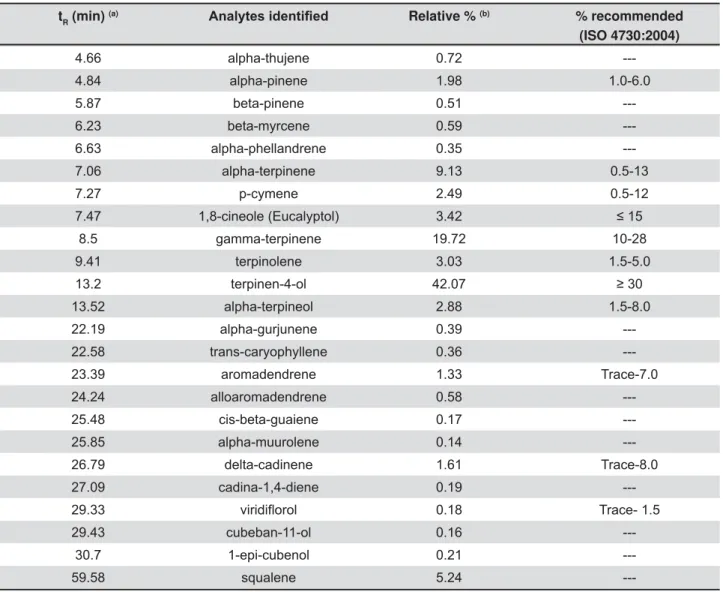
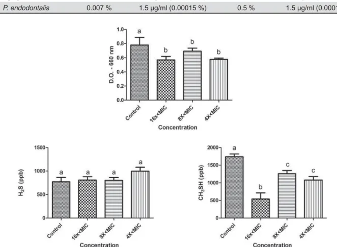
The effects of sub-MIC concentrations of tea tree oil and chlorhexidine on the growth and production of volatile sulphur gases (H2S and CH3SH) are shown P. gingivalis W83, and in Figures 3 and 4 for P. endodont alis. Concentrations tested were lower than the minimum inhibitory concentration (sub-MIC concentrations) and were
The tea tree oil reduced the growth of P. gingivalis
CH3SH at all concentrations tested (16x<MIC, 8x<MIC, and 4x<MIC). Curiously, the 16x<MIC concentration promoted less CH3SH production than higher concentrations (p<0.05). The production of H2S was not altered by the essential oil at any of the concentrations. In the presence of chlorhexidine, P. gingivalis W83 showed a reduction in growth at concentrations of 8x<MIC and 4x<MIC, and they were different from each other (p<0.05). Furthermore, it exhibited a reduction in CH3SH levels for all concentrations tested and in H2S for
Figure 1- Effects of sub-MIC concentrations of tea tree oil on growth (D.O. – 660 nm) and production of volatile sulphur gases (H2S and CH3SH) for P. gingivalis
considered when p<0.05 (ANOVA, Tukey test). Different letters represent differences among groups
Bacterial Strains M. alternifolia Chlorhexidine M. alternifolia Chlorhexidine
P. gingivalis W83 0.007 % 1.5 μg/ml (0.00015 %) 0.007 % 1.5 μg/ml (0.00015 %)
P. endodontalis 0.007 % 1.5 μg/ml (0.00015 %) 0.5 % 1.5 μg/ml (0.00015 %)
Figure 2- Effects of sub-MIC concentrations of chlorhexidine on growth (D.O. – 660 nm) and production of volatile sulphur gases (H2S and CH3SH) for P. gingivalis
considered when p<0.05 (ANOVA, Tukey test). Different letters represent differences among groups
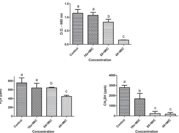
Figure 3- Effects of sub-MIC concentrations of tea tree oil on growth (D.O. – 660 nm) and production of volatile sulphur gases (H2S and CH3SH) for P. endodontalis
8x<MIC and 4x<MIC (p<0.05).
The microorganism P. endodont alis showed a reduction in growth in the presence of tea tree oil for concentrations 8x<MIC and 4x<MIC (p<0.05). Decreased gas production was observed for both H2S (16x<MIC, 8x<MIC, and 4x<MIC) and CH3SH (8x<MIC and 4x<MIC) (p<0.05). However, at the concentrations tested, chlorhexidine showed effects on P. endodont alis growth (p<0.05) but not on VSC production.
DISCUSSION
to halitosis development, many studies involving therapies for the treatment of halitosis have focused on the inhibition and reduction of VSC production. Antimicrobial therapy aims to enhance mechanical treatments and support host defences, reducing the development of microorganisms. Some studies have M. alt ernifolia against oral bacteria7,8,11,24; however, little is known about
its effects on VSC production. Here, we showed that M. alt ernifolia essential oil can reduce growth and VSC levels of P. gingivalis and P. endodont alis, even at sub-MIC concentrations.
GC-MS analysis of the tea tree oil used in this study showed that the composition of this oil was consistent with the International Standard ISO
of M. alt ernifolia oil such as quality requirements. The main components described by the ISO for M. alt er nifolia
20.
All of these compounds were found in the oil used in this study, except for limonene and sabinene. However, these two compounds are generally found in small quantities in tea tree oil (0.5% - 4% and trace – 3.5%, respectively), and their low levels
CG-MS analysis. In contrast, terpinen-4-ol and
activity, were found in satisfactory percentages4,13.
M. alt ernifolia have a mixture of components and their mechanisms of action are not completed elucidated. It is known that the combination of these different substances in the tea tree oil are capable of inducing loss of intracellular material, inhibition of respiration, and alterations in the homeostasis, leading to loss of bacterial membrane integrity and function4.
The tea tree oil showed antimicrobial activity: its MIC value was 0.007% for both bacteria, and MBC values were 0.5% for P. endodont alis and 0.007% for P. gingivalis. In a previous study, the MIC value found for P. gingivalis was 0.13% - 0.25%, and the MBC value was 0.13% - 0.5%22. Although MIC
Figure 4- Effects of sub-MIC concentrations of chlorhexidine on growth (D.O. – 660 nm) and production of volatile sulphur gases (H2S and CH3SH) for P. endodontalis
values differed between studies, MBC values were similar. Differences in MIC values may be due to different strains tested: in this study we used the W83 strain, while Takarada, et al.22 (2004) used
the ATCC 33277, 53977, Su63, and W50 strains. To the best of our knowledge, there are no previous studies in the literature showing the effects of M. alt ernifolia on P. endodont alis.
To evaluate the activity of M. alt ernifolia and chlorhexidine on the production of volatile sulphur compounds, sub-inhibitory concentrations of these substances were used. The sub-MIC concentrations of the tea tree oil affected the growth of both microorganisms, reducing the growth at higher concentrations (8x<, 4x<MIC, and 16x<MIC). Furthermore, at all concentrations tested, tea tree oil reduced CH3SH production in P. gingivalis and both H2S and CH3SH production in P. endodont alis. As not expected, the 16x<MIC concentration promoted a higher reduction of CH3SH than the 8x< and 4x<MIC concentrations. We believe that higher concentrations of tea tree oil (8x and 4x<MIC) could stress the bacteria and stimulate a little the VSC production comparing to the lower concentrations (16x<MIC). However, this hypothesis has to be
The CH3SH and H2S gases are the main VSCs in intra-oral halitosis1, and their toxicity is
associated with the development of periodontal disease15,16,29. Tea tree oil was able to inhibit the
growth of P. gingivalis W83 and P. endodont alis, and the production of VSCs. Thus, this essential oil is a promising substance for treating halitosis. Previously, M. alt er n if olia oil was found to be effective as one of the components of an antiseptic mouth rinse10
in our study, has demonstrated activity against a number of oral pathogens involved in periodontal disease and caries17.
Chlorhexidine is widely used in mouthrinses, causing membrane disruption and inhibition of proteolytic and glycosidic enzimes, leading to growth inhibition and cell death27. Despite being
a potent antimicrobial, it has certain side effects such as altered taste, mucosal desquamation, tooth
and a burning sensation in the oral mucosa19.
Compared with chlorhexidine, tea tree oil showed similar antimicrobial activity, promoting bactericidal and bacteriostatic effects at low concentrations. Thus, tea tree oil could be a good alternative to chlorhexidine in oral hygiene products, mainly mouthrinses. Therefore, the development of new mouthrinses containing tea tree oil and clinical studies testing these products are necessary.
In conclusion, M. a l t e r n i f o l i a oil showed antimicrobial activity against P. gin giv alis W83
and P. endodont alis, reducing the growth and the production of VSCs at sub-MIC concentrations, comparably to chlorhexidine. Future studies can be conducted focusing on the development of pharmaceutical products containing M. alt ernifolia oil for halitosis treatment.
ACKNOWLEDGEMENTS
This study was supported by grants from FAPESP – São Paulo Research Foundation (2009/06037-8 and 2009/14736-3).
REFERENCES
1- Akaji EA, Folaranmi N, Ashiwaju O. Halitosis: a review of the literature on its prevalence, impact and control. Oral Heal Prev Dent. 2014;124(4):297-304.
J Nat Sci Biol Med. 2013;4(1):14-23.
3- Campos Rasteiro VM, Costa AC, Araújo CF, Barros PP, Rossoni RD, Anbinder AL, et al. Essential oil of Melaleuca alt ernifolia for the treatment of oral candidiasis induced in an immunosuppressed mouse model. BMC Complement Altern Med. 2014;14:489. 4- Carson CF Riley TV. Antimicrobial activity of the major components of the essential oil of Melaleuca alt ernifolia. J Appl Bacteriol. 1995;78(3):264-9.
5- Clinical and Laboratory Standards Institute – CLSI. Methods for dilution antimicrobial susceptibility tests for bacteria that grow anaerobically. M11-A7. Wayne: CLSI; 2007.
6- Erovic Ademovski S, Lingström P, Winkel E, Tangerman A, Persson GR Renvert S. Comparison of different treatment modalities for oral halitosis. Acta Odontol Scand. 2012;70(3):224-33.
7- Groppo FC, Ramacciato JC, Simões RP, Flório FM Sartoratto A. Antimicrobial activity of garlic, tea tree oil, and chlorhexidine against oral microorganisms. Int Dent J. 2002;52(6):4337. 8- Hammer KA, Dry L, Johnson M, Michalak EM, Carson CF Riley TV. Susceptibility of oral bacteria to Melaleuca alt ernifolia (tea tree) oil in vit ro. Oral Microbiol Immunol. 2003;18(6):389-92. 9- Hinode D, Fukui M, Yokoyama N, Yokoyama M, Yoshioka M, Nakamura R. Relationship between tongue coating and secretory-immunoglobulin A level in saliva obtained from patients complaining of oral malodor. J Clin Periodontol. 2003;30(12):1017-23.
10- Hur MH, Park J, Maddock-Jennings W, Kim DO, Lee MS. Reduction of mouth malodour and volatile sulphur compounds in intensive care patients using an essential oil mouthwash. Phytother Res. 2007;21(7):641-3.
11- Karbach J, Ebenezer S, Warnke PH, Behrens E, Al-Nawas B. Antimicrobial effect of Australian antibacterial essential oils as alternative to common antiseptic solutions against clinically relevant oral pathogens. Clin Lab. 2015;61(1-2):61-8.
12- Lodhia P, Yaegaki K, Khakbaznejad A, Imai T, Sato T, Tanaka T, et al. Effect of green tea on volatile sulfur compounds in mouth air. J Nutr Sci Vitaminol (Tokyo). 2008;54(1):89-94.
13- Loughlin R, Gilmore BF, McCarron PA Tunney MM. Comparison of the cidal activity of tea tree oil and terpinen-4-ol against
Microbiol. 2008;46(4):428-33.
14- Luqman M. Systemic origin of halitosis : a review. Int J Clin Dent Sci. 2012;3(1):15-9.
16- Nakano Y, Yoshimura M, Koga T. Correlation between oral malodor and periodontal bacteria. Microbes Infect. 2002;4(6):679-83.
17- Park SN, Lim YK, Freire MO, Cho E, Jin D, Kook JK. Antimicrobial
cariogenic bacteria. Anaerobe. 2012;18(3):369-72.
18- Persson S, Claesson R Carlsson J. The capacity of subgingival microbiotas to produce volatile sulfur compounds in human serum. Oral Microbiol Immunol. 1989;4(3):169-72.
19- Prayitno S, Addy M. An in vit ro study of factors affecting the development of staining associated with the use of chlorhexidine. J Periodontal Res. 1979;14(5):397-402.
20- Sciarrone D, Ragonese C, Carnovale C, Piperno A, Dugo P, Dugo G, et al. Evaluation of tea tree oil quality and ascaridole: a deep study by means of chiral and multi heart-cuts multidimensional gas chromatography system coupled to mass spectrometry detection. J Chromatogr A. 2010;1217(41):6422-7.
21- Seemann R, Duarte da Conceição M, Filippi A, Greenman J, Lenton P, Nachnani S, et al. Halitosis management by the general dental practitioner - results of an International Consensus Workshop. Swiss Dent J. 2014;124(11):1205-11.
22- Takarada K, Kimizuka R, Takahashi N, Honma K, Okuda K,
against oral pathogens. Oral Microbiol Immunol. 2004;19(1):61-4. 23- Thaweboon S, Thaweboon B. Effect of an essential oil-containing mouth rinse on VSC-producing bacteria on the tongue. Southeast Asian J Trop Med Public Heal. 2011;42(2):456-62.
24- Thosar N, Basak S, Bahadure RN, Rajurkar M. Antimicrobial in vit ro study. Eur J Dent. 2013;7(1):1-7.
25- Van den Broek A, Feenstra L, de Baat C. A review of the current literature on management of halitosis. Oral Dis. 2008;14(1):30-9. 26- Van Leeuwen MP, Slot DE, Van der Weijden GA. Essential oils compared to chlorhexidine with respect to plaque and parameters
2011;82(2):174-94.
27- Van Leeuwen MP, Slot DE, Van der Weijden GA. The effect of an essential-oils mouthrinse as compared to a vehicle solution
meta-analysis. Int J Dent Hyg. 2014;12(3):160-7.
28- Xu X, Zhou XD, Wu CD. Tea catechin EGCg suppresses the mgl gene associated with halitosis. J Dent Res. 2010;89(11):1304-8. 29- Yaegaki K, Qian W, Murata T, Imai T, Sato T, Tanaka T, et al. Oral malodorous compound causes apoptosis and genomic
2008;43(4):391-9.