Article
0103 - 5053 $6.00+0.00*e-mail: [email protected]
#Present address: Departamento de Química, Mini-Campus, Bloco B, Sala B-13, Universidade Federal do Amazonas, 69077-000 Manaus-AM, Brazil
A New Guaiane Mannoside from a
Eutypa
-like Fungus Isolated from
Murraya paniculata
in Brazil
Afonso D. L. Souza,*
,#,aEdson Rodrigues-Filho,
aAntonia Q. L. Souza,
bFlavio Henrique-Silva
band José O. Pereira
caDepartamento de Química, Universidade Federal de São Carlos, CP 676, 13565-905 São Carlos-SP, Brazil
bDepartamento de Genética e Evolução, Universidade Federal de São Carlos, CP 676, 13565-905 São Carlos-SP, Brazil
cFaculdade de Ciências Agrárias, Universidade Federal do Amazonas, 69077-000 Manaus-AM, Brazil
Um fungo semelhante aos do gênero Eutypafoi isolado da madeira do caule de Murraya paniculata. Cultivado em meio líquido, o fungo produziu o novo sesquiterpeno do tipo guaiano (1R,4S,5S,7R,10R)-10-hidroxiguaianol-10-O- -manopiranosídeo e um diastereômero da harzialactona A, a 3-hidróxi-5-fenilmetil-(3S,5R)-tetraidrofuran-2-ona, obtida pela primeira vez como produto natural. As estruturas desses metabólitos foram elucidadas com base na análise dos seus respectivos dados espectroscópicos.
AEutypa-like fungus was isolated from the stems of Murraya paniculata. The fungus was cultivated in liquid medium and produced the new guaiane-type sesquiterpenoid (1R,4S,5S,7R,10R )-10-hydroxyguaianol 10-O- -mannopyranoside and the 3-hydroxy-5-phenylmethyl-(3S,5R )-tetrahydrofuran-2-one, a diastereomer of harzialactone A, obtained for the first time from a natural source. The structures of these metabolites were elucidated based on analysis of their spectroscopic data.
Keywords:Eutypa-like fungus, endophytic fungus, Murraya paniculata, guaiane mannoside, harzialactone A diastereomer
Introduction
Murraya paniculata (L.) Jack is a small Rutaceae tree
native from Asia and introduced in Brazil for ornamental purposes many years ago. A chemical study of these plant collected in São Paulo State, Brazil, showed a different chemical profile when compared with those studies using plant material collected in Asian countries.1 As part of a
program to investigate this apparent deviation of the plant secondary metabolism, endophytic microorganisms were seasonally gathered from all tissues of four specimens of M. paniculata plants.2 One of these microorganisms, Eupenicillium sp., produces a series of spiroquinazoline
alkaloids containing anthranilic acid residue in their structure. The influence of the presence of that and other fungi on the mentioned deviation has been discussed.3 Several strains of
aEutypa-like fungus were also isolated from M. paniculata
woods.2 Species of Eutypa are pathogenic to many plants.
For example, Eutypa lata is a grapevine fungal pathogen that
causes the “Eutyposis” or “dying-arm disease”, a perennial canker disease that is progressive over several years, and affects vineyards worldwide resulting in a serious economic problem in major wine grape producing areas. Several acetylenic phenols and chromene metabolites were isolated from this fungus and some of them have proved to be toxic.4-6
One of the Eutypa-like isolates, named as FED-3, has been
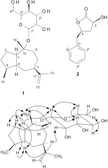
studied to characterize its metabolites. This paper report the identification of the new sesquiterpene (1R,4S,5S,7R,10R
)-10-hydroxyguaianol 10-O- -mannopyranoside (1), and
3-hydroxy-5-phenylmethyl-(3S,5R)-tetrahydrofuran-2-one
(2), isolated from the Eutypa-like strain FED-3.
Results and Discussion
Compound1 was isolated as viscid oil. Its IR spectrum
3400 cm-1, C-O at 1065 and 1019 cm-1, and C=C at 1643
cm-1. ESI mass spectrum in positive ion mode revealed a
low intensity ion at m/z 385 ([M + H]+), and intense ion
peaks at m/z 407 ([M + Na]+), 423 ([M + K]+) and 181
([hexose + H]+). In negative ion mode, ion peaks at m/z 383
([M – H]−), 419 ([M + Cl]−) and 179 ([hexose – H]−) were
recorded. The 1H NMR spectrum of 1displayed signals for
a methyl doublet at G 0.91 (J = 6.9 Hz), a methyl singlet
atG 1.21, a broad olefinic methyl singlet at G 1.68, two vinylic hydrogen multiplets at G 4.55 and 4.67, several low-resolution multiplets at G 1.2-2.6 accounting for 14 protons, and characteristic sugar resonances at G 4.74 (1H,
J= 0.7 Hz) and 3.2-3.9. A total of 21 carbon signals were
detected in the 13C NMR spectrum, including those for a
hexose moiety and an isopropenyl double bond (G 108.5 and 153.7). Combined analysis of these and other NMR, IR and ESI mass spectral data are in agreement with the molecular formula C21H36O6. Furthermore, the tracking of correlations in the 1H_1H COSY, HSQC and HMBC revealed 1 as a
guaiane-type sesquiterpenoid with a hexose moiety at C-10 (Table 1). This hexose was determined as mannose by the coupling constants of 0.7 Hz between H-1´ at G 4.74 (d)
and H-2´ at G 3.84 (dd) and of 3.2 Hz between H-2´ and
H-3´ at G 3.46 (dd), while H-4´ at G 3.57 (t) is coupled
with H-3´ and H-5´ at G 3.19 (ddd) by the same coupling
constant of 9.5 Hz. Further, it was noted the H-1´, H-3´ and H-5´ atoms with NOESY correlations one each other and the absence of such correlation type between the H-2´ and H-4´ atoms, ratifying the axial orientation for all hydrogen atoms at the hexose group, except for H-2´ (Figure 1). The linkage of the mannose with the aglycone moiety was determined by the anomeric H-1´ HMBC correlation with a tertiary carbinolic carbon at G 83.3. Hydrogens of the methyl group at G 1.21 (s) are also in HMBC correlation
with this carbon and with a methylene and a methine ones, at G 31.4 and 55.5, respectively. According to the COSY, the hydrogens connected to these carbons do not couple to each other, shown that the mannose is linked to a carbon of the aglycone of 1 that is connected to a methyl, a methylene and methine carbons. For characterize this aglycone as a guaiane-type structure it was fundamental the
1H_1H COSY correlation. First of all, it was noted that the
hydrogen at G 2.30, with HSQC correlation to the methine carbon at G 55.5, couples with other methine hydrogen at
G 2.12 and two other hydrogens at G 1.54 and 1.72, linked to a methylene carbon at G 26.9. The hydrogen at G 1.54 couples also with other methylene one at G 1.29, which is geminal with another superposed with that at G 1.72 and linked to a carbon at G 31.5. A methine hydrogen at G 2.03, connected to a carbon at G 40.0 (HSQC), couples with these later methylene hydrogens as well with hydrogens
of a methyl group at G 0.91, which are linked to a carbon atG 16.7 (HSQC) and have HMBC correlations with the carbon atoms at G 31.5 and 40.0, as also with a carbon atom at G 47.2. This later is connected to the mentioned methine hydrogen at G 2.12 coupled to that other at G 2.30, as established above. In this way the C-1 to C-5 pentacyclic ring system of 1 was completely characterized, having a
methyl group (C-15) linked to the C-4. The heptacyclic ring system of 1 was typified by similar way, as one can
deduce through correlation data in Table 1.
The relative stereochemistry of the guaiane moiety of 1
was attributed by the NOESY and nOe space interactions (Figure 1). The most important ones are that among H-1, H-4 and H-5, that become apparent the cis-fusion of the guaiane
rings as well as the -orientation of the C-15 methyl group at C-4; between H-5 and H-7, by what the C-11 to C-13 propenyl group at C-7 has -orientation; as well between H-7 and H-2´, more compatible with the mannose linked in -orientation to C-10. Therefore, the assignment of all the hydrogen and carbon signals as well as their respective
Figure 1. NOESY and nOe correlations for compound 1. Arrows with only
correlations (Table 1) revealed 1 as the (1R,4S,5S,7R,10R
)-10-hydroxyguaianol 10-O- -mannopyranoside. While
the opposite configuration is possible, the suggested one was made considering the literature, where guaiane-type sesquiterpenoids have mostly the C-7R configuration.
This was reinforced by papers shown experimental data of biosynthesis of guaianolides from (+)-germacrene A, that is a C-7R result of the farnesyl diphosphate cyclization.7,8
Compound1 is the first sesquiterpene with sugar moiety
isolated from fungi. Furthermore, its novelty is in both the attachment position and type of the sugar moiety, since any sesquiterpenoid with mannose has not been described yet. All reported guaiane-type sesquiterpenoids with sugar moiety were obtained from plants, almost all of them
bearing a glucose unit and presenting in many cases a -lactone involving the 6, 7, 11 and 12 carbons.
The IR spectrum of 2 displays typical absorptions for
hydroxyl and -lactone carboxyl groups at 3352 and 1770 cm-1, respectively. Its full scan ESI mass spectrum revealed
ion peaks at m/z 193 ([M + H]+), 215 ([M + Na]+) and
231 ([M + K]+). These data associated to the NMR data
of 2 are coherent with the molecular formula C11H12O3 and a structure with an aromatic and a lactone ring. From NMR data analysis, 2 was identified as 3-hydroxy-5-phenylmethyltetrahydrofuran-2-one. A emphasis can be give to HMBC correlations of the methylene hydrogen atoms H-4 (G 1.90 and 2.53) with the lactone carbonyl carbon C-2 (G 176.9) and the carbinol carbon atoms C-3 and
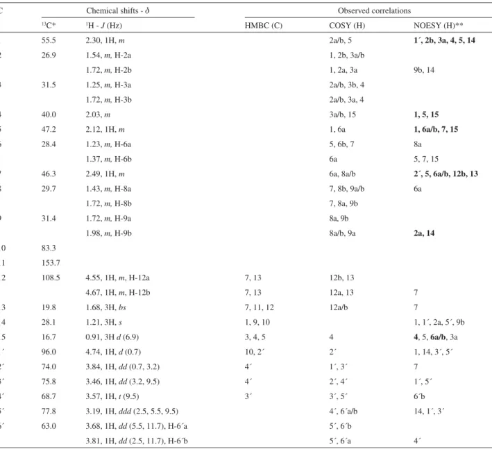
Table 1. NMR data for compound 1 (1R,4S,5S,7R,10R)-10-hydroxyguaianol 10-O- -mannopyranoside (methanol-d4)
C Chemical shifts - G Observed correlations
13C* 1H - J (Hz) HMBC (C) COSY (H) NOESY (H)**
1 55.5 2.30, 1H, m 2a/b, 5 1´, 2b, 3a, 4, 5, 14
2 26.9 1.54,m,H-2a 1, 2b, 3a/b
1.72,m,H-2b 1, 2a, 3a 9b, 14
3 31.5 1.25,m,H-3a 2a/b, 3b, 4
1.72,m,H-3b 2a/b, 3a, 4
4 40.0 2.03,m 3a/b, 15 1, 5, 15
5 47.2 2.12, 1H, m 1, 6a 1, 6a/b, 7, 15
6 28.4 1.23,m,H-6a 5, 6b, 7 8a
1.37,m,H-6b 6a 5, 7, 15
7 46.3 2.49, 1H, m 6a, 8a/b 2´, 5, 6a/b, 12b, 13
8 29.7 1.43,m,H-8a 7, 8b, 9a/b 6a
1.72,m,H-8b 7, 8a, 9b
9 31.4 1.72,m,H-9a 8a, 9b
1.98,m,H-9b 8a/b, 9a 2a, 14
10 83.3
11 153.7
12 108.5 4.55, 1H, m, H-12a 7, 13 12b, 13
4.67, 1H, m, H-12b 7, 13 12a, 13 7
13 19.8 1.68, 3H, bs 7, 11, 12 12a/b 7
14 28.1 1.21, 3H, s 1, 9, 10 1, 1´, 2a, 5´, 9b
15 16.7 0.91, 3H d(6.9) 3, 4, 5 4 4, 5, 6a/b, 3a
1´ 96.0 4.74, 1H, d(0.7) 10, 2´ 2´ 1, 14, 3´, 5´
2´ 74.0 3.84, 1H, dd(0.7, 3.2) 4´ 1´, 3´ 7
3´ 75.8 3.46, 1H, dd(3.2, 9.5) 4´ 2´, 4´ 1´, 5´
4´ 68.7 3.57, 1H, t(9.5) 3´ 3´, 5´ 6´b
5´ 77.8 3.19, 1H, ddd(2.5, 5.5, 9.5) 4´, 6´a/b 14, 1´, 3´
6´ 63.0 3.68, 1H, dd(5.5, 11.7), H-6´a 5´, 6´b
3.81, 1H, dd(2.5, 11.7), H-6´b 5´, 6´a 4´
C-5 (G 68.4 and 77.1, respectively); of the benzyl methylene hydrogen atoms (G 2.90 and 3.09) with the C-4 (G36.5), C-5 and aromatic carbons C-1´ and C-2´/6´ (G 135.5 and 129.3, respectively). The other HMBC correlations as well the COSY ones are all in accord with the structure proposed above for 2. A known metabolite having a such structure is the harzialactone A, a natural product from theTrichoderma harzianum OUPS-N115, a strain isolated
from the sponge Halichondria okadai.9 The stereoisomers
of harzialactone A were obtained only by synthesis.10
By comparison with the NMR data of all those isomers, including harzialactone A, the (3R,5R) isomer,102 NMR spectra were in agreement with either the (3R,5S) or (3S,5R)
enantiomer data, of which the 1H and 13C NMR shifts
for the atoms at the 3, 4 and 5 positions are perceptively unlike from the respective ones in the other stereoisomers. The specific rotation of 2indicated it as the
3-hydroxy-5-phenylmethyl-(3S,5R)-tetrahydrofuran-2-one,10 which is
novel as natural product.
Experimental
General
Optical rotations were measured in MeOH or CHCl3, on a PERKIN-ELMER 241 polarimeter, using a 1 dm path length cell. IR spectra were obtained in a BOMEM MB-102 spectrophotometer, in KBr pellets. NMR spectra, including1H-1H-COSY, HSQC, HMBC, NOESY and nOe
experiments, were recorded on a Bruker DRX spectrometer, operating at 400 MHz for 1H and 100 MHz for 13C, using
d-chloroform or d-methanol as solvent and TMS as the internal standard. ESI-MS data were measured in a low-resolution MICROMASS QUATRO-LC instrument.
Fungus collection and identification
Using a known procedure,11 the fungus FED-3 was
isolated from the wood of the stem of Murraya paniculata
collected in Águas da Prata, São Paulo State, Brazil. The fungus appeared after 30-45 days from pieces of the stem on Petri dish containing PDAY - a potato-dextrose-agar medium with 0.2% yeast extract. Hyphal tips were transferred to other PDAY Petri dish. During the first week growing FED-3 typically release a stink. Its asci and conidia as well as the ITS-1 and ITS-2 rDNA sequences, with 83% sequence identity to Eutypa lata, suggested FED-3
belonging to Eutypagenus. More investigation, however,
is necessary for determine the correct genus and, as well as possible the species of FED-3. The fungus has been studied both for taxonomical classification and metabolites
identification. It is deposited (number LaBioMMi-204) at the Laboratório de Bioquímica Micromolecular de Microorganismos - LaBioMMi - of the Departamento de Química at Universidade Federal de São Carlos, São Carlos, Brazil.
Cultivation of the fungus and metabolites isolation
After about 30 days growing, 3-5 pieces of 8 mm in diameter of the PDAY culture containing FED-3’s mycelium were inoculated in 48 Erlenmeyer flasks (250 mL) each containing 125 mL of PDY (potato-dextrose with 0.2% yeast extract) and incubated at room temperature (25 ± 3 °C) on an opened shaker (130 rpm) during 21 days. The mycelium was separated and the cultured medium, after addition of 2% isopropanol, was partitioned with ethyl acetate. The dried organic extract (180 mg) was fractionated by CC over silica gel with gradient elution with hexane, hexane-ethyl acetate (9.5:5, 9:1, 8:2, 7:3, 4:6 and 0:10), ethyl acetate-methanol (9:1, 6:4 and 0:10) and methanol-water (9:1 v/v). The metabolites1(2 mg; from hexane-ethyl acetate 4:6 eluent)
and2 (4 mg; from ethyl acetate-methanol 6:4 eluent) were
then purified by preparative TLC over Silica gel, eluted with hexane-ethyl acetate-acetone-methanol 10:0:10:0.1 and 65:15:15:5, respectively.
(1R,4S,5S,7R,10R)10Hydroxyguaianol 10O -mannopyranoside (1)
Viscid dark yellow oil; [ ]25
D -22.3 (c 0.0017, MeOH);
IR (KBr) max/cm-1: 3400, 2934, 2870, 1643, 1455, 1380,
1065, 1019, 909; ESIMS, daughter ions of m/z 383, 12 eV:
(R = aglycone) m/z (%) 383 ([M – H]–, 72), 179 ([M – H
– R]–, 37), 161 ([M – H – R – H
2O]–, 16), 149 ([M – H –
R – CH2O]–, 7), 143 ([M – H – R – 2H
2O]–, 9), 131 ([M
– H – R – CH2O – H2O]–, 7), 119 ([M – H – R – 2CH 2O]–,
100), 113 ([M – H – CH2O – 2H2O]–, 11), 107 ([M – H –
R – 4H2O]–, 8), 101 ([M – H – R – 2CH
2O – H2O]–, 22),
89 ([M – H – R – 3CH2O]–, 58), 71 ([M – H – R – 3CH 2O
– H2O]–, 10); NMR spectra see Table 1.
3-Hydroxy-5-phenylmethyl-(3S,5R)-tetrahydrofuran-2-one (2)
Crystalline solid; [ ]25
D -8.0 (c 0.0032, CHCl3)
{literature: [ ]25
D -8.5 (c 0.0011, CHCl3)};10IR (KBr) max/
cm-1: 3352, 1770, 1451, 1197, 1129, 990; ESIMS, daughter
ions of m/z 193, 10 eV: m/z (%) 193 ([M + H]+, 4), 157 ([M
+ H – 2H2O]+, 8), 147 ([M + H – HCO
2H]+, 100), 145 ([M
+ H – H2O – CH2O]+, 9), 129 ([M + H – HCO
2H – H2O]+,
12), 115 ([M + H – CH3CO2H – H2O]+, 17), 91 ([M + H
– -lactone ring]+, 22); NMR spectra of 2 were in complete
Acknowledgments
The authors are gratefull to Fundação de Amparo à Pesquisa do Estado de São Paulo (FAPESP) and to Conselho Nacional de Desenvolvimento Científico e Tecnológico (CNPq) for financial supports. A.D.L.S thanks Coordenação de Aperfeiçoamento de Ensino Superior (CAPES) for student fellowship.
Supplementary Information
1H NMR, 13C NMR, HSQC and HMBC spectra of 1
are available free of charge at http://jbcs.sbq.org.br, as PDF file.
References
1. Ferracin, R. J.; PhD Thesis, Universidade Federal de São Carlos, Brazil, 1996.
2. Souza, A. Q. L.; PhD Thesis, Universidade Federal de São Carlos, Brazil, 2006.
3. Rodrigues-Filho, E.; Barros, F. A. P.; Biochem. Syst. Ecol.2005,
33, 257.
4. Molyneux, R. J.; Mahoney, N.; Bayman, P.; Wong, R. Y.; Meyer, K.; Irelan, N.; J. Agric. Food Chem. 2002,50,1393.
5. Mahoney, N.; Lardner, R.; Molyneux, R. J.; Scott, E. S.; Smith, L. R.; Schoch, T. K.; Phytochemistry2003,64, 475.
6. Kim, J. H.; Mahoney, N.; Chan, K. L.; Molyneux, R. J.; Campbell, B. C.; Curr. Microbiol.2004,49, 282.
7. de Kraker, J.-W.; Franssen, M. C. R.; Joerink, M.; de Groot, A.; Bouwmeester, H. J.; Plant Physiol.2002,129, 257. 8. de Kraker, J.-W.; Franssen, M. C. R.; de Groot, A.; König, W.
A.; Bouwmeester, H. J.; Plant Physiol.1998,117, 1381. 9. Amagata, T.; Usami, Y.; Minoura, K.; Ito, T.; Numata, A.; J.
Antibiot.1998,51, 33.
10. Mereyala, H. B.; Joe, M.; Gadikota, R. R.; Tetrahedron: Asymmetry2000,11, 4071.
11. Souza, A. Q. L.; Souza, A. D. L.; Astolfi-Filho, S.; Pinheiro, M. L. B.; Sarquis, M. I. M.; Pereira, J. O.; Acta Amazônica2004, 34, 185.
Received: October 25, 2007
Web Release Date: August 11, 2008
Supplementary Information
0103 - 5053 $6.00+0.00
A New Guaiane Mannoside from a
Eutypa
-like Fungus Isolated from
Murraya paniculata
in Brazil
Afonso D. L. Souza,*
,#,aEdson Rodrigues-Filho,
aAntonia Q. L. Souza,
bFlavio Henrique-Silva
band José O. Pereira
caDepartamento de Química, Universidade Federal de São Carlos, CP 676, 13565-905 São Carlos-SP, Brazil
bDepartamento de Genética e Evolução, Universidade Federal de São Carlos, CP 676, 13565-905 São Carlos-SP, Brazil
cFaculdade de Ciências Agrárias, Universidade Federal do Amazonas, 69077-000 Manaus-AM, Brazil
Figure S1. 1H NMR spectrum of (1) (methanol-d4, TMS).
*e-mail: [email protected]
#Present Address: Departamento de Química, Mini-Campus, Bloco B,
Figure S2. 13C NMR spectrum of (1) (methanol-d4, TMS).