Article
0103 - 5053 $6.00+0.00
*e-mail: [email protected]
#Dedicated to Prof. Vanderlan da Silva Bolzani, for her outstanding contributions to the development of natural product chemistry and related sciences in Brazil.
Antibacterial Modified Diketopiperazines from two Ascidians of the Genus Didemnum
#Miriam H. Kossuga,a Simone P. Lira,a Shayna McHugh,a Yohandra R. Torres,a,b Bruna A. Lima,c Reginaldo
Gonçalves,c Katyuscya Veloso,d Antonio G. Ferreira,d Rosana M. Rochae and Roberto G. S. Berlinck*,a
aInstituto de Química de São Carlos, Universidade de São Paulo, CP 780, 13560-970 São Carlos-SP, Brazil
bDepartamento de Química, Universidade Estadual do Centro-Oeste, Rua Camargo Varela de Sá, 03, 85040-080 Guarapuava-PR, Brazil
cDepartamento de Diagnóstico Oral, Faculdade de Odontologia de Piracicaba, Universidade Estadual de Campinas, CP 52, 13414-018 Piracicaba-SP, Brazil
dDepartamento de Química, Universidade Federal de São Carlos, 13565-905 São Carlos-SP, Brazil
eDepartamento de Zoologia, Setor de Ciências Biológicas, Universidade Federal do Paraná, Centro Politécnico, Jardim das Américas s/n, CP 19020, 81531-990 Curitiba-PR, Brazil
A investigação química do extrato bruto de uma ascidia do gênero Didemnum levou ao isolamento das dicetopiperazinas modificadas rodriguesinas A (1) e B (2) na forma de uma mistura
de homólogos, os quais puderam ser identificados pela análise de seus dados espectroscópicos inclusive experimentos MS/MS. A investigação de uma segunda ascídia do gênero Didemnum forneceu a N-acetil-rodriguesina A (3) e a N-acetil-rodriguesina B (4). A configuração absoluta
dos compostos 1 e 2 pode ser estabelecida por hidrólise e análise de Marfey e por comparação com dados da literatura do composto 3, previamente obtido como produto de síntese. A mistura
de 1 e 2 apresentou atividade antibiótica moderada contra um isolado clínico de Streptococcus mutans, contra S. mutans UA159 e S. aureus ATCC6538.
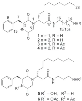
The chemical investigation of the crude extract of an ascidian of the genus Didemnum led to the isolation of the modified diketopiperazine rodriguesines A (1) and (2) as a mixture of homologues,
which could be identified by analysis of spectroscopic data including MS/MS experiments. The investigation of a second Didemnum sp. led to the isolation of N-acetyl-rodriguesine A (3) and
N-acetyl-rodriguesine B (4). The absolute configuration of compounds 1 and 2 could be established by hydrolysis and Marfey’s analysis and comparison with literature data reported for compound 3, previously obtained as a synthetic product. The mixture of 1 and 2 displayed moderate antibiotic
activity against a clinical isolate of Streptococcus mutans and against S. mutans UA159 and Staphylococcus aureus ATCC6538.
Keywords: ascidian, marine, diketopiperazine, absolute configuration, antibiotic
Introduction
Ascidians, or sea-squirts, are cosmopolitan, exclusively marine invertebrates, which constitute a rich source of biologically active secondary metabolites.1 It has been estimated that ca. 85% of ascidian natural products are
derived from amino acids, while the remaining 15% are derived from polyketide and/or terpenoid pathways.2
Ascidians of the family Didemnidae are recognized as a particularly unique source of modified peptides and alkaloids.3 These include the well known tamandarins,4 didemnins, aplidine and related compounds,5 which display potent antitumor and immunosupressive activities, as well as many bioactive alkaloids such as the G2 cell cycle checkpoint inhibitors granulatimide and isogranulatimide.6
encrusting, thin and soft bodied colonies. Two such species were collected during 1999, as red Didemnum spp.
The two Didemnidae species gave crude extracts which displayed antituberculosis and cytotoxic activities. The investigation of these extracts led to the isolation of four modified diketopiperazines, which are the subject of the present report.
Results and Discussion
The hexane-defated MeOH extract of the ascidian
Didemnum sp. (sample BA99ASCI-05) presented
aromatic, nitrogen-bearing derivatives indicated by TLC and 1H NMR analyses. The MeOH extract was separated by chromatography on Sephadex LH-20 and C18 reversed-phase columns. Further HPLC separations using C18-phenyl or cyanopropyl silica columns did not provide rodriguesines A (1) and B (2) as pure compounds, even using TFA, NH4Cl or phosphoric acid buffers in the eluents. Subsequent analysis of 2D NMR and MS/MS analysis indicated that rodriguesines A (1) and B (2) were obtained as an inseparable mixture from the Didemnum sp. sample
BA99ASCI-05.
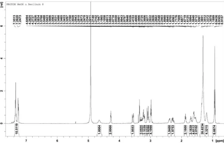
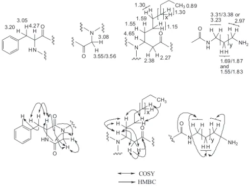
NMR analysis of the fraction containing 1 and 2 indicated the presence of a phenylalanine residue (d Hα
4.27, d CH2-β 3.05 and 3.20, d aromatic Hs 7.24-7.36), a glycine residue (d 3.08 and two doublets at d 3.55/3.56), a linear alkyl β-amino acid and a mono-acylated diamine moiety. These structural features were evident by analysis
of the HSQC, COSY and HMBC spectra, of which key correlations are shown in Figure 1. The linear alkyl β-amino acid moiety was assigned from the carbonyl substituted methylene CH2-18 (d 2.27 and 2.38) coupled in the COSY spectrum to the broad methine signal at d
4.65, itself correlated with a methylene group atd 1.55 and 1.59 (CH2-20) substituted with the alkyl chain (multiplet at d 1.30). The diamino moiety was assigned from the amino-substituted methylene at d 2.97 (CH2-14) which showed couplings with methylene signals at d 1.87 and d
1.69. This methylene (CH2-15 in 1)”. showed a correlation with a methylene group at d 3.31 and 3.28 (CH2-16), while the methylene at d 1.69 (CH2-15 in 2) was coupled to a methylene group at d 1.55 (CH2-15a in 2), itself attached to a methylene at d 3.23 (CH2-16 in 2). Correlations between the methylene at d 3.28 and 3.31 (CH2-16), as well as the methylene CH2-18 (d 2.27 and 2.38), with the carbonyl groups at d 172.9 and 173.6 established the connectivity between the diamino moiety and the β-amino acid residue. Couplings between H-3 and C-2 (d 168.5/168.4) and C-5 (d168.6/168.4), as well as between CH2-6 with the same carbonyl carbons, allowed us to assign the piperazine-2,5-dione moiety. The junction between the diketopiperazine residue with the β-amino acid moiety was established by a correlation observed between the glycyl doublet hydrogens of 1 and 2 at d 3.55/3.56 with C-19.
Considering the planar structures assigned for 1 and 2, a search in MarinLit and SciFinder led us to discover that these compounds were closely related to etzionin (5),
previously isolated from the ascidian Didemnum rodriguesi.
However, the spectroscopic data of etzionin was not very informative, since only broad, poorly-defined NMR signals have been reported for 5.8 A distinctive feature between 1 and 2 and etzionin (5) was that no evidence was obtained in the infrared spectrum of 1 and 2 for the presence of the unusual etzionin hydroxamate group. We observed only a broad νN-H band at 3283 cm-1 but not a band associated with a νO-H at higher wavenumbers. Additionally, since the analysis of the acyl monosubstituted diamino moiety in the mixture of 1 and 2 indicated the presence of two amide carbonyls and two diamine alkyl chains, we suspected that two diamine homologues constituted the fraction isolated from the Didemnum sp. BA99ASCI-05. Indeed, analysis
by HRMS/MS showed that the fraction containing 1 and 2 displayed two quasi-molecular ions [M+H]+ at m/z 459.3335 (C26H43N4O3) and m/z 473.3492 (C27H45N4O3),
which, upon fragmentation, gave an identical fragment ion at m/z 385.2500 (C23H33N2O3) corresponding to the lost of
C3H10N2 (1,3-diaminopropane) from 1 and of C4H12N2 (1,4-diaminobutane) from 2 (Figure 2). The analysis of the MS/MS spectrum also indicated the formation of the corresponding oxazepine and oxazine ions at m/z 238.2181 and 252.2352,
respectively, as well as the formation of a diketopiperazinium at m/z 343.2416 (Figure 2). Therefore, the complete planar
structure of rodriguesine A (1) and B (2) was confirmed. Further support for the structures assigned for both 1 and 2 could be obtained after acetylation of this mixture and separation by HPLC. This procedure gave compounds 3 and 4 (see below) of which compound 3 showed NMR data very similar to that of bis-acetyl etzionin (6).8
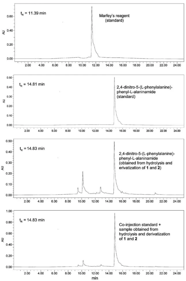
In order to establish the absolute configuration of the phenylalanine residue, the mixture of 1 and 2 was subjected to hydrolysis followed by derivatization with
Marfey’s reagent (1-fluoro-2,4-dinitrophenyl-5-L-alanine
amide) and LC-PDA-MS analysis. Only L-phenylalanine
was detected. Moreover, since the absolute configuration of etzionin (5) was established after the total synthesis of its 4-desoxy-N-acetyl derivative (3),9 we decided to
acetylate the mixture of 1 and 2 in order to separate both
N-acetyl derivatives and compare the specific rotation of
4-desoxy-N-acetyl etzionine (3) derived from 1 with the
literature value reported for synthetic 3. The acetylated mixture of 3 and 4 could be separated by C18 HPLC. Compound 3, obtained after acetylation of 1, showed [α]D−37.0 (c 0.026, CHCl3), very close to the value reported
for synthetic (-)-(3S,19R)-3, [α]D −53.1 (c 0.06, CHCl3).
The 1H NMR spectrum of 3 obtained from the acetylation of 1 was identical to that of synthetic (-)-(3S,19R)-3.9 Since
the absolute configuration of the phenylalanine residue in 3
derived from the acetylation of natural 1 is S, the absolute
configuration of C-19 must be R. After acetylation and
purification, compound 4 obtained from 2 showed [α]D −52 (c 0.04, CHCl3). Therefore, both rodriguesines A (1) and B
(2) have the 3S, 19R absolute configuration.
Sample BA99ASCI-03 of the second Didemnum species
gave, after a similar isolation procedure, small quantities of 3 and 4 as pure compounds. Analysis of the spectroscopic data recorded for N-acetyl rodriguesine A (3) clearly assigned
the presence of an acetyl group (C=O at dC 173.4; CH3 at dC
22.7 and dH 1.92), corroborated by the HRESIMS analysis which showed a [M+H]+ quasi-molecular ion at m/z 501.3483 (C28H45N4O4), indicating an additional C2H2O fragment compared to the formula of 1. Analysis of the NMR data of the diamino moiety in 3 showed a coupling between the methylenes at d 3.18 (t, 8.4 Hz, 6H, CH2-14 and CH2-16) and two carbonyl groups at dC 173.4 and 172.7. Since the methylene CH2-18 (d 2.20 and 2.32) confirmed a long-range coupling with the carbonyl group at dc 172.7 (C-17), and the methyl acetyl group showed a long range coupling with the carbonyl atdC 173.4, the acetyl group could be placed at the diamine terminus of compound 3. Analogous MS and NMR data recorded for N-acetyl rodriguesine B (4)
showed a quasi-molecular ion [M+H]+ at m/z 515.3639 (C29H47N4O4) and quite similar NMR data for the diamide moiety (Tables 1 and 2), except for the presence of an additional methylene group (CH2-15a at dH 1.50 and dC 27.8) in the diamino moiety. Comparison of 1H, 13C NMR and MS data recorded for natural 3 and 4 with data recorded for 3 and 4 derived from the acetylation of 1 and 2 proved them to be identical compounds. Therefore, the structure of the natural, minor N-acetylated compounds 3 and 4, could
be established. Compounds 3 and 4 should have the same absolute configuration as compounds 1 and 2 since their [α]D, HPLC tR, 1H and MS data are practically identical to 3 and 4 obtained from the acetylation of the mixture of 1 and 2.
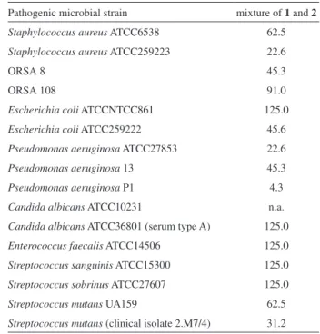
The mixture of 1 and 2 was evaluated in cytotoxic, antituberculosis and antibiotic bioassays against a panel of pathogenic bacteria. Surprisingly, rodriguesines A 1 and B 2 did not present cytotoxic or antituberculosis activities. The mixture of 1 and 2 was weakly active against the majority of the pathogen microbial strains tested (Table 3), except against Pseudomonas aeruginosa P1 (MIC
at 4.3 µg mL-1), obtained from a hospital environment. Interestingly, the mixture of 1 and 2 proved to be more active against antibiotic-resistant strains than against standard ATCC or NTCC strains (Table 3). The strains
Streptococcus mutans clinical isolate 2.M7/4, S. mutans
UA159, Staphylococcus aureus ATCC6538, Escherichia coli NTCC861, Enterococcus faecalis ATCC14506, Streptococcus sanguinis ATCC15300, S. sobrinus
ATCC27607 and Candida albicans ATCC36801(serum
type A) are all associated with muco-bucal diseases.
Conclusion
We reported here the isolation of four new modified diketopiperazines from two ascidians of the genus
Didemnum. The mixture of 1 and 2 displayed moderate
antibiotic activity against several human pathogenic bacteria.
Experimental
General experimental procedures
Optical rotations were measured on a Perkin Elmer 341 MC polarimeter at 20 oC. IR spectra (film on Si plate) were recorded on a FT-IR Bomem MB102 infrared Table 1. 1H data for compounds 1 and 2, 3 and 4 in CD
3OD at 400 MHz [d, multiplicity (J in Hz)]
Position 1 and 2 3 4
3 4.27 (t, 5.0) 4.27 (t, 6) 4.22 (t, 6.5)
6
3.08 (d, 17), 3.55/3.56 (2d, 17 Hz each)
3.00 (d, 22), 3.50 (d, 22)
2.99 (d, 22), 3.50 (d, 22)
7 3.05 (m), 3.20 (m) 3.02 (dd, 5, 17), 3.16 (m) 3.01 (dd, 5, 17), 3.16 (m)
9/13 7.24 (m) 7.21 (m) 7.20 (m)
10/12 7.36 (m) 7.31 (m) 7.28 (m)
11 7.35 (m) 7.30 (m) 7.24 (m)
14 2.97 (t, 7) 3.18 (t, 8.4) 3.16 (m)
15 1.87 (qui, 7) and 1.69
(m) 1.65 (qui, 8.4) 1.50 (m)
15a 1.55 (m) - 1.50 (m)
16 3.28 and 3.31/3.23 3.18 (t, 8.4) 3.16 (m)
18 2.27 (dd, 7, 12), 2.38 (m) 2.20 (dd, 9, 17), 2.32 (dd, 9, 17) 2.18 (dd, 9, 17), 2.32 (dd, 9, 17)
19 4.65 (br) 4.60 (br) 4.57 (br) 20 1.55 (m), 1.59 (m) 1.52 (m) 1.53 (m)
21 1.18 (m) 1.14 (m) 1.14 (m)
22 1.30 (m) 1.26 (m) 1.26 (m)
23 1.30 (m) 1.26 (m) 1.26 (m)
24 1.30 (m) 1.26 (m) 1.26 (m)
25 1.30 (m) 1.26 (m) 1.26 (m)
26 1.30 (m) 1.26 (m) 1.26 (m)
27 1.30 (m) 1.28 (m) 1.28 (m)
28 0.93 (t, 7) 0.89 (t, 9) 0.89 (t, 8)
spectrometer. The NMR spectra were recorded on a Bruker ARX 9.4 T instrument, operating at 400.35 MHz for 1H and 100.10 MHz for 13C, respectively. All NMR spectra were obtained at 25 oC using TMS as an internal reference. Solvents used for extraction and column chromatography were glass distilled prior to use. HPLC-grade solvents were utilized without further purification in HPLC separations. TLC analyses were performed with precoated TLC sheets of Si gel on polyester, eluting with different mixtures of MeOH in CH2Cl2. Plates were observed under a UV lamp (λmax 254 and 365 nm). Semi-preparative HPLC separations were performed with a Waters 600 quaternary pump and a double beam model 2487 UV detector monitored by Waters Millenium 32. High resolution mass spectra obtained for 1 and 2 were recorded on a Micromass Q-ToF Micro, in ES+ mode, using the following experimental conditions:
capillary voltage 3.0 kV; sample cone 42.0 V; sample infused at 10 µL min-1 in 1: 1 MeOH/H
2O. Accurate mass measurements were made using leucine enkephalin as an internal reference at [M+H]+ 556.2771. The MS/MS experiments were recorded using with a range of collision energies from 20 to 30 eV. High resolution mass spectra obtained for compounds 3 and 4 were recorded on a Bruker Daltonics equipment (UltrO-ToF, MA, USA). Samples (0.5 µg mL-1) dissolved in MeOH/H
2O 1:1 were introduced in the electrospray source at 5 µL min-1 with a direct infusion pump. MS/MS and MSn experiments were performed using standard isolation and excitation procedures. Nitrogen was used as nebulising and collision gas with the collision energy set at 4 eV. An accurate-mass calibration was obtained with a post acquisition application of a calibration created from the MS/MS of monensin A [M-18+Na]+ under the same CID and cell conditions. The collision energy was adjusted until the intensity of the parent ion was set between 5 and 20% relative to the base peak. Capillary voltage was set to 3500 V.
Animal material
The ascidians Didemnum sp. BA99ASCI-03 and Didemnum sp. BA99ASCI-05 were collected in August/
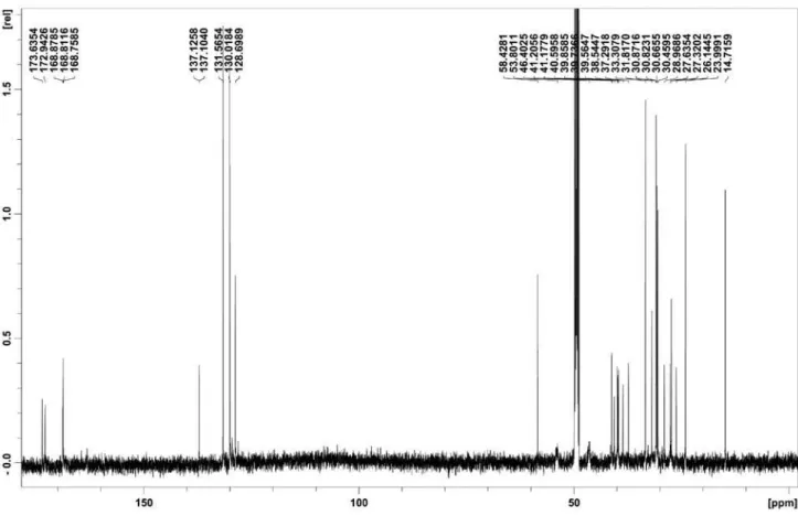
September 1999, from the Baía de Todos os Santos, Salvador, Bahia state, Brazil and immediately frozen. Both samples were deposited in the ascidian collection of the Departamento de Zoologia, Setor de Ciências Biológicas, Table 2. 13C data for compounds 1 and 2, 3 and 4 in CD
3OD at 100 MHz
Position 1 and 2 3 4
2 168.5/168.4 168.5 168.4
3 58.4 58.2 58.2
5 168.6/168.4 168.5 168.4
6 46.4 (br) 46.0 46.3
7 41.1/41.2 41.1 41.0
8 137.12/137.10 136.9 136.8
9/13 130.0 131.4 131.4
10/12 128.6 129.8 129.8
11 128.4 128.5 128.5
14 38.5/40.5 38.0 40.1
15 28.9/26.1 30.2 27.8
15a 27.6 - 27.8
16 37.2/39.8 38.0 40.1
17 172.9/173.6 172.7 172.5
18 39.5a/39.7a 39.6 39.6
19 53.8 (br) 54.1 54.1
20 31.8 31.5 31.6
21 27.3 27.1 27.1
22 27.6 30.7 30.2
23 30.8 30.3 30.4
24 30.6 30.5 30.5
25 30.4 30.6 30.6
26 33.3 33.1 33.0
27 23.9 23.8 23.7
28 14.7 14.5 14.4
CO - 173.4 174.0
Me - 22.7 22.6
a Interchangeable assignments.
Table 3. Antimicrobial activity of 1 and 2 (MIC in µg mL-1)
Pathogenic microbial strain mixture of 1 and 2
Staphylococcus aureus ATCC6538 62.5
Staphylococcus aureus ATCC259223 22.6
ORSA 8 45.3
ORSA 108 91.0
Escherichia coli ATCCNTCC861 125.0
Escherichia coli ATCC259222 45.6
Pseudomonas aeruginosa ATCC27853 22.6
Pseudomonas aeruginosa 13 45.3
Pseudomonas aeruginosa P1 4.3
Candida albicans ATCC10231 n.a.
Candida albicans ATCC36801 (serum type A) 125.0
Enterococcus faecalis ATCC14506 125.0
Streptococcus sanguinis ATCC15300 125.0
Streptococcus sobrinus ATCC27607 125.0
Streptococcus mutans UA159 62.5
Streptococcus mutans (clinical isolate 2.M7/4) 31.2
Universidade Federal do Paraná. BA99ASCI-03: (DZUP DID 210) - colony is reddish orange when alive, but looses the color in formalin, zooids with the sperm duct forming ten coils around the only follicle of the testis, larval trunk oval, 0.5 mm long with four ectodermal ampullae in each side, long and close to each other. The three adhesive papillae are close to each other and present a short stalk.
BA99ASCI-05 (DZUP DID 209) - colony is red when alive, but looses the color in formalin, zooids with sperm duct forming seven coils around the only follicle of the testis, larval trunk 0.4 mm long with four to five ectodermal ampullae in each side, shorter than the ones observed in former voucher. The three adhesive papillae have long stalks.
Extraction and isolation
The frozen ascidian sample BA99ASCI-05 (308.9 g, wet) was freeze dried and exhaustively extracted with MeOH. The MeOH extract was filtered, concentrated to 400 mL and partitioned with hexane (3 × 400 mL). TLC analysis (UV and Dragendorff) of the MeOH fraction indicated the presence of intense UV-absorbing compounds with amine groups. This fraction was evaporated to dryness (5.6 g) before separation by C18 reversed-phase chromatography (gradient of MeOH in H2O) in 0.5-1.0 g portions. This separation yielded eleven fractions (Da-MSP-1 to Da-(Da-MSP-11), of which the eighth (Da-MSP-8, 1.87 g) presented most of the Dragendorff and UV positive compounds. The fraction Da-MSP-8 was then subjected to chromatography on Sephadex LH-20 (MeOH). Seven fractions were obtained (Da-MSP-8A to DA-MSP-8G), of which Da-MSP-8D (0.402 g) and Da-MSP-8E (0.532 g) presented aromatic nitrogen-bearing compounds. Both fractions were similarly purified, firstly by C18 reversed-phase column chromatography (gradient of MeOH in H2O) then by HPLC (phenyl-bonded silica gel, Waters µBondapak 10 µm, 125 Å, 7.8 × 300 mm; eluent: 65:35 MeOH/0.1% TFA in H2O; λmax 220 nm; flow rate: 1 mL min-1; tR 25.6 min) to give 0.148 g of the mixture of 1 and 2 (0.05%, wet).
The frozen ascidian sample BA99ASCI-03 (450.0 g) was freeze dried and exhaustively extracted with MeOH. The MeOH extract was concentrated to 400 mL and partitioned with hexane (3 × 400 mL). The alcoholic fraction was evaporated until an aqueous suspension was obtained. The H2O fraction was first partitioned with EtOAc then with 3:2 CH2Cl2/MeOH. The CH2Cl2/MeOH fraction (2.0 g) was separated by chromatography on Sephadex LH20 (MeOH), to give five fractions (3A to BA99AC-3E). The last fraction obtained (Ba99AC-3E, 214 mg) was
subjected to chromatography on a cyanopropyl-bonded silica cartridge (5 g), with a gradient of EtOAc in CH2Cl2. The first fraction obtained from this separation (BA99AC-3E1, 134 mg) was separated by chromatography on a silica-gel cartridge (2 g) with a gradient of MeOH in CH2Cl2, to give three fractions (Ba99AC-3E1A to BA99AC-3E1C). The second one (BA99AC-3E1B, 38.0 mg) was separated by HPLC using a phenyl-bonded silica column (Inertsil, 250 × 4.6 mm, 5 µm) using 45:55 MeCN/H2O as eluent, to give five fractions. Fractions BA99AC-3E1C2 (7.1 mg) and BA99AC-3E1C2 (4.6 mg) were purified using a C18 reversed phase HPLC column (Phenomenex 5 µm), using 1:1 MeOH/0.1% HCO2H in H2O as eluent, to give N
-acetyl-rodriguesine A (3, tR = 19.8 min, 3.6 mg, 8.0×10⁻⁴%) and N-acetyl-rodriguesine B (4, tR = 21.2 min, 4.6 mg,
1.0×10⁻³%).
Rodriguesines A (1) and B (2)
Colorless, glassy solid; UV (MeOH) λmax 258 nm; 1H NMR (CD
3OD, 400 MHz), see Table 1; 13C NMR (CD3OD, 100 MHz), see Table 2; Positive HRESIMS for 1 m/z 459.3341 [M+H]+, calc. for C
26H43N4O3 459.3335. Positive HRESIMS for 2 473.3499 [M+H]+ calc. for C27H45N4O3 473.3492. MS/MS experiments:
m/z 385.2500, corresponding to C23H33N2O3 (calc.
385.24912); m/z 238.2181, corresponding to C15H28NO
(calc. 238.21654); m/z 252.2352, corresponding to
C16H30NO (calc. 252.23219); m/z 343.2416, corresponding
to C21H31N2O2 (calc. 343.23800).
N-acetyl-rodriguesine A (3)
Colorless, glassy solid; [α]D −37 (c 0.026, CHCl3);
UV (MeOH) λmax 256 nm; 1H NMR (CD3OD, 400 MHz), see Table 1; 13C NMR (CD
3OD, 100 MHz), see Table 2; Positive CID-MS/MS for 1 m/z 501.3483 [M+H]+, calc. for
C28H44N4O4 501.34408.
N-Acetyl-rodriguesine B (4)
Colorless, glassy solid; [α]D −52 (c 0.04, CHCl3);
UV (MeOH) λmax 257 nm; 1H NMR (CD
3OD, 400 MHz), see Table 1; 13C NMR (CD
3OD, 100 MHz), see Table 2; Positive CID-MS/MS for 1 m/z 515.3639 [M+H]+, calc. for C29H47N4O4 515.35918.
Acetylation of rodriguesines A (1) and B (2)
reversed-phase HPLC (Inertsil, ODS 3 250 × 4.6 mm, 5 µm) with a linear gradient of MeCN in H2O, from 7:3 to 95:5 during 30 min, to give 1.7 mg of pure N-acetyl-rodriguesine
A (3) and 3.0 mg of pure N-acetyl-rodriguesine B (4), as
well as 30.7 mg of an intermediate fraction containing both 3 and 4. N-acetyl-rodriguesine A (3) showed [α]D -37.0
(c 0.026, CHCl3) and 1H NMR, HPLC retention time and
MS analysis identical to natural 3. Similarly, N
-acetyl-rodriguesine B (4) showed [α]D −58 (c 0.03, CHCl3) and
1H NMR, HPLC retention time and MS analysis identical
to natural 4.
Hydrolysis and Marfey’s analysis of rodriguesines A (1) and B (2)
Two milligrams of the mixture of 1 and 2 was dissolved in 0.5 mL of 6 mol L-1 HCl and refluxed at 105 oC for 24 h. Then the reaction medium was evaporated until dryness. The hydrolyzed product was dissolved in H2O (40 µL) and a 0.5 mol L-1 NaHCO
3 solution was added, followed by 80 µL of a 1.4 equiv. of Marfey’s reagent (2,4-dinitro-5-fluoro-phenyl-L-alaninamide) in acetone. The reaction was
stirred at 40 oC for 1 h. Afterwards, 16 µL of 0.5 mol L-1 HCl was added to the reaction medium followed by evaporation to dryness. The reaction product was dissolved in 1 mL of MeOH. An identical derivatization procedure was employed for the preparation of a L
-phenylalanine-derivatized standard. The hydrolysed and the standard-derivatized samples were analyzed by HPLC-PDA-MS using the following conditions: Phenomenex C18 Synergi-4 Fusion RP-80 column, 250 × 4.6 mm; eluent: gradient of MeCN in Et3N/H2PO4 aqueous buffer; flow rate: 1 mL min-1. The hydrolyzed and derivatized sample as well as the derivatized phenylalanine standard were injected separately and co-injected. Retention time of 2,4-dinitro-5-(L-phenylalanine)-phenyl-L-alaninamide: 14.85 min.
Antibiotic assay
Procedures for the antimicrobial assays against
Staphylococcus aureus ATCC 6538, S. aureus ATCC
259223, Escherichia coli ATCC861, E. coli ATCC 259222, Pseudomonas aeruginosa ATCC 27853, P. aeruginosa
13, P. aeruginosa P1, Candida albicans ATCC 10231, C. albicans ATCC 36801 (serum type A), Enterococcus faecalis ATCC 14506, Streptococcus sanguinis ATCC
15300, Streptococcus sobrinus ATCC 27607, Streptococcus mutans UA159, S. mutans (clinical isolate 2.M7/4) were
performed using the broth microdilution assay, adapted from the National Committee for Clinical Laboratory Standards (USA) and from Oliveira et al.,10 as it follows.
Briefly, bacteria were grown in BHI (Brain Heart Infusion) liquid medium at 37 °C and suspended, as recommended, to yield final inocula of approximately 106 CFU/mL. Tests were performed in sterile 96-well microplates by dispensing into each well a total volume of 100 µL (40 µL of medium + 20 µL de inocula + 40 µL substance solution diluted in H2O) and incubated 24 h at 37 oC in 10% CO
2. Microorganism growths were determined by absorbance measurement at 550 nm in an automated microplate reader (Molecular Devices, Versa Max). The MIC was defined as the lowest concentration of drug that inhibited growth. As control microbial culture growths without the addition of the tested substance were used and all samples were assayed in replicate.
Acknowledgments
The authors thank Prof. Ian Castro-Gamboa (Instituto de Química, Universidade Estadual Paulista, Araraquara) for fruitful discussions, Michael LeBlanc (Department of Earth and Ocean Sciences, University of British Columbia) and Prof. Louis Barrows (Department of Medicinal Chemistry, University of Utah) for help in the ascidian collection, Dr. Tim S. Bugni and Prof. Chris M. Ireland (Department of Medicinal Chemistry, University of Utah, Salt Lake City, Utah, USA), as well as Prof. Norberto P. Lopes (Faculdade de Ciências Farmacêuticas, Universidade de São Paulo, Ribeirão Preto, Brazil) for assistance in obtaining MS analyses. Prof. Carlos Roque D. Corrêa (Instituto de Química, Universidade Estadual de Campinas) is acknowledged for assistance in the specific rotation measurements. Financial support was provided by the American Society of Pharmacognosy 1998 Research Starter Grant, NIH grant CA 67786 and FAPESP/BIOprospecTA grant 05/60175-2 to R.G.S.B., a CAPES scholarship to M.H.K., a Fulbright scholarship to S.M. and by CNPq scientific research awards to A.G.F., R.M.R. and R.G.S.B.
Supplementary Information
1H NMR, 13C NMR and MS spectra of 1 and 2, 3 and
4 are available free of charge at http://jbcs.sbq.org.br, as a PDF file.
References
1. Blunt, J. W.; Copp, B. R.; Hu, W. P.; Munro, M. H. G.; Northcote, P. T.; Prinsep, M. R.; Nat. Prod. Rep. 2008, 25, 35.
Marine Organisms; Fautin, D. G., ed., California Academy of Sciences: San Francisco, 1988, p. 41; Ireland, C.M.; Molinski, T.F.; Roll, D.M.; Zabriskie, T.M.; McKee, T.C.; Swersey, J.C.; Foster, M. P. In Bioorganic Marine Chemistry; Scheuer, P.
J., ed., Springer-Verlag: Berlin and Heidelberg, 1989, vol. 3, p. 1; Harper, M. K.; Bugni, T. S.; Copp, B. R.; James, R. D.; Lindsay, B. S.; Richardson, A. D.; Schnabel, P. C.; Tasdemir, D.; VanWagoner, R. M.;Verbitski, S. M.; Ireland, C. M. In Marine Chemical Ecology, McClintock, J. B.; Baker, B. J., eds.; CRC Press: Boca Raton, 2001, p. 3.
3. Vera, M. D.; Joullié, M. M.; Med. Res. Rev. 2002, 22, 102. 4. Vervoort, H.; Fenical, W.; Epifanio, R. A.; J. Org. Chem. 2000,
65, 782.
5. Adrian, T. E.; Curr. Pharm. Des.2007, 13, 3417.
6. Roberge, M.; Berlinck, R. G. S.; Xu, L.; Anderson, H.; Lim, L.; Curman, D.; Stringer, C. M.; Friend, S. H., Davies, P.; Vincent, I.; Haggarty, S.; Kelly, M. T.; Andersen, R.J.; Cancer Res. 1998, 58, 5701; Berlinck, R. G. S.; Britton, R.; Piers, E.; Lim,
L.; Roberge, M.; Rocha, R.M.; Andersen, R.J.; J. Org. Chem. 1998, 63, 9850.
7. Seleghim, M. H. R.; Lira, S. P.; Kossuga, M. H.; Batista, T.; Berlinck, R. G. S.; Hajdu, E.; Muricy, G.; Rocha, R. M.; Nascimento, G. G. F.; Silva, M., Pimenta, E. F.; Thiemann, O. H.; Oliva, G.; Cavalcanti, B. C.; Pessoa, C.; Moraes, M. O.; Galetti, F. C. S.; Silva, C. L.; Souza, A. O.; Peixinho, S.; Braz. J. Pharmacogn. 2007, 17, 287.
8. Hirsch, S.; Miroz, A.; McCarthy, P.; Kashman, Y.; Tetrahedron Lett. 1989, 30, 4291.
9. Vaz, E.; Fernandez-Suarez, M.; Muñoz, L.; Tetrahedron:
Asymmetry2003, 14, 1935.
10. Oliveira, J. H. H. L.; Seleghim, M. H. R.; Timm, C.; Grube, A.; Köck, M.; Nascimento, G. G. F.; Martins, A. C. T.; Silva, E. G. O.; Souza, A. O.; Galetti, F. C. S.; Minarini, P. R. R.; Silva, C. L.; Hajdu, E.; Berlinck, R. G. S.; Mar. Drugs2006, 4, 1.
Received: November 11, 2008
Web Release Date: April 24, 2009
Supplementary Information
0103 - 5053 $6.00+0.00*e-mail: [email protected]
#Dedicated to Prof. Vanderlan da Silva Bolzani, for her outstanding con-tributions to the development of natural product chemistry and related sciences in Brazil.
Antibacterial Modified Diketopiperazines from two Ascidians of the Genus
Didemnum
#Miriam H. Kossuga,a Simone P. Lira,a Shayna McHugh,a Yohandra R. Torres,a,b Bruna A. Lima,c Reginaldo
Gonçalves,c Katyuscya Veloso,d Antonio G. Ferreira,d Rosana M. Rochae and Roberto G. S. Berlinck*,a
aInstituto de Química de São Carlos, Universidade de São Paulo, CP 780, 13560-970 São Carlos-SP, Brazil
bDepartamento de Química, Universidade Estadual do Centro-Oeste, Rua Camargo Varela de Sá, 03,
85040-080 Guarapuava-PR, Brazil
cDepartamento de Diagnóstico Oral, Faculdade de Odontologia de Piracicaba, Universidade Estadual de Campinas,
CP 52, 13414-018 Piracicaba-SP, Brazil
dDepartamento de Química, Universidade Federal de São Carlos, 13565-905 São Carlos-SP, Brazil
eDepartamento de Zoologia, Setor de Ciências Biológicas, Universidade Federal do Paraná, Centro Politécnico,
Jardim das Américas s/n, CP 19020, 81531-990 Curitiba-PR, Brazil
Figure S1.1H NMR spectrum of rodriguesines A (1) and B (2) in MeOH-d
Figure S2.13C NMR spectrum of rodriguesines A (1) and B (2) in MeOH-d
4 (100 MHz).
Figure S4. MS/MS analysis of rodriguesine B (2).
Figure S5.1H NMR spectrum of N-acetyl-rodriguesine A (3) in MeOH-d
Figure S6.13C NMR spectrum of N-acetyl-rodriguesine A (3) in MeOH-d
4 (100 MHz).
Figure S7.1H NMR spectrum of N-acetyl-rodriguesine A (4) in MeOH-d
Figure S8.13C NMR spectrum of N-acetyl-rodriguesine A (5) in MeOH-d
![Table 1. 1 H data for compounds 1 and 2, 3 and 4 in CD 3 OD at 400 MHz [ d , multiplicity (J in Hz)] Position 1 and 2 3 4 3 4.27 (t, 5.0) 4.27 (t, 6) 4.22 (t, 6.5) 6 3.08 (d, 17), 3.55/3.56 (2d, 17 Hz each) 3.00 (d, 22), 3.50 (d, 22) 2.99 (d, 22), 3](https://thumb-eu.123doks.com/thumbv2/123dok_br/18993470.461458/4.892.477.821.156.723/table-data-compounds-cd-od-mhz-multiplicity-position.webp)