In Brazil, most cases have been reported in the southern, southeastern and central-west regions. Paracoccidioidomycosis is endemic among rural area populations and affects male individuals in their economically productive years (30-60 years of age); the disease is related
Introduction
Paracoccidioidomycosis is a systemic mycosis originally described by Adolfo Lutz in 1908. Paracoccidioidomycosis is autochthonous to Latin America, and the highest incidence of the disease was registered in South American coun-tries (Brazil, Argentina, Colombia and Venezuela).
Chapter 6 - Paracoccidioidomycosis*
Capítulo 6 - Paracoccidioidomicose
Bodo Wanke, Miguel Abidon Aidê
Abstract
Paracoccidioidomycosis is a systemic mycosis caused by the dimorphic fungus Paracoccidioides brasiliensis. The disease is restricted to Latin America. It is the principal systemic mycosis in Brazil, with higher incidences in the southern, southeastern and central regions. The disease is acquired by inhaling fungal propagules. In endemic areas, the primary infection occurs during childhood and involves the immune system. The most common chronic form of paracoccidioidomycosis in adults is the multifocal form, in which there is dissemination to the lungs, lymph nodes, skin and mucosae. This form of the disease has a chronic progression, and the diagnosis is typically delayed. Cough, dyspnea and weight loss due to cutaneous and mucosal lesions are evident and are the principal complaints reported by paracoccidioidomycosis patients. Chest X-rays reveal diffuse reticulonodular infiltrates, which are more evident in the upper lobes. The etiologic diagnosis is based on the identification of P. brasiliensis in clinical specimens, such as lymph node aspirates or BAL fluid, by direct microscopy and culture. Histopathological testing of tissue samples reveals the thick birefringent cell wall of the fungus and the typical pattern of multiple budding around the mother cell. Double agar gel immunodiffusion is useful for the diagnosis when the fungus cannot be detected through mycological tests. Although paracoccidioidomycosis is most often treated with the sulfamethoxazole-trimethoprim combination, itraconazole is preferable. Amphotericin B is used in severe cases.
Keywords: Paracoccidioidomycosis; Mycosis; Lung diseases, fungal.
Resumo
A paracoccidioidomicose é uma micose sistêmica causada pelo fungo dimórfico Paracoccidioides brasiliensis. A doença é restrita à América Latina. É a principal micose sistêmica no Brasil, com maior frequência nas regiões sul, sudeste e centro-oeste. A doença é adquirida através da inalação de propágulos do fungo. Nas áreas endêmicas, a infecção primária ocorre durante a infância e envolve o sistema imunológico. A forma crônica do adulto mais frequente é de disseminação multifocal, com envolvimento dos pulmões, linfonodos, pele e mucosas. Essa forma tem evolução crônica com diagnóstico tardio. Tosse, dispneia e perda de peso associada a lesões cutâneas e das mucosas são evidentes e constituem as queixas principais da doença. A radiografia simples de tórax apresenta infiltrado reticulonodular difuso mais evidente nos lobos superiores. O diagnóstico etiológico se baseia na achado de P. brasiliensis no exame microscópico direto de espécimes clínicos, tais como aspirado de gânglios ou material de LBA, complementado pelo crescimento do fungo em cultura. O exame histopatológico de amostra de tecidos evidencia a parede espessa e birrefringente do fungo, assim como o aspecto típico de multibrotamento na célula-mãe. A imunodifusão em duplo gel de ágar é muito útil no diagnóstico quando o fungo não é encontrado nos exames micológicos. O tratamento de escolha é realizado com sulfametoxazol e trimetoprima, mas o itraconazol é a melhor droga. A anfotericina B é usada nos casos graves da doença.
Descritores: Paracoccidioidomicose; Micoses; Pneumopatias fúngicas.
* Study carried out at the Universidade Federal Fluminense – UFF, Fluminense Federal University – Niterói, Brazil. Correspondence to: Miguel Abidon Aidê. Rua Uirapuru, 118, Itaipu, CEP 24355-100, Niterói, RJ, Brasil. Tel 55 21 2710-8740. E-mail: [email protected]
Financial support: None.
Classification
According to the 1986 International Colloquium on Paracoccidioidomycosis, cited by the Brazilian Consensus on Paracoccidioidomycosis, paracoccidioidomycosis is classified according to the clinical data and natural history of the disease(3):
1) infection 2) disease
3) acute form in children/subacute form in adolescents
4) chronic form in adults: unifocal or multifocal
5) residual form
Clinical forms
Acute/subacute (children/adolescents)
The acute/subacute form of paracoccidioid-omycosis is the clinical form of the disease in children, adolescents and adults up to 35 years of age. This form accounts for 3-5% of the cases of paracoccidioidomycosis. The principal manifes-tations of the disease are as follows: superficial lymph node enlargement; deep lymph node enlargement; digestive symptoms; cutaneous symptoms; and osteoarticular symptoms. Other manifestations include anemia, fever and weight loss, the overall health of children deteriorating rapidly. Pulmonary involvement is rare.(3)
Chronic unifocal/multifocal form in
adults
The chronic unifocal/multifocal form is the most common form of paracoccidioidomycosis, accounting for 90% of the cases; males are predominantly affected. The unifocal form is characterized by a chronic progression, predom-inantly accompanied by weakness, weight loss, cough, dyspnea, reticulonodular infiltrate (generally in the two upper thirds of the lungs) and distal hypertransparent areas at both lung bases.
The multifocal form is characterized by the involvement of extrapulmonary sites, such as the skin, the oral mucosa (mulberry-like stoma-titis), the pharyngeal mucosa or the laryngeal mucosa (or a combination of the two) and the apices of teeth. The symptoms of the multifocal form include pain during mastication, sialorrhea to agricultural activities. The etiologic agent is
a thermally dimorphic fungus (Paracoccidioides brasiliensis).(1,2)
Paracoccidioidomycosis constitutes a serious public health problem because it is quite poten-tially disabling and causes premature deaths.
Epidemiology
Paracoccidioidomycosis is a disease for which reporting is not compulsory, and there is a lack of precise data regarding its incidence in Brazil.
It is believed that the annual incidence of paracoccidioidomycosis in endemic rural areas ranges from 3-4 new cases/1,000,000 population to 1-3 new cases/100,000 population. Paracoccidioidomycosis is considered to be the third leading cause of death from chronic infectious disease, and the mortality rate for paracoccidioidomycosis is 1.65 cases/1,000,000 population.(3)
The incidence of paracoccidioidomycosis is higher among males (1:10-15 males) in the 30-50 year age bracket. In individuals younger than 14 years of age, paracoccidioidomycosis affects both genders equally but is, in general, uncommon.(4)
Pathophysiology (natural history of
paracoccidioidomycosis)
acid-Schiff staining of tissue biopsy specimens are imperative to visualize the fungus. Culture on Sabouraud agar can aid in the diagnosis of paracoccidioidomycosis and therefore should always be ordered, although it does not yield rapid results.(1-3,5)
In addition to aiding in the diagnosis, sero-logic tests can evaluate treatment response and disease recurrence. In these cases, elevated anti-body titers usually precede clinical recurrence. Agar gel double immunodiffusion is the test that is most readily available in clinical practice, and its sensitivity and specificity are greater than 80% and 90%, respectively. Agar gel double immun-odiffusion should always be titrated so that the interpretation of the therapeutic response is more accurate.(7)
and odynophagia. Wasting is common due to a delay in seeking medical assistance. A chest X-ray reveals the same lesions as those observed in the unifocal form.
Other sites affected by paracoccidioidomy-cosis include the adrenal glands, the central nervous system, the cervical lymph nodes, the submandibular lymph nodes, the intestines, the osteoarticular system, the epididymis, the liver and the spleen.(3-5)
Differential diagnosis
The principal differential diagnosis is pulmonary tuberculosis, which is quite similar to paracoccidioidomycosis with regard to the radiological alterations and clinical manifesta-tions (Chart 1). The diagnosis is confirmed by the presence of the fungus (paracoccidioidomy-cosis) or of Koch’s bacillus (tuberculosis) in the specimens analyzed. Tuberculosis and paracoc-cidioidomycosis can affect the same individual, and this association occurs in 5.5-19% of the cases, which makes the diagnosis of the two diseases more difficult.(4,6)
Initial investigation
The initial investigation should include the following(3):
• history taking focusing on the clinical
forms in adults and in children
• chest X-ray • blood workup • ESR
• liver function tests
• urea, creatinine, sodium and potassium
Specific investigation
The definitive diagnosis (gold standard) is based on the identification of the fungus in clinical specimens or in tissue biopsy specimens.
The 10% potassium hydroxide test applied to smear samples (on slides coverslipped for direct examination) is a highly effective and inexpen-sive method of screening for the fungus. This method can be used for various specimens, such as sputum, scrapings (from cutaneous lesions or from mucosae) and lymph node aspirate, as well as for material obtained through fiberoptic bronchoscopy. The Grocott-Gomori methen-amine-silver stain technique and the periodic
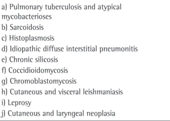
Chart 1 - Principal differential diagnoses for
paracoccidioidomycosis.
a) Pulmonary tuberculosis and atypical mycobacterioses
b) Sarcoidosis c) Histoplasmosis
d) Idiopathic diffuse interstitial pneumonitis e) Chronic silicosis
f) Coccidioidomycosis g) Chromoblastomycosis
h) Cutaneous and visceral leishmaniasis i) Leprosy
j) Cutaneous and laryngeal neoplasia
Figure 1 - Anteroposterior chest X-ray: bilateral and
Severe disease
In severe cases, the following can occur(1):
a) reduction of more than 10% in the body mass index
b) difficulty in swallowing
c) respiratory failure: PaO2/FiO2 < 250 d) neurological manifestations and central
nervous system involvement e) adrenal gland involvement
Treatment regimens: mild/moderate
forms
The following regimens have been used(1,2,10):
1) sulfamethoxazole-trimethoprim combi-nation, capsules of 80-400 mg and of 160-800 mg
• initial dose: 3 capsules (80-400 mg)
every 12 h for 21 days
• 2 capsules every 12 h for 21 days • 1 capsule every 12 h for 2 years
• Children: 8-10 mg • kg−1 • day−1 of
trimethoprim or 40-50 mg • kg−1 • day−1 of sulfamethoxazole every 12 h
• > 80% of cure
2) Ketoconazole, capsules of 200 mg
• dose: 400 mg/day for 3 months, followed
by 200 mg/day for 9 months
• taken with a full meal
• side effects: gynecomastia, decreased
libido, liver disease, teratogenesis 3) Itraconazole, capsules of 100 mg
• dose: 200 mg/day at large meals for
6-9 months
• side effects: digestive disorders,
teratogenesis
• children < 30 kg of weight or > 5 years of age: 5-10 mg • kg−1 • day−1
4) Fluconazole, capsules of 50 mg; 100 mg; 150 mg
• dose: 400 mg/day for 3-6 months • maintenance: 100 to 200 mg/day for
6-12 months
• children: 3-6 mg • kg−1 • day−1 5) Voriconazole(10)
• dose: 200 mg every 12 h
• side effects: decreased visual acuity and
blurred vision
6) Amphotericin B, 50 mg/bottle
• indication: severe forms; allergy, resis -tance or intolerance to sulfonamides
• dose: 1 mg • kg−1 • day−1; total of 25-35 mg/kg (until 1-2 g)
Radiological findings
A simple chest X-ray (Figure 1) can reveal asymmetric reticulonodular infiltrate, predomi-nantly in the two upper thirds of both lungs, accompanied by hypertransparent areas at the lung bases.(1,2,4)
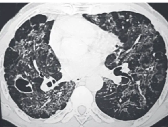
A HRCT scan (Figure 2) can reveal nodules, ground-glass opacities, tree-in-bud pattern, acinar lesions, parenchymal bands, peribroncho-vascular interstitial thickening, cavities, reticular pattern, “reversed halo” sign, scar-related emphy-sema and traction bronchiectasis.(8,9)
Treatment
General considerations
The following points should be observed: a) Support measures should be adopted in
cases of clinical complications.
b) Itraconazole is the best option for mild/ moderate forms.
c) The sulfamethoxazole-trimethoprim combination is the alternative that is most widely used in the outpatient treatment of paracoccidioidomycosis.
d) The consumption of cigarettes and alcohol should be limited.
e) Intestinal parasitosis (particularly strongyloidiasis), quite common in para-coccidioidomycosis patients, should be treated.
f) Long-term treatment is required.
g) Patients should be monitored until the criteria for cure are met.
Figure 2 - HRCT scan of the chest. Ground-glass
Criteria for cure
(10)The criteria for cure are as follows(10):
• clinical, radiological and mycological
improvement
• stabilization of agar gel double immunodi -ffusion results at 1:2 or negative conversion of two samples within a 6-month interval after the treatment
• protein electrophoresis, ESR and muco -proteins showing normal results for 3 consecutive months, as an alternative to agar gel double immunodiffusion
References
1. Tarantino AB, Gonçalves AJR, Capone D, Aide MA, Lazera MS, Wanke B. Micoses Pulmonares. In: Tarantino AB, editor. Doenças Pulmonares. Rio de Janeiro: Guanabara Koogan; 2002. p. 416-50.
2. Wanke B, Lazer MS, Capone D. Paracoccidioidomicose. In: Sociedade de Pneumologia e Tisiologia do Estado do Rio de Janeiro, Aidé MA, editors. Pneumologia aspectos práticos e atuais. Rio de Janeiro: Revinter; 2001. p. 147-52.
3. Shikanai-Yasuda MA, Telles Filho FQ, Mendes RP, Colombo AR, Moretti MA. Consenso de paracoccidioidomicose. Rev Soc Bras Med Trop. 2006;39:297-310.
4. Paniago AM, Aguiar JI, Aguiar ES, Cunha RV, Pereira GR, Londero AT, et al. Paracoccidioidomicose: estudo clínico e epidemiológico de 422 casos observados no Estado do Mato Grosso do Sul. Rev Soc Bras Med Trop. 2003;36(4):455-459.
5. Londero AT. Paracoccidioidomicose: patogenia, formas clínicas, manifestações pulmonares e diagnóstico. J Pneumol. 1986;(12):41-60.
6. Quagliato Júnior R, Grangeia Tde A, Massucio RA, De Capitani EM, Rezende Sde M, Balthazar AB. Association between paracoccidioidomycosis and tuberculosis: reality and misdiagnosis. J Bras Pneumol. 2007;33(3):295-300.
7. Valle AC, Costa RL, Monteiro PC. Interpretation and clinical correction of serological test in paracoccidioidomycosis. Medical Mycology. 2001;39:373-7.
8. Muniz MAS, Machiori E, Magnago M, Moreira LB, Almeida Jr JG. Paracoccidioidomicose pulmonar: aspectos na tomografia computadorizada de alta resolução. Radiol Bras. 2002;35:147-54.
9. Souza AS Jr, Gasparetto EL, Davaus T, Escuissato DL, Marchiori E. High-resolution CT findings of 77 patients with untreated pulmonary paracoccidioidomycosis. AJR Am J Roentgenol. 2006;187(5):1248-52.
10. Yasuda MA. Pharmacological management of paracoccidioidomycosis. Expert Opin Pharmacother. 2005;6(3):385-97.
• maintenance with a sulfonamide for
1-3 years
• side effects: azotemia, ↑↓ potassium, anemia, fever, chills and phlebitis
• other forms of presentation:
• amphotericin B in colloidal dispersion: 1 mg • kg−1 • day−1
• liposomal amphotericin B: 3-5 mg •
kg−1 • day−1
• amphotericin B lipid complex: 5 mg •
kg−1 • day−1
7) Rifampin (severe, disseminated forms; used in combination with amphotericin B)
• dose: 600 mg/day + amphotericin B, 25 mg • kg−1 • day−1, three times a week
Drug interaction between azoles and
sulfonamides
The interaction between azoles and sulfon-amides increases the concentration of the following drugs: aminophylline; calcium channel blockers; coumarins; hypoglycemic agents; and protease inhibitors.(3,10)
It should be highlighted that voriconazole cannot be combined with rifampin or with rifabutin.
Sulfonamides decrease the effect of contra-ceptives and increase methotrexate-induced bone marrow suppression.(3,10)
Sequelae of paracoccidioidomycosis
The principal sequelae of paracoccidioid-omycosis are as follows(10):
• worsening of COPD
• adrenal gland dysfunction (15-50% of the
cases)
• dysphonia or laryngeal obstruction, or a
combination of the two
• reduced mouth opening
• epilepsy or hydrocephalus, or a combina -tion of the two (6-25% of the cases)
About the authors
Bodo Wanke
Physician Researcher in the Department of Mycology. Evandro Chagas Hospital Research Center, Fundação Oswaldo Cruz – Fiocruz, Oswaldo Cruz Foundation – Rio de Janeiro, Brazil.
Miguel Abidon Aidê