ABSTRACT
Objective: To describe the implementation of a robotic thoracic surgery program at a public tertiary teaching hospital and to analyze its initial results. Methods: This was a planned interim analysis of a randomized clinical trial aimed at comparing video-assisted thoracoscopic surgery and robotic surgery in terms of the results obtained after pulmonary lobectomy. The robotic surgery program developed at the Instituto do Câncer do Estado de São Paulo, in the city of São Paulo, Brazil, is a multidisciplinary initiative involving various surgical specialties, as well as anesthesiology, nursing, and clinical engineering teams. In this analysis, we evaluated the patients included in the robotic lobectomy arm of the trial during its irst three months (from April to June of 2015). Results: Ten patients were included in this analysis. There were eight women and two men. The mean age was 65.1 years. All of the patients presented with peripheral tumors. We performed right upper lobectomy in four patients, right lower lobectomy in four, and left upper lobectomy in two. Surgical time varied considerably (range, 135-435 min). Conversion to open surgery or video-assisted thoracoscopic surgery was not necessary in any of the cases. Intraoperative complications were not found. Only the irst patient required postoperative transfer to the ICU. There were no deaths or readmissions within the irst 30 days after discharge. The only postoperative complication was chest pain (grade 3), in two patients. Pathological examination revealed complete tumor resection in all cases. Conclusions: When there is integration and proper training of all of the teams involved, the implementation of a robotic thoracic surgery program is feasible and can reduce morbidity and mortality.
Keywords: Pneumonectomy; Robotic surgical procedures; Thoracic surgery; Minimally invasive surgical procedures; Lung neoplasms.
Robotic pulmonary lobectomy for lung
cancer treatment: program implementation
and initial experience
Ricardo Mingarini Terra1, Pedro Henrique Xavier Nabuco de Araujo2, Leticia Leone Lauricella2, José Ribas Milanez de Campos1,
Herbert Felix Costa2, Paulo Manuel Pego-Fernandes1
Correspondence to:
Ricardo Mingarini Terra. Avenida Dr. Enéas de Carvalho Aguiar, 44, bloco II, 2º andar, sala 9, CEP 05403-900, São Paulo, SP, Brasil. Tel./fax: 55 11 2661-5248. E-mail: [email protected]
Financial support: None.
INTRODUCTION
Over the past 20 years, minimally invasive surgery has developed rapidly. Beginning in the 1990s, video-assisted technology came to be widely used for surgery, thus playing a decisive role in increasing the prominence of minimally invasive surgery. Video-assisted technology also had an impact on thoracic surgery, pleural procedures and easily performed resections having rapidly come to be performed by means of video-assisted thoracoscopic surgery (VATS) in many countries, including Brazil.(1,2)
Studies published in the last decade contributed to consolidating the role of VATS in resections that are more complex, such as lobectomy and pneumonectomy.(3,4) More
recently, robotic surgery has emerged as an alternative to video-assisted surgery, its objective being to increase the amplitude and accuracy of intracavitary maneuvers and movements, as well as to provide better visualization via three-dimensional imaging. Studies have shown that robotics can be applied to thoracic surgery, being particularly useful for mediastinal tumors and anatomic lung resections, such as pulmonary lobectomy.(5-8)
The real role of robotics in thoracic surgery has yet to
be deined. Although large case series have shown good
results regarding intraoperative morbidity, intraoperative mortality, and length of hospital stay,(9) retrospective
studies involving databases have raised questions regarding the costs and complications of the new method.(10) The
results of an analysis of a US hospital database including 15,502 patients undergoing lung resection via VATS or robot-assisted thoracic surgery showed that the latter had
signiicantly higher costs and longer operative times.(10)
In this setting of uncertainty, the implementation of a robotic surgery program is particularly challenging, and, in addition to risk minimization, attention should be given to structural issues and costs. The objectives of the present study were to describe the implementation of a robotic thoracic surgery program at the University of São Paulo School of Medicine Hospital das Clínicas Instituto do Câncer do Estado de São Paulo (ICESP, São Paulo State Cancer Institute), in the city of São Paulo, Brazil, and to analyze its initial results.
1. Disciplina de Cirurgia Torácica, Instituto do Coração, Faculdade de Medicina, Universidade de São Paulo, São Paulo (SP) Brasil.
2. Instituto do Câncer do Estado de São Paulo – ICESP – Hospital das Clínicas, Faculdade de Medicina, Universidade de São Paulo, São Paulo (SP) Brasil.
Submitted: 1 September 2015. Accepted: 25 February 2016.
Study carried out in the Disciplina de Cirurgia Torácica and at the Instituto do Câncer do Estado de São Paulo – ICESP – Hospital das Clínicas, Faculdade de Medicina, Universidade de São Paulo, São Paulo, Brazil.
METHODS
This was a planned interim analysis of a randomized clinical trial that is currently under way at our institution and that is aimed at comparing VATS and robotic surgery in terms of the results obtained after pulmonary lobectomy. In this analysis, we evaluated the patients included in the robotic lobectomy arm of the trial during
its irst three months (from April to June of 2015), i.e., after the surgical team had been certiied (in March
of 2015). All of the patients who were included in the study gave written informed consent, and the study was approved by the local research ethics committee. In addition to the presence of primary lung cancer or lung metastasis and written informed consent, the criteria for inclusion in the randomized clinical trial were as follows, having been evaluated during the clinical staging phase:
• eligibility for the treatment of lung cancer or lung metastasis by pulmonary lobectomy
• presence of a tumor of less than 5 cm in diameter at its widest point
• absence of hilar or mediastinal lymphadenopathy • absence of tumor invasion of the chest wall, the
mediastinum, or another lung lobe
• absence of tumor invasion of a main bronchus or a lobar bronchus less than 1 cm from the secondary carina
• Clinical and anesthetic evaluation results showing that the patient was able to undergo the proposed procedure
The exclusion criteria were as follows:
• having previously undergone a thoracic surgical procedure in the hemithorax that was to be operated on
• being unable to remain on single-lung ventilation during the procedure
In the present analysis, the following variables were evaluated: operative time; length of hospital stay; complications; patient-reported pain; extent of lymph node dissection; and number of lymph nodes removed during lymph node dissection. Intraoperative and postoperative complications (up to postoperative
day 30) were recorded and classiied on the basis
of the Common Terminology Criteria for Adverse Events, version 4.0.(11) The magnitude of the systemic inlammatory response was assessed by measuring
serum creatine phosphokinase and C-reactive protein levels on postoperative day 2. Pain was assessed by a visual analog pain scale (a Likert scale)—which was administered in the morning on postoperative days 1, 2, and 3 and during follow-up visits on postoperative days 15 and 30—and by the duration of use and dose of opioids. In the early postoperative
period, opioids were administered at ixed times;
subsequently, they were administered as needed. The
date of opioid discontinuation was deined as the day
on which patients received their last dose of opioids. The extent of lymph node dissection was determined by counting the resected lymph nodes. The resected lymph nodes were counted by using a procedure that
has been standardized by the ICESP Department of Anatomic Pathology and that is consistent with the current literature.
The robotic surgery program developed at the ICESP is a multidisciplinary initiative involving various surgical specialties, as well as anesthesiology, nursing, and clinical engineering teams. All involved received
speciic training in operating the robot. The thoracic
surgery team training consisted of an online course on how the robotic surgical system works; 20 hours of virtual reality simulation in order to familiarize participants with the movements of the robot; and lobectomy simulation in animal models. The process
of certiication lasted 2 days, having taken place in a
specialized center abroad and having involved animal
models and human cadavers. After certiication,
we participated as observers in various procedures performed at centers of excellence in robotic surgery.
Before the irst procedure, simulations were performed
with the participation of the entire multidisciplinary team. All surgical procedures were performed with the use of selective intubation and an epidural catheter for
postoperative analgesia. We used a slightly modiied
version of a robotic lobectomy technique that was originally described by Dylewski et al.(12) Patients are
placed in the lateral decubitus position with pads under their axillae, the robot being placed over their heads. A total of four ports are used: three for the robotic arms
and one for the assistant surgeon (Figure 1). The irst
incision is made in the 6th intercostal space at the anterior axillary line. After insertion of a 5-mm trocar,
carbon dioxide insuflation is initiated. With the aid of a
5-mm endoscope, the locations of the remaining ports are determined. Initially, the diaphragm insertion on the chest wall at the level of the 10th intercostal space
is identiied, and a 12-mm trocar is inserted at that
site, the trocar being used by the assistant surgeon for exposure, aspiration, stapling, introduction/removal of materials (such as gauze), and removal of specimens for pathological examination. Subsequently, two other ports are placed in the 7th or 8th intercostal space at the midaxillary and posterior axillary lines, respectively. The robotic camera is introduced through the midaxillary line trocar, and the robotic graspers are introduced through the remaining two ports. It is extremely important that
these ports are caudal to the oblique issure.
The surgical procedure was systematized in order to minimize intraoperative lung manipulation. In all
cases, the irst step was to section the pulmonary
ligament. Next, in a posterior and superior direction, paraesophageal and subcarinal lymph nodes were dissected. Right interlobar lymph nodes or those located between the pulmonary artery and the left main bronchus were then resected. In cases of
right or left lower lobectomy, the oblique issure is
artery branches and, subsequently, the pulmonary vein
are then divided, the bronchus and oblique issure
remaining to be divided last. In cases of right or left lower lobectomy, the bronchus and pulmonary vein are dissected after the pulmonary artery has been sectioned, being stapled sequentially. The procedure is completed with dissection of right paratracheal lymph nodes and left para-aortic lymph nodes. A 28-Fr chest tube is then introduced through the lower port.
Two aspects of our robotic surgical technique are noteworthy. First, the robotic ports are closed in order to allow the use of carbon dioxide, which, in addition to increasing the workspace by lowering the diaphragm and reducing visual interference from the “smoke” from cauterization, facilitates dissection of hilar
structures and the oblique issure. Second, removal
of the surgical specimen is an important step in our robotic surgical technique. To that end, the lower port is used as originally described by Dylewski et al.(12)
Given that the lower port is located at the transition between the diaphragm and the chest wall and below the 10th rib, the resected lobe can be removed without the limitation imposed by the ribs, although the same is not true for the remaining ports. Larger specimens can be removed this way as well, resulting in less pain. In the postoperative period, patients are habitually transferred to the hospital ward. Elderly patients with multiple comorbidities or patients with intraoperative complications are admitted to the ICU. Postoperative analgesia includes oral dipyrone every 6 h and patient-controlled epidural anesthesia (local anesthetics and opioids), which is discontinued immediately after
chest tube removal. Anti-inlammatory drugs and oral
opioids are administered as needed.
The data in the present study were prospectively
collected and stored with the aid of speciic software.
Continuous variables are expressed as mean and
standard deviation or as median and interquartile range. Categorical variables are expressed as absolute numbers and proportions.
RESULTS
During the irst three months of our program, ten
patients underwent robot-assisted pulmonary lobectomy for the treatment of lung cancer. Patient demographic data are detailed in Table 1. During the same period, seven patients were randomly allocated to the VATS arm of our comparative study, but they were not included in the present interim analysis.
All patients had peripheral tumors. In nine, lung adenocarcinoma was found to be the most common histological type. Of those nine patients, four had acinar predominant adenocarcinoma, four had lepidic predominant adenocarcinoma, and one had papillary predominant adenocarcinoma. One patient was diagnosed with large cell carcinoma. Of the ten patients included in the present analysis, four underwent mediastinoscopy: two did because of suspicion of mediastinal lymph node involvement; one did because of the presence of a tumor > 3 cm in size; and one did because of a history of surgery for brain metastasis. Pathological examination showed no hilar or mediastinal lymph node involvement in any of the patients.
Operative times varied considerably across patients and are detailed in Figure 2. It is of note that, in the cases of patients 2 and 6, intraoperative complications
signiicantly prolonged operative times. In the case
of patient 2, the patient had received a stab wound, which required chest tube drainage at the time. The patient did not disclose that information during the process of selection and randomization. However, in the intraoperative period, a large quantity of
pleuro-pulmonary adhesions were identiied, most of which
A B
C
were lysed during the VATS procedure performed prior to robot-assisted surgery. In the case of patient 6, the selective tube was displaced during the procedure and
compromised the surgical ield, given that the operated lung inlated. An attempt was made to reposition the
tube with the patient in the lateral decubitus position; however, after several unsuccessful attempts, the robot was disconnected and the tube was repositioned with the patient in the supine position. After the tube was
conirmed to be in the correct position, the robot was
reconnected and the surgical procedure was completed as originally planned.
There were no intraoperative complications, and the mean quantity of bleeding was 49.1 ± 35.7 mL. None of the patients required blood transfusion. Only
the irst patient required postoperative transfer to
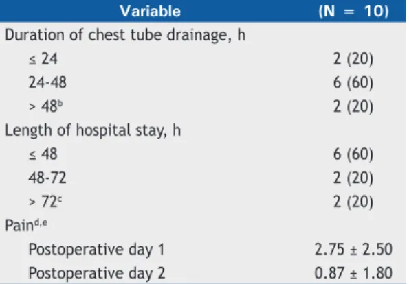
the ICU, because of prolonged operative time; the remaining patients were taken to the recovery room and, subsequently, to a ward bed. Data regarding duration of chest tube drainage, length of hospital stay, postoperative pain, and markers of systemic
inlammation are summarized in Table 2. There were no deaths or readmissions within the irst 30 days
after discharge. The only postoperative complication was chest pain (grade 3), which was observed in two patients and prolonged their hospital stay by 1 and 2 days. For pain control, patients received additional doses of intravenous morphine.
Pathological examination revealed complete tumor resection in all cases. The mean number of resected
lymph nodes was 9.5 ± 3.5. The number of resected lymph nodes increased with experience; in the last two patients, 12 lymph nodes (7 mediastinal lymph nodes and 5 hilar lymph nodes) and 15 lymph nodes (9 mediastinal lymph nodes and 6 hilar lymph nodes)
were resected, whereas, in the irst two patients, 5
lymph nodes (2 mediastinal lymph nodes and 3 hilar lymph nodes) and 7 lymph nodes (4 mediastinal lymph nodes and 3 hilar lymph nodes) were resected.
DISCUSSION
The present study showed that, when there is an institutional program and proper training of the multidisciplinary team, robotic thoracic surgery can be implemented with satisfactory results from the very beginning. Although operative times were long,
particularly in the irst patients, there were no signiicant
complications, and 80% of the patients were discharged
within the irst 72 h after surgery. Two patients had
pain that was more severe, and this prolonged their hospitalizations by 1 and 2 days; however, mean daily pain scores were low, especially in comparison with the results obtained on postoperative day 2.
The results of the present study are very encouraging, especially if we take into account that they refer to our initial experience. Morbidity was found to be very low
(two cases of grade 3 pain); this inding is consistent
with those of large studies, such as those conducted by Nasir et al.(9) and Meli et al.,(13) who reported mortality Table 1. Characteristics of the patients studied.a
Variable (N = 10)
Gender
Male 2 (20)
Female 8 (80)
Age, yearsb 64 (55-80)
Comorbidities
Systemic arterial hypertension 7 (70)
Diabetes mellitus 1 (10)
COPD 2 (20)
BMIc, kg/m2 27.8 ± 4.5
Affected lobe
LUL 2 (20)
RUL 4 (40)
RLL 4 (40)
Clinical stage (TNM staging system)
T1aN0M0 3 (30)
T1bN0M0 4 (40)
T2aN0M0 1 (10)
T2bN0M0 1 (10)
T1bN0M1b 1 (10)
Tumor diameter*, cmb,d 2.5 (1.2-4.7)
LUL: left upper lobe; RUL: right upper lobe; RLL: right lower lobe; and TNM: tumor-node-metastasis. aValues expressed as n (%), except where otherwise indicated. bValues expressed as median (interquartile range).cValue
expressed as mean ± SD. dTumor diameter measured at its widest point on CT scans of the chest (lung window).
1 2 3 4 5 6 7 8 9 10
500
450
400
350
300
250
200
150
100
50
0 minutes
RUL
RUL RUL
RUL LUL
LUL RLL
RLL RLL
RLL
patients
Conclusion Console Docking Trocars
rates of less than 0.5% and complication rates of 27% and 33%, respectively. The length of hospital stay in our patients constitutes further evidence that they
responded well to robotic surgery. Our inding that 80% of our patients were discharged within the irst
72 h after surgery is consistent with those of other studies, in which the length of hospital stay ranged from 2 days to 4 days.(9,10,13)
The fact that operative times were long during our initial experience with robotic surgery is a cause for concern. Our mean operative time was 277.3 min,
slightly longer than that for the irst 60 procedures performed by Meli et al.,(13) i.e., 237 min. However,
results from a large multihospital database in the USA show a mean operative time of 269 min for robotic lobectomy.(10) In addition, despite the small number of
patients, our operative times were found to decrease
with increasing experience, a inding that is consistent
with those of other studies.(13)
The technique used in order to perform a robotic lobectomy has changed over time. Initially, the procedure was divided into the robotic dissection phase, in which the robot is used in order to dissect the vessels and bronchi, and the VATS lobectomy phase, in which the robot is removed and the surgeon returns to the operating table in order to staple the vessels and remove the surgical specimen; the procedure involved incisions that resembled those used for VATS.(5)
Subsequently, a total endoscopic robotic video-assisted approach involving three robotic arms and the use of
carbon dioxide in order to increase the surgical ield
was developed.(12) Finally, completely portal robotic
lobectomy with four arms emerged, providing surgeons with greater autonomy, given that the fourth arm allows surgeons to retract the lung for themselves.(6) Although
the use of a total endoscopic robotic video-assisted approach is clearly advantageous, no differences have been reported between the three-arm approach and the four-arm approach. We chose the former because it is easier to learn and less costly, given that fewer forceps are used. Given that the assistant surgeon can
easily retract the lung, we believe that the advantage of using a fourth robotic arm is relative.
The advantages of robotic surgery over conventional surgery have been demonstrated by Cerfolio et al.,(6)
who, in a comparative study including propensity score analysis, found a lower rate of postoperative complications (27% vs. 38%) and a shorter hospital stay (median length of hospital stay, 2 days vs. 4 days) in patients undergoing robotic surgery. Similar results were reported in another study, in which data from a large US database were analyzed.(14) The authors of the aforementioned study found signiicant
reductions in mortality, length of hospital stay, and overall complication rates in the group of patients undergoing robotic surgery.(14)
The advantages of robotic lobectomy over VATS lobec-tomy are less clear. Studies in which large databases and propensity scores were used in order to compare the two techniques showed no differences regarding morbidity, mortality, or length of hospital stay.(10,14) Randomized studies are needed in order to conirm the aforementioned indings and compare the two methods
in terms of long-term survival. Although VATS is an excellent procedure in experienced hands, the learning curve for it is steep because of limitations inherent to the use of new instruments and approaches. The da Vinci robotic surgical system was primarily developed to overcome such limitations.(15) The learning curve for
robotic lobectomy when performed by surgeons who are experienced in video-assisted thoracic surgery has been estimated at 18 ± 3 cases.(16)
We believe that robotic surgery was successfully implemented at our institution because of institutional investment and the intensive training of all teams involved; therefore, we believe that our results are generalizable to specialized tertiary institutions adopting the same policy. The main limitation of the present
study is the number of patients, which is insuficient to
ensure the safety of the method in Brazil. Therefore,
more cases are needed in order to conirm the low
rate of complications observed in the present study. In conclusion, robotic thoracic surgery can be safely implemented in a tertiary hospital provided that all teams involved participate in the process. Our initial results with robotic lobectomy are very encouraging,
and we hope to publish deinitive comparative data on
robotic lobectomy and VATS lobectomy at our institution.
ACKNOWLEDGMENTS
We would like to thank Dr. Ricardo Abdalla for his commitment, expertise, and dedication to tutoring us in all of the cases reported in the present study. In addition, we would like to thank Professor Ivan Ceconello for his unconditional support and for making it possible to implement a robotic surgery program at the ICESP. Finally, we would like to thank Evelise Zaidan and the ICESP Clinical Research Center for their operational support and the collection of the study data.
Table 2. Robotic lobectomy results in the patients studied.a
Variable (N = 10)
Duration of chest tube drainage, h
≤ 24 2 (20)
24-48 6 (60)
> 48b 2 (20)
Length of hospital stay, h
≤ 48 6 (60)
48-72 2 (20)
> 72c 2 (20)
Paind,e
Postoperative day 1 2.75 ± 2.50
Postoperative day 2 0.87 ± 1.80
aValues expressed as n (%), except where otherwise
indicated. bChest tube removed on postoperative days
3 and 5, respectively. cDischarge on postoperative
days 4 and 6, respectively. dVisual analog pain scale (Likert scale). eValue expressed as mean ± SD.
REFERENCES
1. Terra RM, Waisberg DR, Almeida JJ, Devido MS, Pego-Fernandes PM, Jatene FB. Does videothoracoscopy improve clinical outcomes when implemented as part of a pleural empyema treatment algorithm? Clinics (Sao Paulo). 2012;67(6):557-64. http://dx.doi. org/10.6061/clinics/2012(06)03
2. Cirino LM, Milanez de Campos JR, Fernandez A, Samano MN, Fernandez PP, Filomeno LT, et al. Diagnosis and treatment of mediastinal tumors by thoracoscopy. Chest. 2000;117(6):1787-92. http://dx.doi.org/10.1378/chest.117.6.1787
3. McKenna RJ Jr, Houck W, Fuller CB. Video-assisted thoracic surgery lobectomy: experience with 1,100 cases. Ann Thorac Surg. 2006;81(2):421-5; discussion 425-6. http://dx.doi.org/10.1016/j. athoracsur.2005.07.078
4. Flores RM, Park BJ, Dycoco J, Aronova A, Hirth Y, Rizk NP, et al. Lobectomy by video-assisted thoracic surgery (VATS) versus thoracotomy for lung cancer. J Thorac Cardiovasc Surg. 2009;138(1):11-8. http://dx.doi.org/10.1016/j.jtcvs.2009.03.030
5. Gharagozloo F, Margolis M, Tempesta B, Strother E, Najam F. Robot-assisted lobectomy for early-stage lung cancer: report of 100 consecutive cases. Ann Thorac Surg. 2009;88(2):380-4. http://dx.doi. org/10.1016/j.athoracsur.2009.04.039
6. Cerfolio RJ, Bryant AS, Skylizard L, Minninch DJ. Initial consecutive experience of completely portal robotic pulmonary resection with 4 arms. J Thorac Cardiovasc Surg. 2011;142(4):740-6. http://dx.doi. org/10.1016/j.jtcvs.2011.07.022
7. Louie BE, Farivar AS, Aye RW, Vallières E. Early experience with robotic lung resection results in similar operative outcomes and morbidity when compared with matched video-assisted thoracoscopic surgery cases. Ann Thorac Surg. 2012;93(5):1598-604; discussion 1604-5. http://dx.doi.org/10.1016/j.athoracsur.2012.01.067
8. Park BJ, Meli F, Mussi A, Maisonneuve P, Spaggiari L, Da Silva RK, et al. Robotic lobectomy for non-small cell lung cancer (NSCLC): long-term oncologic results. J Thorac Cardiovasc Surg. 2012;143(2):383-9.
http://dx.doi.org/10.1016/j.jtcvs.2011.10.055
9. Nasir BS, Bryant AS, Minnich DJ, Wei B, Cerfolio RJ. Performing robotic lobectomy and segmentectomy: cost, proitability, and outcomes. Ann Thorac Surg. 2014;98(1):203-8; discussion 208-9. http://dx.doi.org/10.1016/j.athoracsur.2014.02.051
10. Swanson SJ, Miller DL, McKenna RJ Jr, Howington J, Marshall MB, Yoo AC, et al. Comparing robot-assisted thoracic surgical lobectomy with conventional video-assisted thoracic surgical lobectomy and wedge resection: results from a multihospital database (Premier). J Thorac Cardiovasc Surg. 2014;147(3):929-37. http://dx.doi. org/10.1016/j.jtcvs.2013.09.046
11. National Cancer Institute [homepage on the Internet]. Bethesda: National Institutes of Health [cited 2014 Oct 6]. Common Terminology Criteria for Adverse Events (CTCAE) version 4.0 [Adobe Acrobat document, 196p.]. Available from: http://evs.nci.nih.gov/ftp1/ CTCAE/CTCAE_4.03_2010-06-14_QuickReference_5x7.pdf 12. Dylewski MR, Ohaeto AC, Pereira JF. Pulmonary resection using
a total endoscopic robotic video-assisted approach. Semin Thorac Cardiovasc Surg. 2011;23(1):36-42. http://dx.doi.org/10.1053/j. semtcvs.2011.01.005
13. Meli FM, Fanucchi O, Davini F, Romano G, Lucchi M, Dini P, et al. Robotic lobectomy for lung cancer: evolution in technique and technology. Eur J Cardiothorac Surg. 2014;46(4):626-30; discussion 630-1. http://dx.doi.org/10.1093/ejcts/ezu079
14. Kent M, Wang T, Whyte R, Curran T, Flores R, Gangadharan S. Open, video-assisted thoracic surgery, and robotic lobectomy: review of a national database. Ann Thorac Surg. 2014;97(1):236-42; discussion 242-4. http://dx.doi.org/10.1016/j.athoracsur.2013.07.117
15. Kumar A, Asaf BB, Cerfolio RJ, Sood J, Kumar R. Robotic lobectomy: The irst Indian report. J Minim Access Surg. 2015;11(1):94-8. http:// dx.doi.org/10.4103/0972-9941.147758
16. Meyer M, Gharagozloo F, Tempesta B, Margolis M, Strother E, Christenson D. The learning curve of robotic lobectomy. Int J Med Robot. 2012;8(4):448-52. http://dx.doi.org/10.1002/rcs.1455