Copyright © 2007 by Sociedade Brasileira de Pediatria
R
EVIEWA
RTICLEHypothyroidism in children: diagnosis and treatment
Nuvarte Setian*Abstract
Objective:To present relevant and updated information on the status of hypothyroidism in the pediatric
popula-tion (newborn infants to adolescents).
Sources:Original and review articles and books containing relevant updated data.
Summary of the findings:This review addressed data on the etiopathogeny of hypothyroidism and on the
importance of screening for congenital hypothyroidism to assure early diagnosis and treatment of the newborn. We point out the difficulties experienced in the handling of subclinical hypothyroidism; we also address the importance of diagnosing autoimmune Hashimoto’s thyroiditis, the high incidence of the disease among adolescents, mainly females, and the occurrence of a severe neurological condition, Hashimoto’s encephalopathy. We indicate situations in which severe hypothyroidism may lead to puberty disorders (precocious or delayed puberty) and describe the importance of transcription factors in thyroid embryogenesis. Diagnostic and therapeutic criteria are also addressed.
Conclusion:Thyroid hormones are necessary for normal growth and development since fetal life. Insufficient
production or inadequate activity on the cellular or molecular level lead to hypothyroidism. These hormones are neces-sary for the development of the brain in the fetus and in the newborn infant. Neonatologists and pediatricians deal with child development issues in their practice, and many of these issues start during intrauterine life. Currently, with neonatal screening, neonatologists and pediatricians can prevent irreversible damage through early treatment. They should also be alert for dysfunctions such as subclinical hypothyroidism and Hashimoto’s thyroiditis, which may provoke dam-age not only to growth, but also to the neurological and psychological development of these children and adolescents.
J Pediatr (Rio J). 2007;83(5 Suppl):S209-216:Hypothyroidism, thyroid hormones, thyropathies, thyroid failure, pediatric hypothyroidism, thyroid deficiency.
Introduction
Deficiency in the production or in the activity of thyroid hormones (TH) leads to hypothyroidism, one of the most fre-quent hormone diseases in children. The first known descrip-tion of this syndrome dates back to 1874, by Gull; the name myxedema was defined by Ord in 1878. The term myxedema was used for several years to refer to the disease, although Haliburton, in 1893, emphasized the fact that many patients did not present that sign.1Clinical conditions resulting from TH deficiency will depend on the degree and duration of the deficiency, and will affect basically all tissues to a lower or greater extent. However, it is during intrauterine life that the lack of adequate TH production determines more damaging consequences, since these hormones have a fundamental role in normal fetal brain development.2The advent of molecular
biology brought significant advances regarding information on the disease, including elucidations regarding its etiology, which may have an origin during intrauterine life. In the past 3 decades, knowledge about the otogenesis, pathophysiol-ogy and early diagnosis of hypothyroidism have grown strongly, and early diagnosis has allowed for intervention on the first days of life of newborn infants (NB), thus preventing damage to neuropsychomotor development. For an adequate TH production, it is important that the hypothalamic-pituitary-thyroid axis be maintained whole so as to ensure the sequence of activities of the hypothalamic releasing hormone (thyrotropin-releasing hormone – TRH) over the pituitary gland, producing thyroid-stimulating hormone (TSH), which in turn acts on the thyroid, producing TH. Deficiencies in these stages lead to tertiary (hypothalamic), secondary (pituitary) or primary (thyroid) hypothyroidism.3,4
* Professora associada, Faculdade de Medicina, Universidade de São Paulo (USP), São Paulo, SP, Brazil.
This study was carried out at Unidade de Endocrinologia Pediátrica, Instituto da Criança, Hospital das Clínicas, Faculdade de Medicina, Universidade de São Paulo (USP), São Paulo, SP, Brazil.
Suggested citation:Setian N. Hypothyroidism in children: diagnosis and treatment. J Pediatr (Rio J). 2007;83(5 Suppl):S209-216. doi 10.2223/JPED.1716
Physiology
Iodine is an essential element for the synthesis of TH, the only substances in our body that contain iodine in their con-figuration. Dietary sources of iodine include bread, iodized salt and dairy products. The recommended daily intake of iodine is of at least 75 µg/day, which corresponds to 10 g of iodized salt, according to recommendations of the World Health Orga-nization (one part of sodium iodide in 100,000 parts of NaCl).5
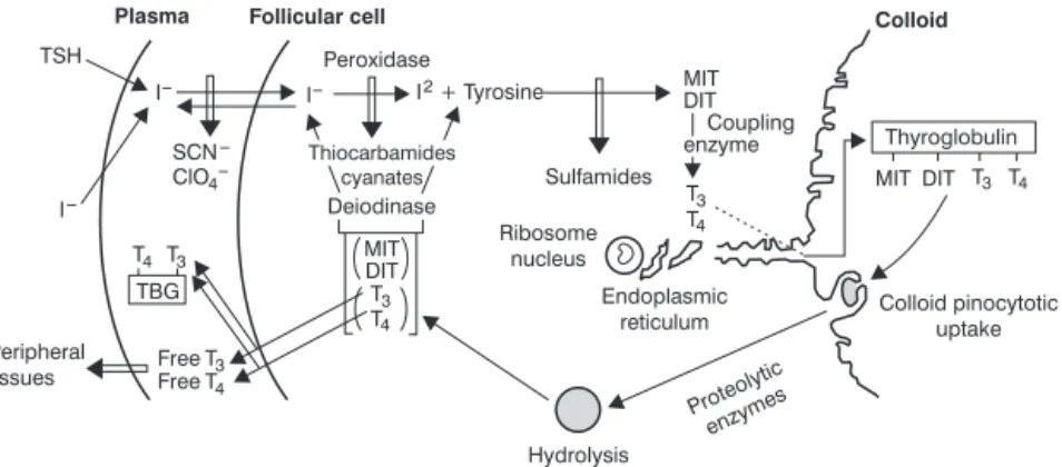
Inorganic iodine present in circulation enters the thyroid follicular cells, where it is organified. This transport depends on the TSH and on a sodium-iodide symporter (NIS), which is located in the membrane of the thyroid cells. In general, an increase in the organic iodine content inside the follicular cells decreases iodide transport; in addition, transport can also be inhibited by some anions, such as perchlorate and thiocyan-ate. Human NIS has been already identified in breast, colon and ovary cells, and tissues such as salivary glands and gas-tric mucosa are also capable of concentrating iodide.3 Pen-drin, a protein of the Pendred syndrome gene, was described after the performance of studies with patients carrying the syndrome, which consists of an association between hypothy-roidism and hearing and speaking impairment. Pendrin also acts in the transport of iodide into follicular cells. Once inside the cell, iodide binds to tyrosine, a thyroglobulin residue. Such iodization is catalyzed by hydroxygen peroxid or peroxidase, whose source is unknown. This passage may be inhibited by thiocarbamides and cyanates.
The thyroid has a limited capacity to use iodides.3In nor-mal conditions, thyroid iodide clearance rates are higher than organification rates (iodide incorporation into amino acids). Progressively higher concentrations of extracellular iodide increase its transport into the cell until organification reaches its maximum rate; then a sudden decrease is observed, a short-duration phenomenon known as Wolff-Chaikoff effect.
After tyrosine organification, the formation of monoiodotyrosines (MIT) and diiodotyrosines (DIT) already
incorporated into thyroglobulin will take place. These hor-mones will couple to form two main TH: triiodothyronine (T3) and tetraiodothyronine (T4). Thyroglobulin is a large soluble protein with a molecular weight of 660 kd that is present in the light of the thyroid follicle (colloid). Only three to four T4 molecules are formed in each thyroglobulin molecule, and the thyroid gland usually produces a significantly greater quan-tity of T4 than T3. The T4 to T3 ratio is 15:1 in normal thyro-globulin. MIT and DIT formation may be inhibited by sulfamides (Figure 1).
Thyroglobulin releases TH by the action of lysosomal pro-teases inside the follicular cell. Colloid droplets then form on the apical surface of the cell via endocytosis, stimulated by TSH; finally, lysosomes release proteolytic enzymes which will in turn release TH. Considerable amounts of circulating thy-roglobulin are only found when the thyroid cell has been dam-aged. Excess iodide inhibits the release of TH. The treatment of severe hyperthyroidism usually benefits from this effect.3
The TH released in the circulation will bind to carrier molecules: globulin (thyroxine-binding globulin – TBG); tran-sthyretin (TTR), previously called prealbumin (thyroxine-binding prealbumin – TBPA); and albumin. TBG binds 70% of T4 and 80% of T3.6,7
Reverse T3 (rT3) derives from the peripheral monodeio-dination of T4.
Mechanism of action of thyroid hormones
Most part of the biological effects associated with TH are determined by interactions between T3 and their specific nuclear receptors. The binding of TH with their nuclear tors allows the transcription of specific mRNA (nuclear recep-tors are transcription facrecep-tors). Nuclear receprecep-tors have a high affinity for T3, and their affinity for T4 is 15 times lower. In the normal animal, about 85% of the total iodothyronine that is bound to the nucleus of hepatic and renal cells are of the T3
DIT = diiodotyrosines; MIT = monoiodotyrosines; T3 = triiodothyronine; T4 = tetraiodothyronine; TBG = thyroxine-binding globulin; TSH = thyroid-stimulating hormone.
type, and only 15% are T4. TH stimulate Na+, K+-ATPase in the cell membrane, increasing the consumption of oxygen.
TH may actually be considered a growth factor, and TH deficiency impairs child growth and development, even when the growth hormone (GH) is present. TH act in practically all tissues of the body and influence enzyme concentration and activity, the metabolism of substrates, vitamins and mineral salts, basal metabolism or calorigenesis; they also stimulate the consumption of oxygen and act in other endocrine sys-tems.1,3
TH stimulate the synthesis and degradation of proteins. The influence of TH on growth is related to its activity in pro-tein synthesis. When TH reach significantly high levels, they accelerate protein catabolism and increase nitrogen excre-tion.
TH alter the metabolism of carbon hydrates. By increas-ing the action of epinephrine, they stimulate glycogenolysis and neoglucogenesis and also improve insulin action in gly-cogen synthesis and glucose use. Low levels of TH increase glycogen synthesis in the presence of insulin, whereas high levels stimulate glycogenolysis. TH also increase the rate of intestinal glucose absorption and its uptake in the adipose and muscular tissues.
TH act on lipid metabolism. In cases of TH insufficiency, a decrease in cholesterol synthesis and its metabolic conver-sion is observed; however, since the degradation is more affected than the synthesis, blood cholesterol levels become high. The opposite is observed in cases of excess TH, when cholesterol, phospholipid and triglyceride levels are low. One mechanism that may contribute to an increase in cholesterol metabolism in response to TH is the ability of TH to increase the number of low-density lipoprotein receptors on the cell surface. By increasing lipolysis in the adipose tissue, TH affect the metabolism of fatty acids.1,3
TH are essential for the development of the central ner-vous system, and deficiency of these hormones during fetal and newborn life extends tissue immaturity, leads to hypopla-sia of cortical neurons, delayed myelinization and reduced vascularization. If hormone replacement therapy is not car-ried out soon after birth, lesions will become irreversible, and the child’s neuropsychomotor development will be damaged.
Effects of TH deficiency: clinical features of hypothyroidism
The early recognition of clinical features in a case of TH deficiency is of fundamental importance and is considered a pediatric emergency in newborn care. Early signs include: pro-longed or recurrent jaundice, delay in umbilical cord separa-tion and umbilical hernia. Crying is hoarse, and sounds emitted are low. In the first months of life, other signs become present: feeding difficulty, insufficient weight gain, noisy breathing, nasal congestion, respiratory disorders, obstipa-tion, lethargy, dry, cold and pale skin, withlivedo reticularis.
However, these signs and symptoms are not always evident, and a precious time can sometimes be wasted before treat-ment is started. This is why the performance of laboratory tests in the nursery room is so important.
Delayed neuropsychomotor development and growth are observed, body proportions are abnormal, and the lower limbs are short if compared to the trunk.
When hypothyroidism is acquired at a later stage, mental retardation may be less evident, but growth will be affected, and these children will present a delay in bone maturation or bone age. In adolescents, hypothyroidism clinical features may show a slower evolution, with tiredness, difficulties at school, intestinal obstipation, dry skin and hair, hair loss, brittle nails, intolerance to cold weather and decreased appe-tite (it is important to emphasize that obesity is not a charac-teristic of hypothyroidism). Girls may present menstrual irregularities, and an increase in menstrual cycle periods are more common than amenorrhea.1
When hypothyroidism remains untreated, more signifi-cant physical alterations may be observed in the long term. The skin becomes cereous, pale or yellowish due to carotene impregnation. Myxedema may occur due to the high concen-tration of mucopolysaccharides in the subcutaneous cell tis-sue and in other tistis-sues. Movements and bone-tendon reflexes are slow. Some children with severe muscle myxe-dema show muscular pseudo-hypertrophy and slow muscle action. Myxedema may affect the cardiac musculature, pos-sibly increasing its volume and finally causing stroke.1
Other endocrine alterations may also be observed in hypothyroidism. Some adolescents may present sexual infan-tilism, and paradoxically, some others may present preco-cious puberty. In the long term, thyrotroph hypertrophy may be observed, with an increase in the pituitary gland and in the sella turcica.8
Differential diagnosis
Differential diagnosis should include Down syndrome, Beckwith syndrome, mucopolysaccharidoses, chondrodys-trophies, hypopituitarism, and obesity. It is always important to take into consideration that hypothyroidism is very rarely associated with obesity.
Congenital hypothyroidism: classification
Table 1 presents the classification and prevalence of con-genital hypothyroidism according to Fisher.9
Hypothyroidism manifestations in practically all tissues do not depend on its etiology, but rather on the degree of hor-mone deficiency.
Neonatal screening
Neonatal screening (heel prick test) should be performed in the nursery room, ideally between 3 and 5 days after birth. Many mothers are discharged from hospital before the third day after delivery; dosages performed before the ideal time increase the prevalence of NB with high levels of TSH, due to the physiological increase of this hormone, and may lead to false-positive results. One drop of blood is collected on a filter paper card. Currently, T4 and TSH dosages may also be car-ried out. TSH values are considered significant when around 20 to 25 µU/mL. Taking into consideration that primary hypothyroidism is the most frequent manifestation of the dis-ease, elevated TSH values allow for early diagnosis and treat-ment. Normal male NB may present low total T4 levels and normal TSH levels. In these cases, free T4 and TBG carrier values should also be assessed. If free T4 values are normal in the presence of TBG deficiency, this means that the boy is normal, or that the diagnosis of congenital hypothyroidism is discarded. The prevalence of TBG deficiency is 1:5,000 to 1:12,000, and its genetic transmission is related to the X gene.15
Dyshormonogenesis
Inborn errors of metabolism correspond to about 15% of the causes of congenital hypothyroidism and are associated
with enzyme defects of autosomal recessive genetic transmis-sion. When the cascade reactions to TH synthesis are ana-lyzed, it is possible to observe that each inefficient enzyme action alters the cascade, causing deficient hormone produc-tion and hypothyroidism. Except for the absence of TSH response, all other forms progress with goiter, which may or may not be present from birth. Several types of hypothyroid-ism have similar clinical features, and their distinction is only possible based on laboratory tests. The only exception is Pen-dred’s syndrome, an organification defect associated with both hypothyroidism and hearing and speaking impair-ment.16,17
Hypothalamic-pituitary hypothyroidism
Central hypothyroidism is relatively rare among NB. Its prevalence is between 1:50,000 and 1:150,000. Up to the 1990s, the dysfunction was considered to be a consequence of trauma associated with delivery. The finding of TH defi-ciency and image exams revealing posterior pituitary ectopia started to suggest that central hypothyroidism might be part of a broader set of pituitary hormone deficiencies, linked to gene mutations of transcription factors involved in hypothalamic-pituitary embriogenesis. POU1F1 (previously Pit1) gene mutations are associated with a subtype of pan-hypopituitarism that evolves with GH, prolactin and TSH defi-ciency. In spite of the severity resulting from the presence of
Table 1- Classification and prevalence of congenital hypothyroidism9
Dysgenesis 1:4,000
Agenesis
Hypogenesis
Ectopia
Dishormonogenesis 1:30,000
Absence of TSH response
Defect in iodide transport or uptake
Defect in organification
Defect in thyroglobulin synthesis
Defect in iodotyrosinase deiodinase
Hypothalamic-pituitary 1:100,000
Hypothalamic-pituitary anomaly
Pan-hypopituitarism
TSH deficiency alone
Resistance to TH
Transitory hypothyroidism 1:40,000
Drug-induced
Induced by maternal antibodies
Pregnant women treated with antithyroid or irradiation drugs
Idiopathic
these multiple hormone deficiencies, they are rarely diag-nosed in the neonatal period. Diagnostic suspicion may occur based on neonatal screening tests showing low levels of T4 and TSH.14,18
Resistance to thyroid hormones
Resistance to TH (RTH) may reveal two different conditions: hypothyroidism, in which all tissues are affected, also known as generalized RTH syndrome; and hyperthyroid-ism, which affects the pituitary more severely, also known as pituitary RTH syndrome. There is consensus in that the phe-notype of these two defects does not correspond to two dif-ferent syndromes, but rather reflects a continuum spectrum of a similar molecular defect with variable tissue resistance. TH receptor proteins are coded by two genes:αgene, located in chromosome 17, andβgene, located in chromosome 3. The molecular defect of the cases studied so far involves theβ1 receptor of chromosome 3. Inheritance is considered to be autosomal dominant, with 15 to 20% of sporadic cases. RTH patients usually present increased serum levels of T3 and T4 and normal or increased TSH results. Newborn screening pro-grams that primarily assess TSH may detect the condition, since TSH can be slightly or moderately high, and increased T4 levels associated with nonsuppressed TSH levels may sug-gest RTH.14The prevalence of RTH has been found to be of approximately 1:100,000 NB in some countries. Patients usu-ally present with delayed growth and goiter, and hyperactiv-ity and attention deficit may be associated. Hearing impairment has also been observed in RTH patients, as a result of the mutation in theβTH receptor,19which is related to recur-rent otitis, with a negative impact on cochlear function.
T3 analog 3,5,3’triiodoacetic acid (Triac), at an initial dose of 1 to 2 mg/day, has been used empirically in the treatment of RTH, and has been observed to improve symptoms and thy-roid function parameters (TSH and T4 levels are low, whereas T3 remains high).19
Subclinical hypothyroidism
This denomination applies to asymptomatic patients pre-senting normal T3 and T4 levels and slightly high TSH levels. This type of hypothyroidism is considered to be mild and to represent a risk factor for evolution to overt hypothyroidism and other dysfunctions. Diagnostic implications start with the definition of normal TH levels, more specifically TSH levels. The accepted cut-off point for normal TSH levels is 4 to 5 mU/L, which has been conventionally used to diagnose high concen-trations of TSH.20Some studies have considered lower cut-off points, of 2 to 2.5 mU/L; however, justifications for adopting such numbers were considered insufficient, and as a result it has been recommended that normal TSH levels be maintained at 0.4-4 mU/L. The classification of results between 2 and 4 mU/L as abnormal and the consequent intro-duction of medication in these cases would most likely have more disadvantages than advantages.
Subclinical hypothyroidism is considered a risk factor for some cardiovascular diseases, hypothyroidism, alterations in lipid and carbohydrate metabolism, neuromuscular symp-toms, and decreased energy metabolism. When limits con-sidered to be normal are exceeded, patients should be assessed for signs that justify treatment with levothyroxine: goiter, presence of antiperoxidase and thyroglobulin antibod-ies, manic-depressive disorders, fertility problems, preg-nancy or anticipation of delivery, autoimmune thyroiditis patients (risk for progression of thyroid dysfunction) and chil-dren and adolescents with or without goiter (to avoid possible side effects on growth and development).
TSH may return to normal levels spontaneously, without medication, in about 40% of the cases, which explains the ori-gin of controversies about the treatment of subclinical hypothyroidism: cardiovascular risk factors have not been totally proved; there is not a defined standard for TSH normalization; treatment cost and noncompliance are rel-evant issues; and T4 overdoses may worsen osteoporosis. TSH levels should be monitored carefully to prevent them from going below normal, since T3 and T4 stimulate bone resorp-tion and increase the number of osteoblasts.
If parameters contraindicating treatment are found, it is recommended that clinical and laboratory assessments be carried out every 6 months.
Transient hypothyroidism
In this situation, hormone levels behave similarly as in pri-mary hypothyroidism, that is, low T4 levels and high TSH lev-els will be observed. The prevalence of transient hypothyroidism varies according to different geographical regions, is related to the intake of iodine and is higher at lower gestational ages. Premature newborns require higher levels of iodine than term newborns in order to maintain a positive balance of iodine and an adequate production of T4 in extra-uterine life; therefore, in areas that are geographically poor in iodine, newborns may develop neonatal iodine deficiency. Transient hypothyroidism manifests itself in the first or sec-ond week of life, usually associated with transient hypothy-roxinemia of prematurity. Treatment is recommended, as this form of hypothyroidism may persist for several months.
Transient hypothyroxinemia
These patients are usually premature newborns with clini-cal features similar to those of tertiary or hypothalamic hypothyroidism. The condition is transient and resolves spon-taneously by the 10th week of life. Treatment is not neces-sary, except if TSH levels are high.15
Other causes
intake of drugs containing iodine is rare, but should also be considered.
Chronic lymphocytic thyroiditis or autoimmune/ Hashimoto’s thyroiditis
In 1912, Hashimoto first described chronic lymphocytic thyroiditis in women with asymptomatic goiter. After surgical removal of the gland, the author classified them asstruma lymphomatosa. Later on, in 1938, diagnosis was made in chil-dren presenting goiter with lymphocytic infiltrate. Up to 1956, when antibodies were detected, it was considered a rare dis-ease in pediatrics, but since then incidence numbers have been increasing. Currently, Hashimoto’s thyroiditis is consid-ered to be the most frequent thyroid disease in pediatric patients when compared to other autoimmune thyroid dis-eases.
Etiopathogeny
Chronic lymphocytic thyroiditis (CLT) is basically deter-mined by immunological mechanisms and can be detected in the blood by the presence of antithyroglobulin and peroxi-dase antibodies. CLT and Graves’s disease are controlled by altered autoimmune processes, and sometimes it is difficult for pathologists to differentiate between both conditions. Cases of patients with classic histological manifestations of CLT and classic clinical manifestations of Graves’s disease have been described. Both processes may appear in the same family and share HLA haplotypes. Major histocompatibility complex (MHC) genes are responsible for different immuno-logical responses, including thyroid auto-antigens. The high incidence of CLT in the female sex at any age group suggests the participation of mutant dominant X chromosome genes, or even an influence by the absence of chromosome Y, with changes in genetic susceptibility potentially associated with chromosomes X and 21. This could explain the high incidence of CLT in Turner and Down syndromes (trisomy 21). There have been reports of families with homozygote twins where one child has CLT and the other Graves’s disease. Such fami-lies very frequently are found to carry autoimmune diseases, and sometimes cases of diabetes mellitus, pernicious ane-mia, myasthenia gravis, rheumatoid arthritis and Addison’s disease are also detected. Although genetic predisposition plays its part in CLT etiopathogeny, few patients evolve to a clinically evident manifestation, and the great majority of patients will most likely remain in a subclinical status, which the authors denominate immunological surveillance status.
Incidence
CLT is considered to be the most common thyropathy among children and adolescents, and it is recognized as the main cause of nontoxic goiter. In an American population with age between 11 and 18 years, five new cases were detected out of 1,000 adolescents screened every year. The incidence is higher among girls, varying from 4:1 to 8:1 depending on
the geographical region covered. The disease is rare before 4 years of age and is frequent between 10 and 11 years.
Clinical features
The presence of goiter is one of the main complaints. The gland presents a diffuse increase in volume (two to five times its normal size) and is generally not nodular. The natural his-tory of the disease is as follows: 1) toxic, transient, self-limited thyroiditis; 2) euthyroid goiter; 3) hypothyroidism with/without goiter. However, children may be in any of these phases on the first medical consultation, since there is not a fixed duration for each stage. The clinical course of toxic thy-roiditis may vary from weeks to months. In this phase, labo-ratory data (TH and antibodies) may be confounded with hypothyroidism data. Therefore, it is often difficult to estab-lish a clear clinical profile. Many children may remain euthy-roidic for some years and then present the clinical features of hypothyroidism.21Children and adolescents with low stature or a progressively lower growth rate, delayed bone age, dry skin and other hypothyroidism-related aspects, even in the absence of goiter, may present a more severe form of hypothy-roidism, in which the gland has become fibrotic. Therefore, patients with CLT should be reassessed periodically, with spe-cial attention to the finding of nodules on ultrasound, which may require a biopsy puncture to prevent the development of tumor (10 to 25% of these nodules may be carcinomas).22,23
Hashimoto’s encephalopathy, which consists of the involvement of the central nervous system in an encephal-opathy status, should be considered in cases of unknown eti-ology. Adolescents with a positive history for the presence of antibodies, even in euthyroid situations (normal T4 and TSH), and who present a progressive cognitive decline should be assessed. Although the etiology is unknown, a good response to steroid medications suggests an inflammatory or autoim-mune dysfunction.22,23Antibodies are considered important markers for the identification of patients who will benefit from an efficient treatment with glucocorticoids.
Laboratory features of hypothyroidism
The diagnosis of congenital hypothyroidism can be con-firmed based on T4 and TSH dosages. In the neonatal period, i.e., between 1 and 4 weeks of life, T4 levels < 6.5 µg/dL and TSH levels > 10 mU/L are suggestive of congenital hypothy-roidism.
Hypothalamic-pituitary hypothyroidism is characterized by low levels of T4 and normal or even low levels of TSH. Decreased TSH response during the TRH test suggests a diag-nosis of central hypothyroidism.
Especially after 4 years of age, in addition to T4 and TSH values, antithyroglobulin and antiperoxidase antibodies should also be assessed to diagnose Hashimoto’s thyroiditis.
The presence of thyroglobulin in serum indicates paren-chymal lesion and may be a tumor marker.22,23
Thyroid ultrasound will always be an important labora-tory test for the purposes of diagnosis and follow-up. Images showing an irregular texture in the parenchyma are sugges-tive of thyroiditis. The presence of nodules or cysts deserves special attention in order to discard the possibility of carcino-mas.24,25
Two and 24-hour thyroidal radioactive isotope uptake tests with99mTc or123I are carried out to diagnose ectopic glands, agenesis or thyroid dysgenesis.26
Treatment
In the nursery room, screening can ensure early diagno-sis and treatment (in the first 3 to 4 weeks of life), thus guar-anteeing an adequate neuropsychomotor development for the NB.
TH replacement is the simplest among all hormone replacement therapies. The drug of choice is levothyroxine (L-T4 sodium salt), which allows measuring serum T4 levels to assess the efficacy of treatment and adjusting doses. Levothyroxine has a mean life of 7 days, and the maximum response is reached in the second week of treatment, when great part of T3 will have been converted. It is administered once a day in the morning. Table 2 shows the recommended doses for different age groups.
These doses may change according to laboratory varia-tions. They should be adjusted whenever signs of overdose are observed: irritability, inability to sleep, red areas on the skin, diarrhea, tachycardia, and sweatiness. Breastfed infants submitted to high doses of levothyroxine may develop cran-iostenosis.
Since these children may present some degree of psycho-motor disorder, they should be followed by professionals from the areas of speech therapy, physical therapy and psichope-dagogy.27
References
1. Setian N. Hipotireoidismo congênito. In: Setian N, editora. Endocrinologia pediátrica: aspectos físicos e metabólicos do recém-nascido ao adolescente. São Paulo: Sarvier; 2002. p. 259.
2. Morreale de Escobar G. The role of thyroid hormone in fetal neurodevelopment. J Pediatr Endocrinol Metab. 2001;14 Suppl 6:1453-62.
3. Griffin JE. The thyroid. In: Griffin JE, Ojeda SR, editors. Textbook of endocrine physiology. New York: Oxford University Press; 2004. p. 294-318.
4. Foley TP. Disorders of the thyroid in children. In: Sperling MA, editor. Pediatric endocrinology. Philadelphia: W B Saunders; 1996. p. 171-94.
5. Dunn JT. Endemic goiter and cretinism: an update on iodine status. J Pediatr Endocrinol Metab. 2001;14:1469-73.
6. Dai G, Levy O, Carrasco N. Cloning and characterization of the thyroid iodide transporter. Nature. 1996;379:458-60.
7. Ingenbleek Y, Young V. Transthyretin (prealbumin) in health and diseases nutritional implications. Annu Rev Nutr. 1994;14:495-533.
8. Anasti JN, Flack MR, Froehlich J, Nelson LM, Nisula BC. A potential novel mechanism for precocious puberty in juvenile hypothyroidism. J Clin Endocrinol Metab. 1995;80:276-9.
9. Fisher DA. Thyroid disease in the neonate and childhood. In: De Groot LJ, editor. Endocrinology. Philadelphia: Saunders; 1981. p. 733.
10. Clifton-Bligh RJ, Wentworth JM, Heinz P, Crisps MS, John R, Lazarus JH, et al. Mutation of the gene encoding human TTF-2 associated with thyroid agenesis, cleft palate and choanal atresia. Nat Genet. 1998;19:399-401.
11. Sheng HZ, Moriyama K, Yamashita T, li H, Potter SS, Mahon KA, et al. Multistep control of pituitary organogenesis. Science. 1997; 278:1809-12.
12. Damante G. Thyroid defects due to Pax 8 gene mutation. Eur J Endocrinol. 1998;139:563-6.
13. Macchia PE, Lapi P, Krude H, Pirro MT, Missero C, Chiovato L, et al. PAX8 mutations associated with congenital hypothyroidism caused by thyroid dysgenesis. Nat Genet. 1998;19:83- 6.
14. Kambe E, Seo H. Thyroid specific transcription factors. Endocr J. 1997; 44:775-84.
15. Ares S, Escobar-Morreale, HF, Quero J, Duran S, Presas MJ, Herruzo R, et al. Neonatal hypothyroxinemia: effects of iodine intake and premature birth. J Clin Endicrinol Metab. 1997; 82:1704-12.
16. Medeiros-Neto G, Bunduki V, Tominori E, Gomes S, Knobel M, Martin R, et al. Prenatal diagnosis and treatment of dyshormonogenetic fetal goiter due to defective thyroglobulin synthesis. J Clin Endocrinol Metab 1997;82:4239-42.
17. van Tijn DA, de Vijlder JJ, Verbeeten Jr B, Verkerk PH, Vulsma T. Neonatal detection of congenital hypothyroidism of central origin. J Clin Endocrinol Metab. 2005;90:3350-9.
18. Kunitake JM, Hartman N, Henson LC, Lieberman J, Williams DE, Wong M, et al. 3,5,3’ – triiodothyroacetic acid therapy for thyroid hormone resistance. J Clin Endocrinol Metab. 1989;69: 461-6.
19. Yoh SM, Chatterjee VK, Privalski ML. Thyroid hormone resistance syndrome manifests as an aberrant interaction between mutant T3 receptors and transcriptional corepressors. Mol Endocrinol. 1997;11:470-80.
Table 2- Recommended levothyroxine doses for children and ado-lescents
Age Dose (µg/kg/day)
0 to 3 months 10 to 15
3 to 12 months 6 to 10
1 to 3 years 4 to 6
3 to 10 years 3 to 5
20. Brabant G, Beck-Peccoz P, Jarzab B, Laurberg P, Orgiazzi J, Szabolcs I, et al. Is there a need to redefine the upper normal limit of TSH ? Eur J Endocrinol. 2006;154:633-7.
21. Radetti G, Gottardi E, Bona G, Corrias A, Salardi S, Loche S; Study Group for Thyroid Diseases of the Italian Society for Pediatric Endocrinology and Diabetes (SIEDP/ISPED). The natural history of euthyroid Hashimoto’s thyroiditis in children. J Pediatr. 2006; 149:827-32.
22. Saravanan P, Dayan CM. Thyroid autoantibodies. Endocrinol Metab Clin North Am. 2001;30:315-37, viii.
23. Janes SE, Santosh B, Thomas D, Vyas H. Hashimoto’s encephalopathy: an unusual cause of seizures in the intensive care unit. Pediatr Crit Care Med. 2004;5:578-81.
24. Hegedüs L. Bonnema SJ, Bennedbaek FN. Management of simple nodular goiter: current status and future perspectives. Endocr Rev. 2003;24:102-32.
25. Bubuteishvili L, Garel C, Czernichow P, Leger J. Thyroid abnormalities by ultrasonography in neonates with congenital hypothyroidism. J Pediatr. 2003;143;759-64.
26. Meier DA, Kaplan MM. Radioiodine uptake and thyroid scintiscanning. Endocrinol Metab Clin North Am. 2001;30:291-13, viii.
27. Bongers-Schokking JJ, de Muinck Keiser-Schrama SM. Influence of timing and dose of thyroid hormone replacement on mental psychomotor, and behavioral development in children with congenital hypothyroidism. J Pediatr. 2005;147:768-74.
Correspondence: Nuvarte Setian