Copyright © 2007 by Sociedade Brasileira de Pediatria
R
EVIEWA
RTICLENot every diabetic child has type 1 diabetes mellitus
Thais Della Manna*Abstract
Objective:Although it is type 1 diabetes mellitus of autoimmune origin that is most prevalent in childhood and adolescence, other forms of diabetes can also affect this population, resulting in different prognosis and treatment.
Sources:Information was obtained by means of a bibliographic review, carried out by running searches for scientific articles in the MEDLINE and LILACS databases, in addition to classic publications on the subject, with the most representative being chosen.
Summary of the findings:This article discusses the pathophysiological mechanisms, clinical presentation and treatment of the various forms of diabetes that affect the pediatric age group, such as type 1 diabetes mellitus, type 2 diabetes mellitus, maturity-onset diabetes of youth, neonatal diabetes, mitochondrial diabetes, diabetes of general-ized lipodystrophy, diabetes secondary to other pancreatic diseases, diabetes secondary to other endocrine diseases, diabetes associated with infections and cytotoxic drugs and diabetes related to certain genetic syndromes.
Conclusions:Recognition of the primary pathophysiologic mechanism of the form of diabetes presented can guide specific treatment, optimizing metabolic control and minimizing complications over the long term.
J Pediatr (Rio J). 2007;83(5 Suppl):S178-183:Diabetes mellitus, differential diagnosis, child, diabetes mellitus neonatal, syndromes.
Introduction
Diabetes mellitus (DM) is a metabolic disease of multiple etiologies. It is characterized by chronic hyperglycemia result-ing from disorders in the metabolism of carbohydrates, pro-teins and fats, secondary to insufficient and/or absent insulin secretion, and also to defects in its action on target tissues (liver and muscle and adipose tissue).1
The current classification of DM is based on pathophysi-ologic knowledge, and includes four clinical classes: type 1 DM, type 2 DM, other specific types of DM and gestational DM.
Type 1 diabetes mellitus
Type 1 DM is the most common form among children and adolescents, caused by partial or total destruction of the beta cells of the Langerhans islets, resulting in progressive inca-pacity to produce insulin. This aggression is generally of an autoimmune nature, resulting both from environmental and genetic processes. There is a great propensity to diabetic ketoacidosis, which is a severe state of diabetic decompensa-tion with immediate risk of death. Insulin is always necessary
for treatment of type 1 DM, and should be initiated as soon as diagnosis is confirmed.
The serological markers of immunological destruction of pancreatic beta cells are the autoantibodies for islet cells (ICA), for insulin (IAA), for glutamic acid decarboxylase (GAD 65) and for tyrosine phosphatase (IA 2). Frequently, more than one of these antibodies is present in 90% of individuals at the point of diagnosis.2
Type 2 diabetes mellitus
Type 2 DM is the result of insulin resistance mechanisms associated with defects in the secretion of this hormone, and is the most prevalent form of diabetes worldwide. It can have onset at any age, but it is most frequently diagnosed after 40 years of age. The increase in the number of cases of type 2 DM in the young has followed the increase in prevalence of childhood obesity. Currently, type 2 DM accounts for a consid-erable proportion of recently diagnosed cases of diabetes in the pediatric population of some clinics in North America, pri-marily adolescents from minority populations, such as His-panic Americans, African-Americans, and the Natives of North
* Doutora em Ciências, Faculdade de Medicina, Universidade de São Paulo (USP), São Paulo, SP, Brazil. Médica assistente, Unidade de Endocrinologia Pedi-átrica, Instituto da Criança, Hospital das Clínicas, Faculdade de Medicina (USP), São Paulo, SP, Brazil.
Suggested citation:Della Manna T. Not every diabetic child has type 1 diabetes mellitus. J Pediatr (Rio J). 2007;83(5 Suppl):S178-183. doi 10.2223/JPED.1714
American and Canada and of certain islands in the Pacific ocean. The changes in lifestyle that took place during the last century, such as changes in the diet and the dramatic reduc-tion in physical activity, together with fetal exposure to hyper-glycemia, in the form of gestational diabetes and glucose intolerance of pregnancy, have determined the impact of this phenomenon.3
There are no specific markers for type 2 DM. Low birth weight and obesity during the prepubescent phase are risk factors for insulin resistance and diabetes.3,4
Type 2 diabetes mellitus generally affects young people in the intermediate phase of puberty with mean age of 13.5 years, affecting more girls than boys, at proportions from 1.6:1 to 3:1. Obesity is present in the great majority of patients, who frequently exhibit body mass index (BMI) over the 85th percentile for their age and sex. In 60 to 95% of cases there isacanthosis nigricans, a heavily pigmented and vel-vety cutaneous lesion that appears primarily on flexible sur-faces such as the neck and armpits. A family history of type 2 DM is very important, and there are generally several mem-bers of the family affected in different generations.5Children with type 2 DM tend to exhibit milder degrees of hyperglyce-mia, more elevated levels of insulin and C-peptide and lower levels of ketonuria and metabolic acidosis. The relationship between insulin resistance and hyperinsulinemia is present from an early age in high-risk populations, indicating suscep-tibility to type 2 DM.
Recent evidence has shown that type 2 DM does not have a favorable prognosis, causing an enormous physical, psycho-logical, economic and social impact. Diabetic children from a group of Pima Indians in North America exhibited a high preva-lence of risk factors for cardiovascular disease, such as severe obesity (85%), hypercholesterolemia (7%), arterial hyper-tension (18%) and microalbuminuria (22%).3
Maturity-onset diabetes of youth (MODY)
Approximately 10% of cases diagnosed as type 1 DM and 5% of cases of type 2 DM are, in fact, maturity-onset diabetes
of youth (MODY), which is a form of diabetes mellitus that is inherited genetically, with early onset, caused by defects in beta cell function and exhibiting an autosomal dominant transmission pattern. In type 1 DM, the parents are generally not diabetic; in MODY, one of the parents is generally affected; and in type 2 DM, both parents have type 2 DM or impaired glucose tolerance.6
With MODY, a discreet level of hyperglycemia will gener-ally go unnoticed, being discovered only during intercurrent diseases or pregnancy. A suspicion of MODY should be aroused when a patient presents with non-insulin dependent diabe-tes, with early onset, a history of diabetes in at least one fam-ily member with onset at less than 25 years and also by the presence of three successive generations suffering from the disease.
At least five genes have been linked to subtypes of MODY:
- Hepatocyte nuclear factor - 4αgene (MODY 1)
- Glucokinase gene - (MODY 2)
- Hepatocyte nuclear factor - 1αgene (MODY 3)
- Insulin Promoter Factor - 1 (IPF-1) gene (MODY 4)
- Hepatocyte nuclear factor - 1βgene (MODY 5)
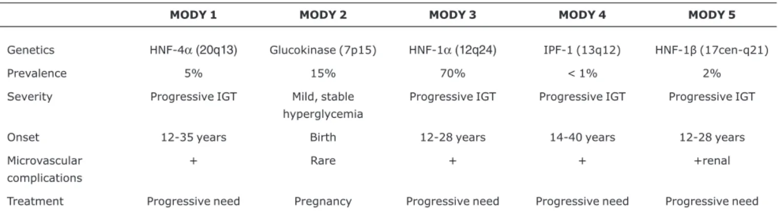
Mutations to the glucokinase gene (MODY 2) and to nuclear transcription factors (MODY 1, 3, 4 and 5) provoke beta cell dysfunction and varying degrees of insulin defi-ciency. Both sexes are affected, and there is no association with obesity. The principal clinical and genetic characteristics of the MODY subgroups are listed in Table 1. Differential diag-nosis between MODY and types 1 and 2 DM is illustrated in Table 2.
Other forms of diabetes mellitus in childhood and adolescence
Other pathological conditions can also involve childhood DM.
Table 1- Genetic and clinical characteristics of MODY subgroups
MODY 1 MODY 2 MODY 3 MODY 4 MODY 5
Genetics HNF-4α(20q13) Glucokinase (7p15) HNF-1α(12q24) IPF-1 (13q12) HNF-1β(17cen-q21)
Prevalence 5% 15% 70% < 1% 2%
Severity Progressive IGT Mild, stable
hyperglycemia
Progressive IGT Progressive IGT Progressive IGT
Onset 12-35 years Birth 12-28 years 14-40 years 12-28 years
Microvascular complications
+ Rare + + +renal
Treatment Progressive need Pregnancy Progressive need Progressive need Progressive need
Neonatal DM is extremely rare, occurring with a fre-quency of 1/400,000 to 1/600,000 live births. It manifests as a hyperglycemic newborn with episodes of dehydration, diffi-culty gaining weight and, frequently, with a history of intrau-terine growth restriction. Ketoacidosis is rarer, but may occur if treatment with insulin is not initiated.
This rare form of insulin-dependent DM manifests within the first month of life and is associated with rare monogenic diseases, and may be transitory, have a transitory and recur-rent (generally in adolescence) course, or can be perma-nent.7,8
The permanent forms can be related to mutations in one of three principal groups of genes:
a) genes involved in the development of pancreatic Langer-hans islets, such as mutations to the PDX-1 or IPF-1 (13q12.1) genes, responsible for synthesizing a transcrip-tion factor that is important for the development of the pancreas, causing neonatal DM associated with exocrine pancreatic insufficiency. Mutations to the EIF2AK3 (2p12) gene determine Wolcott-Rallison syndrome, which progresses with DM of neonatal onset, multiple epiphy-seal dysplasia, convulsions, retarded development, short stature, liver disease and nephropathy.
b) genes involved in insulin synthesis, such as mutations causing loss of glucokinase enzyme function (7p15-p13), with homozygous inheritance. Mutations to the KCNJ11 (11p15.1) gene, which is responsible for changes in the Kir6.2 protein, a component of the adenosine triphos-phate (ATP) sensitive potassium channel of pancreatic beta cells, cause DM with neonatal onset, and may involve epilepsy, retarded development and minor dysmor-phisms. Recently, cases of neonatal DM were described that were caused by mutations to the KCNJ11 gene and where therapeutic success was achieved with an oral sul-phonylurea, glibenclamide, rather than insulin, even in the pediatric age range.8
c) genes related to autoimmunity, such as mutations to the FOXP3 (Xp11.23-q13.3) gene which causes a state of immune dysregulation, polyendocrinopathy with neona-tal DM, enteropathy and X-linked thrombocytopenia (IPEX syndrome).
The transitory form, when associated with intrauterine growth restriction, macroglossia and umbilical and inguinal hernias, can be caused by abnormalities in the distal portions of the long arm of chromosome 6 (6q24). Activating muta-tions of the ABCC8 gene, responsible for alteramuta-tions to the SUR1 protein, which is also a component of the ATP sensitive potassium channel of pancreatic beta cell, cause neonatal onset DM, but more frequently exhibit a transitory clinical course with recurrence at a more advanced age.
Mitochondrial diabetes is the result of transmission of a mutated mitochondrial DNA (mtDNA) with maternal inherit-ance.8,9Patients with mtDNA mutations exhibit a clinical syn-drome that involves relatively simple to recognize mitochondrial dysfunctions, as follows:
Melas
Mitochondrial encephalopathy, lactic acidosis, episodes of cerebral vascular accidents (CVA), short stature, bilateral pro-gressive deafness, DM with onset in adolescence and encephalopathy with onset in the third or fourth decade of life.
Kearns-Sayre syndrome
Sporadic systemic disease, with progressive external oph-thalmoplegia, retinal dystrophy, heart block, cerebellar ataxia and elevated protein levels in cerebrospinal fluid. Diabetes mellitus is a relatively frequent finding and may have onset in childhood.
Pearson syndrome
Sideroblastic anemia, exocrine pancreatic dysfunction, lactic acidosis. Generally DM will have onset early on in child-hood. Affected children do not generally survive beyond ten years.
Table 2- Differential diagnosis between type 1 diabetes mellitus, type 2 diabetes mellitus and MODY
DM type 1 DM type 2 MODY
Beta cell failure Beta cell dysfunction and insulin resistance Beta cell dysfunction
< 24% overweight 85% overweight General/and not obese
5% with family history 75-100% with family history Autosomal dominant
Insulin > 95% Insulin 17-37% Hypoglycemic/diet oral/insulin
F = M F > M F = M
Peak at 10-14 years Peak at 12-14 years Onset at birth
70-80% ICA +85-98% GAD + ICA negative GAD may be + ICA and GAD negative
The forms of diabetes that are associated with genetic defects of insulin activity are characterized by the presence of major resistance to the action of insulin, such as congenital generalized lipodystrophy or Berardinelli syndrome, which has autosomal recessive inheritance, and combines complete absence of adipose tissue with muscular hypertrophy, gross facial features,acanthosis nigricansin flexible skin regions, accelerated growth, hypertriglyceridemia and DM, which gen-erally emerges from the second decade of life on.10
Primary diseases of the exocrine pancreas may involve DM, as with cystic fibrosis and acute and chronic pancreatitis or after pancreatectomies.
The primary cause of cystic fibrosis-related diabetes is deficient insulin secretion; however, insulin resistance sec-ondary to the infectious processes and to certain medica-tions, such as bronchodilators, glucocorticoids, can contribute to hyperglycemia during acute phases of the disease. The appearance of cystic fibrosis related diabetes is a signal of poor prognosis and generally affects adolescents and young adults, compromising even further catabolism and immunoresponse to infections.
Screening is recommended for cystic fibrosis-related DM from 10 years of age onwards, by means of fasting glycemia or by annual oral glucose tolerance tests. Initially, treatment with insulin may be necessary during acute periods and with progression intensive insulin replacement may be indi-cated.11,12
Approximately 50% of cases of acute pancreatitis may exhibit mild to severe hyperglycemia as a complication, and just 1 to 15% of them will progress to permanent DM. In con-trast, up to 85% of cases of chronic pancreatitis will develop DM within 15 years.11
Diabetes mellitus results from all cases of total pancreate-ctomy and from 40-50% of cases of distal pancreatepancreate-ctomy. Since pancreatic alpha cells that produce glucagon are also removed, ketoacidosis is infrequent, although there is great sensitivity to exogenous insulin.11
Other endocrine diseases may also progress with second-ary DM. Approximately 25% of Cushing’s syndrome patients exhibit DM without ketonemia, and which responds to treat-ment of the primary cause of hypercortisolism. In addition to increasing hepatic glucose production by stimulating neo-glucogenesis, glucocorticoids also compromise peripheral glu-cose uptake by insulin target tissues.
Acromegaly, hypersecretion of growth hormone (GH) in adults, frequently caused by adenoma of the pituitary, causes insulin resistance leading to impaired glucose tolerance or even to DM in 15-30% of these patients after 5 to 10 years of the disease. With normalization of GH levels after treatment, glucose tolerance tends to normalize. Growth hormone
replacement therapy given to patients deficient in the hor-mone or to carriers of Turner syndrome can cause minor changes to glycemia and fasting insulinemia.13
Hyperthyroidism provokes insulin resistance and can inhibit secretion of insulin by means of sympathetic mimetic effects of the thyroid hormone. Generally, it causes impaired glucose tolerance, and can worsen the metabolic control of diabetic patients.
Around 75% of pheochromocytoma patients exhibit impaired glucose tolerance as a result of the effect of cat-echolamines stimulating glycogenolysis and lypolisis and inhibiting insulin secretion.
Certain pharmaceuticals and chemical agents can induce DM, generally transitory. The most common situations are those associated with the use of glucocorticoids in high doses, immune suppressors and chemotherapy. Oncological proto-cols that employ L-asparaginase associated with doses of glu-cocorticoids can provoke transitory DM.14In transplantation, the use of cyclosporine and tacrolimus can cause permanent DM due to destruction of Langerhans islet cells.15
Pancreatic beta cells are the target of several viruses which can cause injury by direct cytolytic effects (coxsackievirus) or by activation of autoimmunity. Approximately 12 to 20% of cases of congenital rubella will develop DM in a timeframe of 5 to 20 years. Etiology is probably autoimmune, since around 50 to 80% of those affected are positive for anti-islet and anti-insulin autoantibodies.16
Diabetes mellitus is associated with several hereditary syndromes of varying etiologies, and their recognition has important implications both for specific treatment and prog-nosis and genetic counseling.
Wolfram syndrome (DIDMOAD) is characterized by the presence of diabetes insipidus, DM, atrophy of the optic nerve and sensorineural deafness and has autosomal recessive inheritance associated with non-immunological degenera-tion of pancreatic beta cells. The WSF-1 gene linked to the syndrome is located on chromosome 4.17Clinical manifesta-tions include:
- DM (mean age 8.2 years);
- Atrophy of the optic nerve (mean age 13.1 years);
- Diabetes insipidus(mean age 14.1 years);
- Sensorineural deafness (mean age 15 years);
- Neurological degeneration (atrophy of the central nervous system seen on magnetic resonance imaging);
- Psychiatric disorders;
- Death (mean age 28 years).
syndrome than in the general population, with an estimated prevalence of 1.4 to 10.6%.18Turner syndrome, classically associated with the 45,X karyotype, occurs in approximately 1 in every 2,500 live female births. Impaired glucose toler-ance is reported in 10 to 43%, and DM in 4 to 8%, with unknown etiology. Although autoimmune thyroiditis is highly frequent in this syndrome, there is no evidence that type 1 DM is more prevalent than in the general population.19Growth hormone is often employed in the treatment of short stature in Turner syndrome, and may contribute to increased insulin resistance and hyperglycemia.20
Klinefelter syndrome (47,XXY) appears with a frequency of around 1 in every 1,000 live male births, exhibiting an increased risk for glucose intolerance, probably related to obe-sity in adulthood.21
Certain genetic syndromes that involve obesity are also associated with forms of DM with insulin resistance, gener-ally in adolescence, such as Prader-Willi, Bardet-Biedl and Alström syndromes.
The Prader-Willi syndrome is the most common genetic cause of obesity. It is a rare disease and it has complex etiol-ogy linked to mutations of chromosome 15 (15q11-13). It is clinically characterized by neonatal hypotonia, mental retar-dation, hypogonadism, hypopigmentation, short stature, facial characteristics, small hands and feet and early devel-opment of obesity associated with abnormal carbohydrate metabolism. Approximately 7 to 20% of the patients with this syndrome develop DM, and the etiopathogenesis of this has been attributed to the obesity, through mechanisms similar to those of type 2DM; on the other hand, there are studies that point to differences between Prader-Willi syndrome DM and type 2 DM.22
Bardet-Biedl syndrome has autosomal recessive inherit-ance, and exhibits highly variable clinical status. Its principal characteristics are polydactyly, centripetal obesity, mental retardation, hypogonadism, retinal dystrophy, renal anoma-lies, short stature, speech disorders and dental anomalies. The DM that may be associated with this syndrome is associ-ated with insulin resistance.23
Alström syndrome is a rare autosomal recessive disease that affects multiple organs. These patients may develop early retinal pigment degeneration, sensorineural deafness and metabolic disorders, such as insulin resistance, type 2 DM, hypertriglyceridemia, centripetal obesity, hypogonadism in males, hypothyroidism, accelerated skeletal maturity, which results in short stature, kyphoscoliosis and low levels of GH.24
Treatment
The natural history of DM includes increased risk of chronic microvascular complications, such as retinopathy, nephropa-thy and peripheral neuropanephropa-thy, and also of macroangiopa-thies, such as cardiovascular disease and peripheral vascular
disease. The most effective way to reduce this risk is by main-taining normoglycemia for the greatest percentage of time possible throughout life.
The forms of DM that exhibit total or partial insulin secre-tion deficiency should be given hormone replacement with exogenous insulin. In contrast, in the forms of DM where the primary pathophysiologic mechanism is insulin resistance, control of the conditions causing this resistance should receive priority treatment, such as, for example, withdrawal of a hyperglycemic drug, treatment of obesity and lifestyle changes. In some cases treatment with insulin will be neces-sary for acute control of these disorders.25
References
1. Lebowitz HE. Goals of treatment. In: Lebowitz HE, editor. Therapy for diabetes mellitus and related disorders. 3rd ed. Alexandria: American Diabetes Association; 1998. p. 1-4.
2. Sociedade Brasileira de Diabetes. Tratamento e acompanhamento do diabetes mellitus: Diretrizes da Sociedade Brasileira de Diabetes - 2006. Marins N, editor. Rio de Janeiro, RJ: Diagraphic; 2006. p. 8-11.
3. Dabelea D, Hanson RL, Bennett PH, Roumain J, Knowler WC, Pettitt DJ. Increasing prevalence of Type II diabetes in American Indian children. Diabetologia. 1998;41:904-10.
4. Gautier JF, Wilson C, Weyer C, Mott D, Knowler WC, Cavaghan M, et al. Low acute insulin secretory responses in adult offspring of people with early onset type 2 diabetes. Diabetes 2001; 50:1828-33.
5. Hannon TS, Rao G, Arslanian SA. Childhood obesity and type 2 diabetes mellitus. Pediatrics. 2005;116:473-80.
6. Barret TG. Nonautoimmune forms of diabetes. In: Sperling MA, editor. Type 1 diabetes: etiology and treatment. New Jersey: Humana Press; 2003. p. 163-78.
7. Babenko AP, Polak M, Cavé H, Busiah K, Czernichow P, Scharfmann R, et al. Activating mutations in ABBC8 gene in neonatal diabetes mellitus. N Engl J Med. 2006;355:456-66.
8. Pearson ER, Flechtner I, Njolstad PR, Malecki MT, Flanagan SE, Larkin B, et al; Neonatal Diabetes International Collaborative Group. Switching from insulin to oral sulfonylurea in patients with diabetes due to Kir6.2 mutations. N Engl J Med. 2006;355:467-77.
9. Alcolado JC, Laji K, Gill-Randall R: Maternal transmission of diabetes. Diabet Med. 2002;19:89-98.
10. Setian N. Síndromes associadas a peso e/ou estatura elevados. In: Setian N, editor. Endocrinologia pediátrica. 2ª ed. São Paulo: Sarvier: 2002. p. 149-60.
11. O’Toole P, Lombard M. Pancreatic disease and diabetes mellitus. In: Pickup J, Williams G editors. Textbook of diabetes. 2nd ed. Oxford, UK: Blackwell Science; 1997 p. 24.1-24.12.
12. Della Manna T. Avaliação do comprometimento endócrino do pâncreas em crianças e adolescentes portadores de fibrose cística [tese]. São Paulo: Faculdade de Medicina da Universidade de São Paulo; 2005.
14. Pui CH, Burghen GA, Bowman WP, Aur RJ. Risk factors for hyperglycemia in children with leukemia receiving L-asparaginase and prednisone. J Pediat. 1981;99:46-50.
15. Drachenberg CB, Klassen DK, Weir MR, Wiland A, Fink JC, Bartlett ST, et al. Islet cell damage associated with tacrolimus and cyclosporine: morphological features in pancreas allograft biopsies and clinical correlation. Transplantation. 1999;68:396-402.
16. Yoon JW. Environmental factors in the pathogenesis of insulin-dependent diabetes mellitus. In: Pickup J, Williams G, editors. Textbook of diabetes. 2nd ed. Oxford, UK: Blackwell Science; 1997. p. 14.1-14.14.
17. Barrett TG, Bundey SE, Macleod AF. Neurodegeneration and diabetes: UK nationwide study of Wolfram (DIDMOAD) syndrome. Lancet. 1995;346:1458-63.
18. Anwar AJ, Walker JD, Frier BM. Type 1 diabetes mellitus and Down's syndrome: prevalence, management and diabetic complications. Diabet Med. 1998;15:160-3.
19. Veld PAI, Bruining J. Genetic syndromes. In: Pickup J, Williams G, editors. Textbook of diabetes. 2nd ed. Oxford, UK: Blackwell Science: 1997. p. 28.1-28.11.
20. Wilson DM, Frane JW, Sherman B, Johanson AJ, Hintz RL, Rosenfeld RG. Carbohydrate and lipid metabolism in Turner syndrome: effect of therapy with growth hormone, oxandrolone, and a combination of both. J Pediatr. 1998;112:210-7.
21. Aoki N. Klinefelter's syndrome, autoimmunity, and associated endocrinopathies. Intern Med. 1999;38:838-9.
22. Schuster DP, Osei K, Zipf W. Characterization of alterations in glucose and insulin metabolism in Prader-Willi subjects. Metabolism. 1996;45:1514-20.
23. Beasles PL, Elcioglu N, Woolf AS, Parker D, Flinter FA. New criteria for improved diagnosis of Bardet-Biedl syndrome: results of a population survey. J Med Genet. 1999;36:437-46.
24. Marshall JD, Bronson RT, Collin GB, Nordstrom AD, Maffei P, Paisey RB et al. New Alström syndrome phenotypes based on the evaluation of 182 cases. Arch Intern Med. 2005;165:675-83.
25. Della Manna T, Damiani D, Dichtchekenian V, Setian N. Diabetes mellitus na infância e adolescência. In: Setian N, editor. Endocrinologia pediátrica. 2ª ed. São Paulo: Sarvier; 2002. p. 195-231.
Correspondence: Thais Della Manna
Instituto da Criança – HC-FMUSP Av. Dr. Enéas de Carvalho Aguiar, 647 CEP 05403-900 – São Paulo, SP – Brazil Tel.: +55 (11) 3069.8536