Ultrasound evaluation of obstructive uropathy and its
hemodynamic responses in southwest Nigeria
_______________________________________________
I. N. Apoku
1, O. O. Ayoola
1, A. A. Salako
2, B. M. Idowu
11 Department of Radiology, Obafemi Awolowo University Teaching Hospitals Complex, Ile - Ife, Osun
state, SouthWest Nigeria; 2 Department of Surgery, Obafemi Awolowo University Teaching Hospitals Complex, Ile - Ife, Osun state, SouthWest Nigeria
ABSTRACT
ARTICLE
INFO
______________________________________________________________ ______________________
Purpose: To determine the renal arterial hemodynamic changes induced by obstructive uropathy using Doppler ultrasonography.
Materials and Methods: 60 adult subjects with suspected obstructive uropathy and 60 asymptomatic apparently healthy controls with normal renal ultrasound features were evaluated.
B-mode sonography of the kidneys and spectral Doppler examination of the renal interlobar arteries of all the participants were performed. The mean resistive indices (mRI) of both interlobar arteries were obtained and compared to that of the controls. The mRI of bilaterally obstructed kidneys were also compared with the mRI of unila-terally obstructed kidneys.
Results: The mRI of the right and left kidneys of subjects were 0.72±0.04 and 0.69±0.06 while those of the controls were 0.64±0.04 and 0.63±0.03 respectively. The mRI for the grades of caliectasis increased from grade I (0.72±0.03) to grade II (0.73±0.03) and grade III (0.73±0.02) but fell within the most severe levels of obstruction (0.69±0.07). There was no statistically significant relationship between the grades of caliectasis and unilateral or bilateral obstruction for both kidneys. The results show a sensitivity and specificity of 86.7% and 90% respectively when mRI≥0.7 was used to determine presence of obstruction.
Conclusion: Renal duplex sonography is highly sensitive and specific for diagnosis of obstructive uropathy. Increased resistive index of the obstructed kidney may be a useful diagnostic tool in situations where intravenous urography cannot be done or is contraindicated.
Key words:
Urologic Diseases; Renal Artery; Hemodynamics;
Ultrasonography
Int Braz J Urol. 2015; 41: 556-61
_____________________
Submitted for publication: April 20, 2014
_____________________
Accepted after revision: September 28, 2014
INTRODUCTION
Obstructive uropathy is a commonly en-countered problem in clinical practice. It refers to structural impedance to the flow of urine anywhe-re along the urinary tract leading to pelvicalyceal dilatation. Renal parenchymal damage resulting from obstructive uropathy is termed “obstructive nephropathy” (1). Obstructive uropathy is often secondary to anatomical pathologies of the
MATERIALS AND METHODS
This prospective, non-randomized, case--control study was carried out in a tropical uni-versity teaching hospital from July, 2011 to June, 2012. Approval for the study was obtained from the Ethics and Research Committee of the hospital. Verbal informed consent was obtained from all participants. Sixty adults aged 18 years and abo-ve, referred for ultrasonography with symptoms of obstructive uropathy along with 60 age-and sex-matched apparently healthy control subjects were recruited consecutively by the urologist and referred to the sonologist who was blinded to the clinical status of the subjects to minimize bias. The sonologist who carried out the examination was a third year resident at the Radiology department of the institution while the urologist had been prac-ticing as a specialist for 15 years. All subjects had no clinical symptoms and signs, or sonographic features of urinary tract infection. Any participant with clinical symptoms (dysuria, flank pain), signs (renal angle or suprapubic tenderness), or sono-graphic features (thickened urinary bladder wall, urine containing mobile low-level internal echoes) suggestive of urinary tract infection were exclu-ded from the study. Pregnant women, sickle cell disease patients, diabetic patients, subjects with renal artery stenosis, smokers, hypertensive pa-tients, subjects with congenital renal anomalies, and subjects with elevated urea and creatinine values were excluded from the study. Their age, sex, symptom duration and prospective diagno-sis were documented. Subjects who presented as emergency were examined immediately while the others were examined after at least eight hours fasting to reduce intra-abdominal gas and enhan-ce visualization of the renal arteries.
Renal sonography was done in both supine and prone positions using 3.5-5MHz transducer of MINDRAY® Real time Ultrasound Machine: Model
DC-6 (Shenzhen Mindray Bio-medical Electronics, Nanshan, Shenzhen, China). Both kidneys of each subject were initially examined by ultrasonogra-phy to rule out congenital anomalies and renal artery stenosis and then to assess for caliectasis. The presence of hydronephrosis was documented for each subject on B-mode and graded as
descri-bed by Sehata et al. (3) Thereafter, Doppler stu-dies (color and spectral) of the interlobar arteries was done as previously described (4). The Doppler mode was activated with the Doppler angle adjus-ted to 60 degrees or less and kept parallel to the vessel wall. The sample gate size used was 1mm. The interlobar artery resistive indices were recor-ded for each kidney in subjects and controls by taking three reproducible wave forms from each kidney and averaging them to derive the mRI of the kidney. Finally, a general abdominopelvic ul-trasound was done to determine the possible cau-se of obstruction. Independent samples t test was used to compare the mean age and mRI of sub-jects and controls. One-way analysis of variance (ANOVA) was used to compare means of variables where there were three or more groups. Chi square test was used to evaluate categorical variables e.g. grades of caliectasis in the proportions of male and female patients. The significance level was set at p<0.05. Data analysis was done using the Statistical Package for Social Sciences (SPSS Inc., Chicago, IL, USA), version 17.0 for Windows.
RESULTS
There were 120 participants comprising 60 study subjects and 60 controls. The study subjects comprised 27 (45%) men and 33 (55%) women, while the controls comprised 32 (53%) men and 28 (47%) women; p=0.465. The mean age of the study subjects was 48.68±16.69 years (mean±SD) while that of controls was 36.97±11.19 years.
Of the 60 subjects, 13.3% were below 30 years of age, while 23.3%, 20% and 8.3% were in their 4th, 5th and 6th decades of life
respective-ly. Twenty-one (35%) subjects were aged 60 years and above. Sixteen (26.7%) controls were younger than 30 years of age, while 40%, 16.7% and 10% were in their 4th, 5th and 6th decades of life, res-pectively. Four (6.7%) controls were aged 60 years and above.
The median symptom duration in subjects was 8 months with a range of 1 week to 5 years.
ova-rian cysts, cervical carcinoma, rectal carcinoma, ureteral stricture and vesico-ureteral obstruction respectively.
Twenty (33.3%) subjects had unilateral obstruction (either left or right kidney), of which 19 (31.7%) had only right renal obstruction whi-le 1 had only whi-left renal obstruction. Thirty-three (55%) subjects had bilateral obstruction while 7 (11.7%) subjects had no obstruction at all on B--mode sonography.
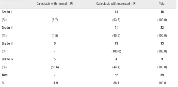
Based on the assessment of the right kid-ney, 15 subjects (7 men and 8 women) had grade I caliectasis, 22 (12 men and 10 women) had gra-de II caliectasis, 14 (five men and nine women) had grade III caliectasis while nine (3 men and 6 women) had grade IV caliectasis (Table-1). The mRI of the right kidney of subjects was 0.72±0.04,
while that of their left kidney was 0.69±0.06 (p=0.000). In the controls, the mRI of the right kidney was 0.64±0.04 while that of the left kidney was 0.63±0.03 (p=0.598), (Table-2).
The mRI of the right kidney in male jects was 0.72±0.04 while that of the female sub-jects was 0.73±0.04 (p=0.703).
Based on a cut-off mRI value of >0.70, the presence of obstructive nephropathy as documen-ted on B-mode ultrasound was predicdocumen-ted with a sensitivity of 86.7% indicating 52 of the 60 study subjects were identified correctly. The specificity at the same value was 90% indicating 54 of the 60 controls were predicted correctly as not having obstructive nephropathy as adjudged by absence of caliectasis. Fifty-two (86.7%) subjects had ri-ght renal obstruction (mRI>0.70) while 8 (33.3%)
Table 1 - Grades of right renal caliectasis and mRI in subjects.
Caliectasis with normal mRI Caliectasis with increased mRI Total
Grade I 1 14 15
(%) (6.7) (93.3) (100.0)
Grade II 1 21 22
(%) (4.5) (95.5) (100.0)
Grade III 0 13 13
(% ) - (100.0) (100.0)
Grade IV 5 4 9
(%) (55.6) (44.4) (100.0)
Total 7 52 59
% 11.9 88.1 100.0
Pearson x2=19.69, df=3, p=0.000
Table 2 - Mean Resistive Index (mRI) in the study participants.
Right kidney Left kidney p Value Note on analysis
Subjects, n=60 0.72±0.04 0.69±0.06 0.000 paired t test, r = 0.4; p=0.000
had normal right kidney (mRI<0.70). Thirty-four subjects (56%) had left renal obstruction while 26 (43.3%) had normal left kidneys.
Analysis of the relationship between gra-des of caliectasis and mRI of the right kidney showed that in grade I, 1 subject had normal mRI while 14 subjects had increased mRI. In grade II, 1 subject had normal mRI while 21 subjects had increased mRI. All the subjects in grade III had increased mRI. In grade IV, 5 subjects had normal mRI while 4 had increased mRI. The mRI by gra-des of obstruction increased steeply from grade I to III but fell rapidly with the most severe level of obstruction (Table-3). This change was statisti-cally significant (p=0.030). In other words, renal mRI rose with increasing severity of obstruction
up to certain level above which it was less rema-rkable (Figure-1).
When the grades of caliectasis in unila-teral and bilaunila-teral obstruction were compared, there were no statistically significant differences in the proportion of subjects with unilateral or bilateral obstruction across each grade of caliec-tasis (p=0.798).
Comparison of right renal mRI of the uni-laterally and biuni-laterally obstructed right kidney showed no significant difference between the two groups (p=0.171).
One way ANOVA comparing right and left kidney mRI values in cases of bilateral obstruc-tive nephropathy across the grades of caliectasis showed that there was no significant difference
Table 3 - One way ANOVA for right renal mean resistive index by severity of obstruction.
N Mean mRI Std. Deviation 95% Confidence Interval
Lower Bound Upper Bound
Grade I 15 0.7220 0.0336 0.70 0.74
Grade II 22 0.7341 0.0314 0.72 0.75
Grade III 13 0.7346 0.0161 0.72 0.74
Grade IV 9 0.6911 0.0683 0.64 0.74
Total (All subjects) 59 0.7246 0.0395 0.71 0.73
in the mRI values (p=0.270 for the right kidney, and 0.480 for the left kidney). There was a mode-rate positive correlation between the age of stu-dy subjects and right renal mRI (r=0.4, p=0.006). This correlation was also found with the right mRI of the controls (p=0.000). This means that the right renal mRI increased with age in both subjects and controls.
DISCUSSION
Ultrasonography is cheap, readily availa-ble, and also easily identifies the cause of obstruc-tive uropathy. In this study, uterine fibroid was the commonest cause of obstructive uropathy in women while enlarged prostate was the commo-nest cause in men. This is similar to the findings by Soyebi et al. (5).
This study showed that most study sub-jects had raised right renal mRI, which may in-dicate the presence of obstructive uropathy. This was supported by the accompanying calyceal di-latation on grey-scale examination. With urina-ry obstruction, there is elevation of the pressure within the intrarenal collecting system. This can induce reduction in renal blood flow as a result of increased renovascular resistance. An increase in intra-renal vascular resistance diminishes diasto-lic blood flow velocity in intrarenal arteries and subsequently, the RI is increased (6).
The subjects had significantly higher right and left renal mRI than the controls (p=0.000).
This means that renal artery hemodynamics change in response to urinary tract obstruction.
Therefore, intra-renal duplex Doppler so-nography can provide physiologic information re-flecting the renal hemodynamic status by means of the RI (7).
Our sensitivity and specificity using the re-nal mRI cut-off of 0.70 in the diagnosis of obstruc-tion were 86.7% and 90% respectively. This is close to what Platt and his coworkers (8) reported. They had a sensitivity of 92% and a specificity of 88% and an overall accuracy of 90% in diagnosing the presence or absence of obstruction in an adult po-pulation. Gottlieb and colleagues (9) also reported a sensitivity of 100% in a smaller group of adult
sub-jects. However, a similar study carried out by Chen and coworkers (10) reported a low sensitivity of 53%. The discordance in results could be due to the diffe-rences in the degrees of urinary tract obstruction and the potential vasodilatory effects of non-steroidal medications typically used for the treatment of pain associated with obstructive uropathy (11, 12).
This study also showed that the mRI by grades of obstruction increased steeply from grade I to grade III but fell rapidly with the most se-vere levels of obstruction. This observation was also reported by Platt et al. (13) where a markedly hydronephrotic kidney did not show an increase in RI (i.e. RI<0.70) despite the presence of what was thought to be obvious severe obstruction. This lack of response is believed to be caused by a ma-rked decrease in absolute blood flow in chronically high grade obstruction (grade IV), and decreased filtration pressure produced by minimally func-tioning renal cortex or elevated compliance in a capaciously dilated collecting system (14). Rawa-shdesh et al. (15) also observed that in chronic renal obstruction with marked parenchymal loss, the RI does not change. This was attributed to the probable absence of vasoconstriction at that stage.
CONCLUSIONS
Mean resistive index (mRI) effectively de-monstrates the hemodynamic changes in the renal artery and values ≥0.70 are highly sensitive and specific for identifying an obstructed kidney. The-refore, it may be a very useful tool in patients in whom excretory urography cannot be done or is contraindicated. A limitation of this study is that no other investigative modality was used to cor-roborate the diagnosis or exclusion of obstructive uropathy in the participants.
ABBREVIATIONS
mrI = Mean Resistive Index/Indices
CONFLICT OF INTEREST
REFERENCES
1. Walsh PC, Retnik AB, Vaughan ED, Wein AJ. Pathophysiology of urinary tract obstruction. In: Campbell’s Urology. 7th Ed. Philadelphia: WB Saunders. 1998; pp. 343-60.
2. Klahr S. Obstructive nephropathy; pathophysiology and management. In: Schrier RW (Ed). Renal electrolyte disorder. 5th Ed. Philadelphia: Lippincott-Raven. 1997; pp. 554-89.
3. Sehata AA, Azmy TM, El Beblawy MA, Hassn A, Motawe M. Duplex sonography in obstructive Uropathy. Suez Canal Univ Med J. 2003; 6: 277-90.
4. Norris CS, Pfeiffer JS, Rittgers SE, Barnes RW. Noninvasive evaluation of renal artery stenosis and renovascular resistance. Experimental and clinical studies. J Vasc Surg. 1984;1:192-201.
5. Soyebi K.O, Awosanya G.O.G. Causes of obstructive uropathy at the Lagos University Teaching. Niger Q J Hosp Medicine.1996;6:173-7.
6. Klahr S, Pukerson ML. The pathophysiology of obstructive nephropathy: the role of vasoactive compounds in the hemodynamic and structural abnormalities of the obstructed kidney. Am J Kidney Dis. 1994;23:219-23.
7. Karadeniz T, Topsakal M, Eksioğlu A, Ariman A, Basak D. Renal hemodynamics in patients with obstructive uropathy evaluated by color Doppler sonography. Eur Urol. 1996;29:298-301.
8. Platt JF, Rubin JM, Ellis JH. Distinction between obstructive and nonobstructive pyelocaliectasis with duplex Doppler sonography. AJR Am J Roentgenol. 1989;153:997-1000. 9. Gottlieb RH, Luhmann K 4th, Oates RP. Duplex ultrasound
evaluation of normal native kidneys and native kidneys with urinary tract obstruction. J Ultrasound Med. 1989;8:609-11.
10. Chen JH, Pu YS, Liu SP, Chiu TY. Renal hemodynamics in patients with obstructive uropathy evaluated by duplex Doppler sonography. J Urol. 1993;150:18-21.
11. Sjödin JG. Clinical experience of indomethacin in pain from ureteral stone. Scand J Urol Nephrol Suppl. 1983;75:35-6. 12. Kmetec A, Peskar-Babnik D, Buturovic-Ponikvar J.
Time-dependent changes of resistive index in acute renal obstruction during nonsteroidal drug administration. BJU Int. 2002;89:847-50.
13. Patti G, Menghini ML, Todini AR, Marrocco G, Calisti A. The role of the renal resistive index ratio in diagnosing obstruction and in the follow-up of children with unilateral hydronephrosis. BJU Int. 2000;85:308-12.
14. Kapoor R, Saha MM, Mandal AK. Antenatal sonographic detection of Wolffian duct cyst. J Clin Ultrasound. 1989;17:515-7.
15. Rawashdeh YF, Djurhuus JC, Mortensen J, Hørlyck A, Frokiaer J. The intrarenal resistive index as a pathophysiological marker of obstructive uropathy. J Urol. 2001;165:1397-404.