Impact of retrograde flexible ureteroscopy and
intracorporeal lithotripsy on kidney functional outcomes
_______________________________________________
Nicolas Hoarau
1, Francois Martin
1, Souhil Lebdai
1, Denis Chautard
1, Thibaut Culty
1, Abdel Rahmene
Azzouzi
1, Pierre Bigot
11 Département d’urologie, Hôpital universitaire d’Angers, Angers, France
ABSTRACT
ARTICLE
INFO
______________________________________________________________ ______________________
Objective: The aim of the study was to evaluate renal function and to identify factors associated with renal function deterioration after retrograde intrarenal surgery (RIRS) for kidney stones.
Materials and Methods: We retrospectively analyzed patients with renal stones trea-ted by RIRS between January 2010 and June 2013 at a single institute. We used the National Kidney Foundation classification of chronic kidney disease (CKD) to classify Glomerular Filtration Rate (GFR) in 5 groups. The baseline creatinine level was syste-matically pre-operatively and post-operatively evaluated. All patients had a creatinine blood measurement in June 2013. A change toward a less or a more favorable GFR group following RIRS was considered significant.
Results: We included 163 patients. There were 86 males (52.8%) and 77 females (47.3%) with a mean age of 52.8±17 years. After a mean follow-up of 15.5±11.5 mon-ths, median GFR was not significantly changed from 84.3±26.2 to 84.9±24.5 mL/min (p=0.675). Significant renal function deterioration occurred in 8 cases (4.9%) and sig-nificant renal function amelioration occurred in 23 cases (14.1%). In univariate analy-sis, multiple procedures (p=0.023; HR: 5.4) and preoperative CKD (p=0.011; HR: 6.8) were associated with decreased renal function. In multivariate analysis these factors did not remain as predictive factors.
Conclusion: Stone management with RIRS seems to have favorable outcomes on kid-ney function; however, special attention should be given to patients with multiple procedures and preoperative chronic kidney disease.
Key words:
kidney stones, holmium laser lithotripsy, renal function, retrograde intrarenal surgery, urolithiasis
Int Braz J Urol. 2015; 41: 920-6
_____________________
Submitted for publication: August 08, 2014
_____________________
Accepted after revision: December 04, 2014
INTRODUCTION
Retrograde intrarenal surgery (RIRS), com-bined with Holmium laser lithotripsy is widely ap-plied for the management of intra-renal stones. For large or multiple stones, RIRS may represent an alternative therapy to percutaneous nephro-lithotomy (PNL) even if multiple procedures are often required (1, 2). It is accepted that stone re-moval can improve renal function; however, a
stone-removing procedure may negatively impact the kidney parenchyma (3). Recently, El-Tabey et al. reported that Percutaneous Nephrolithotomy (PNL) for calculi in solitary kidneys provided sig-nificant improvement in renal function at long--term follow-up (4).
under water pressure. Irrigation during endosco-py cool the tip of energy-delivering devices and helps to maintain a clear visual field by displacing blood, stone fragments, and cellular debris. Howe-ver, this leads to prolong renal calices distension. Moreover, delivering laser energy next to or di-rectly onto renal tissue may cause damage to the renal papillae. Thus, one could advocate neither prolonging nor multiplying the surgical procedure to avoid renal function deterioration and to de-crease post-operative complications. The type and the time of post-operative ureteral catheterization could also protect from renal dysfunction.
The aim of this study was to evaluate renal function and identify factors associated with renal function deterioration or amelioration after RIRS.
MATERIALS AND METHODS
Patients
After approval from an institutional re-view board, we retrospectively analyzed 163 pa-tients undergoing 205 RIRS for intra-renal stones between January 2010 and June 2013 in an aca-demic department of Urology. All patients with a unique radio-opaque stone were previously trea-ted by at least one procedure of Shock Wave Li-thotripsy (SWL) without result. Patients were pre and post-operatively evaluated by computed to-mography. All procedures were conducted under sterile urine and verified by a urine culture seven days before the RIRS. Confirmed urinary infection or bacteriurias were systematically treated du-ring at least five days with adapted antibiothe-rapy. We recorded patient age, gender, body mass index, number of stones, diameter of the largest stone, cumulative stone diameter, and stone com-position. The perioperative parameters analyzed were the use of pre-operative stenting, the mean operative time, the use of a UAS (Ureteral Access Sheath), the presence of a post-operative ureteral stent or a JJ-stent, the number of procedures and the mean follow-up. The stone free (SF) status was defined when no residual fragments (<2mm) were seen on a non-contrast enhanced tomography performed 1 to 2 months after the last retrograde flexible ureteroscopy session. We performed ano-ther RIRS in case of significant persistent stone
(>4mm) on the post-operative tomography. RIRS were performed between 2 and 3 months after the previous procedure.
Surgical procedure
The procedures were performed by 10 di-fferent surgeons from the same institution. All procedures were done under general anesthesia in a standard flexible ureteroscopy installation. A 0.035 inch polytetrafluoroethylene coated wire was placed in the upper urinary tract under visu-al and fluoroscopic control through a rigid cys-toscope. A safety wire was routinely used. The use of a UAS (Ureteral Access Sheath with AQ, Cook Medical, Spencer, USA) was done under the assessment of the surgeon, as well as its inser-tion method. When no UAS was used, the flexible ureteroscope (Flex-X2 TM, Karl Storz endocopy, Tuttlingen, Germany) was inserted in a monorail way over the second wire. Normal saline irriga-tion was performed at a pressure of 60 cm H2O through the same channel used for working ins-truments. If necessary a transient water pressure was carried out using a hand pump. A holmium--YAG laser was used at an energy varying betwe-en 0.6 to 0.8 Joules, and at a frequbetwe-ency of 8 to 10 Hertz. A 270µm or 400µm laser fibre was used for delivering laser energy. At the end of the pro-cedure a JJ stent or a ureteral stent was inserted under the assessment of the surgeon.
Renal function evaluation
The evaluation of the Glomerular Fil-tration Rate (GFR) was derived from the Modi-fication of Diet in Renal Disease (MDRD) study group equation (5). The baseline creatinine level was systematically pre-operatively and post-op-eratively (day one) evaluated. All patients had a creatinine blood measurement in June 2013.
Statistical analysis
Pair t-test was used for comparisons of GFR before and after RIRS. Independent-sample t-test and chi-square tests were used for compa-risons of means and proportions, respectively. Univariate and multivariate regression models were used to assess the influence of different variables on renal function outcomes. Only fac-tors that were significant in univariate analysis were considered for multivariate analysis. All tests were done using SPSS® version 10.
RESULTS
Patient and stone characteristics
We included 163 patients. There were 86 males (52.8%) and 77 females (47.3%) with a mean age of 52.8±17 years. The mean BMI was 26.2±5.9 cm/kg. Patients presented a GFR greater than 60 mL/min/1.73m2 in 128 (78.5%) cases. Patients presented 3a, 3b and 4 preope-rative CKD stage in 27 (16.6%), 7 (4.3%) and 1 (0.6%) cases, respectively. The mean GFR was 84.30±26.2 mL/min/1.73m2.
Multiple stones were present in 73 pa-tients (44.7%). The mean diameter of the lar-gest stone was 12.9±5.7mm, and the cumulative stone diameter was 15±8.6mm. Stone compo-sition was calcium oxalate monohydrate, cal-cium oxalate dehydrate, uric acid, carbapatite, and unknown in 56 (34%), 8 (5%), 13 (8%), 27 (16.6%) and 86 (53%) cases, respectively. A pre-operative stent was inserted in 46 (63.8%) cases. The mean operative time was 96.4±40.78 mi-nutes. A ureteral access sheath was used in 144 (88.3%) procedures and a postoperative ureteral JJ stent was left in 115 (70.6%) cases. Multiple procedures were performed in 29 (17.8%) pa-tients 24 (14.7%) papa-tients had two procedures and 5 (3%) patients had 3 procedures). At the end of the follow-up, 121 (74.2%) patients were stone free.
Perioperative complications occurred in 16 (9.8%) patients (5 pyelonephritis, 3 macros-copic hematuria, 6 pains with the necessity of grade II analgesia, and 1 urinoma). Patient and stone characteristics are reported in the Table-1.
Table 1 - Characteristics of patients and Calculi.
N=163 patients Sex, male (n,%) 86 (54.6 %) Age (y) 52.8±17 BMI (cm/kg2) 26.2±5.9
Multiple stones (n,%) 73 (44.7%) Diameter of the largest stone 12.9±5.7 Cumulative stone diameter (mm) 15±8.6
Stone composition
Calcium oxalate monohydrate (n,%) 56 (34%) Calcium oxalate dehydrate (n,%) 8 (4.9%) Uric acid (n,%) 13 (8%) Carbapatite (n,%) 27 (16.6%) Unclear (n,%) 59 (36.1%) Preoperative stenting (n,%) 46 (63.8%) Mean operation time (min) 96.4±40.78 Ureteral Access Sheath (n, %) 144 (88.3%) Postoperative ureteral JJ stent 115 (70.6%) Multiple procedures (n, %) 29 (17.8%) Stone free rate (n,%) 121 (74.2%) Follow-up (months) 15.5±11.5
Analysis of the immediate post-operative renal function
The median GFR after procedure was 89.45 mL/min/1,73m2. An acute kidney injury, defined as any GFR less than 15 mL/minute/1.73m2, oc-curred in one (0.06%) patient and no patients ne-eded dialysis. Significant renal function change occurred in 18 cases (11%) with 13 (7.9%) amelio-ration and 5 (3%) deterioamelio-ration.
Analysis of long-term renal function
change towards a worse or a better CKD group was observed in 8 (4.9%) and 23 (14.1%) cases, respectively (Table-2).
Analysis of predicting factors of renal function changes
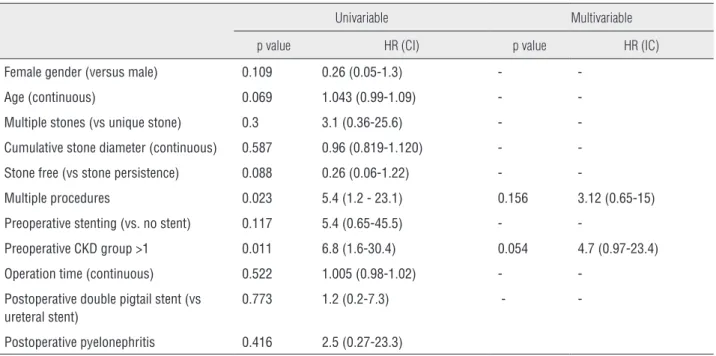
The univariate Cox regression analysis sho-wed only two significant factors for renal deterio-ration: presence of multiple procedures and pre--existing chronic kidney disease (p=0.023; HR=5.4 and p=0.011; HR=6.8 respectively). In multivariate analysis these factors did not remain as predicti-ve (p=0.156; HR=3.12 and p=0.054; HR=4.7) (Ta-ble-3). None of the analysed factors were predicti-ve of renal function amelioration (Table-4).
DISCUSSION
In this study, we intended to evaluate the incidence of CKD after RIRS for stone manage-ment. A key finding of this study is that RIRS se-emed to have a small impact on kidney function and was associated with 14.1% of long-term im-provement.
Ureteroscopy is recommended by the Euro-pean Association of Urology guidelines as a first--line treatment for proximal ureteral stones gre-ater than 1 cm, but they do not recommend this procedure as a first-line therapy for intra-renal stones (7). This is essentially due to the efficiency and the non-invasive nature of SWL. Moreover,
complications of RIRS are often arguments for its detractors. Nonetheless, if infection or urete-ral injuries have been widely reported and stu-died, the renal functional outcomes after RIRS are unknown (8-10).
As we know, the use of pressurized water during RIRS leads to a dilatation of renal cavities. However, the consequences of these high intrarenal pressures have not been studied. What we do know is that excessive renal pressure is the main factor of kidney destruction during acute obstructions. Tubular function is threatened by acute excessive urinary pressure. The renal tubular cells stretched by the hydrostatic pressure leads to a tubular in-terstitial inflammation with macrophages proli-feration and myoblasts accumulation. Alteration of the tubular cells associated with macrophages and myoblasts infiltration lead to the production of cytokines and growth factors which are respon-sible for renal tubular cell apoptosis. This results in chronic obstructive nephropathy with tubular atrophy and loss of nephrons which are replaced by interstitial fibrosis (11-13).
Concerning evaluation of the GFR, we focused on pre-operative, early post-operative and long-term blood creatinine level measure-ment. Even if the mean GFR before and after RIRS was not significantly different, we found a trend towards GFR improvement. This can be explained by numerous factors. The true preva-lence of obstructive nephropathy is unknown but it
Table 2 - Analysis of renal function after ureteroscopy and intracorporeal lithotripsy for intrarenal stones.
Preoperative Postoperative p
CKD group, n (%)
GFR>60 mL/min/1.73m2 128 (78.5%) 141 (86.5%) 0.003
45≤GFR<59 mL/min/1.73m2 27 (16.6%) 14 (8.6%) 0.018
30≤GFR<45 mL/min/1.73m2 7 (4.3%) 8 (4.9%) 0.529
15≤GFR<30 mL/min/1.73m2 1 (0.6%) 0 (0%) 0.319
GFR<15 mL/min/1.73m2 0 (0%) 0 (0%) 1
Change to a worse CKD group 8 (4.9%) Change to a better CKD group 23 (14.1%)
Table 3 - Univariable and multivariable Cox regression analysis of predicting factors for renal function deterioration in 163 patients treated by retrograde flexible ureteroscopy and intracorporeal lithotripsy for intrarenal stones.
Univariable Multivariable
p value HR (CI) p value HR (IC)
Female gender (versus male) 0.109 0.26 (0.05-1.3) - -Age (continuous) 0.069 1.043 (0.99-1.09) - -Multiple stones (vs unique stone) 0.3 3.1 (0.36-25.6) - -Cumulative stone diameter (continuous) 0.587 0.96 (0.819-1.120) - -Stone free (vs stone persistence) 0.088 0.26 (0.06-1.22) -
-Multiple procedures 0.023 5.4 (1.2 - 23.1) 0.156 3.12 (0.65-15) Preoperative stenting (vs. no stent) 0.117 5.4 (0.65-45.5) -
-Preoperative CKD group >1 0.011 6.8 (1.6-30.4) 0.054 4.7 (0.97-23.4) Operation time (continuous) 0.522 1.005 (0.98-1.02) -
-Postoperative double pigtail stent (vs ureteral stent)
0.773 1.2 (0.2-7.3) -
-Postoperative pyelonephritis 0.416 2.5 (0.27-23.3)
HR=Hazard ratio; CI=Confidence interval
Table 4 - Univariable Cox regression analysis of predicting factors for renal function amelioration in 163 patients treated by retrograde flexible ureteroscopy and intracorporeal lithotripsy for intrarenal stones.
Univariable
p value HR (CI) Female gender (versus male) 0.251 1.7 (0.7- 4.1) Age (continuous) 0.051 1.028 (0.99- 1.056) Multiple stones (vs unique stone) 0.546 0.75 (0.29-1.9) Cumulative stone diameter (continuous) 0.445 0.97 (0.9-1.05) Stone free (vs stone persistence) 0.121 2.1 (0.82-5.35) Multiple procedures 0.546 0.75 (0.295 - 1.9) Preoperative stenting (vs. no stent) 0.765 0.872 (0.35-2.1) Operation time (continuous) 0.311 1.006 (0.99-1.02) Postoperative double pigtail stent (vs ureteral stent) 0.087 3. (0.85-10.6)
HR=Hazard ratio; CI=Confidence interval
is known that nephrolithiasis duration, subsequent urinary tract infections, and size of stones are factors that influence renal function (14-16). As a conse-quence, removing these factors may reasonably in-fluence GFR positively.
fact that patients with multiple procedures often have larger stones and more advanced nephroli-thiasis disease. Interestingly, operative time was not a predictive factor of renal function loss. In this context, kidney function did not seem to be a good argument for limiting the RIRS procedu-re time. Moprocedu-reover, if a UAS is known to decprocedu-rease intra-renal pressure, we did not find any influen-ce on post-operative renal functional outcomes (17). The univariate cox regression analysis also showed that pre-existing renal dysfunction was another factor correlated with renal function. This result is not surprising considering that renal insufficiency is known as a risk factor for intra and post-operative complications in general sur-gery and, more particularly, in renal sursur-gery (18). Only a 63 years old man with previous stage 3b CKD (37 mL/min) had an acute renal failure (13 mL/min) after the second procedure (120 min) for multiple renal stones (cumulative stone diameter of 15mm). He did not need hemodyalisis. Three months after RIRS and with hyperhydratation, his renal function was 28 mL/min.
Considering the superior number of pa-tients changing to a better CKD group, we rese-arched predictive factors of renal function impro-vement but no significant result clearly appears. However, our result confirmed that removal of a stone improves postoperative renal function (3).
To our knowledge, there is no study asses-sing the renal functional outcomes after flexible ureteroscopy. Nevertheless, this point is essential in the therapeutic decision for the management of kidney stone diseases. PNL is considered as a more invasive procedure and is indicated for lar-ger stone diameters (more than 2 cm). For these larger stones, if RIRS is performed, multiple pro-cedures might be required, whereas usually only a single session is required for PNL. Moreover, PNL is known to have a good functional outcome even on solitary kidneys (3, 19). A kidney functional approach in stone management is primordial. In-deed, nephrolithiasis is considered as a chronic disease, and patients often undergo multiple pro-cedures. Thus, the management of nephrolithiasis must take into consideration the kidney functional outcomes especially in fragile patients. According to our data, we believe that the improvements of
laser and flexible ureteroscope technologies raise the possibility of mini-invasive approaches.
There are several limitations to this study. First, this is a retrospective review from a single institution. The results are based on a relatively small sample size and we could not confirm that multiple procedures and preoperative kidney di-sease were prognostic factors of kidney function deterioration in multivariate analysis. Moreover, there might have been a confusing factor due to the contralateral compensation of the non-treated kidney for all patients with two kidneys. However, a solitary kidney model could have been more in-formative but it is rare and represents a very diffe-rent clinical situation. Moreover, kidney function deterioration could also be linked to urolithiasis disease and it is difficult to know the part played by either natural history of the disease or RIRS. Larger cohorts and longer periods of follow-up will be necessary to consolidate these data and to confirm the identified predictive factors.
CONCLUSIONS
After RIRS, 8 (4.9%) patients had a decre-ase and 23 (14.1%) patients had an improvement of their renal function. Stone management with RIRS seems to have favorable outcomes on kid-ney function; however, special attention should be given to patients with multiple procedures and preoperative chronic kidney disease.
CONFLICT OF INTEREST
None declared.
REFERENCES
1. Breda A, Ogunyemi O, Leppert JT, Lam JS, Schulam PG. Flexible ureteroscopy and laser lithotripsy for single intrarenal stones 2cm or greater--is this the new frontier? J Urol. 2008;179:981-4.
2. Breda A, Ogunyemi O, Leppert JT, Schulam PG. Flexible ureteroscopy and laser lithotripsy for multiple unilateral intrarenal stones. Eur Urol. 2009;55:1190-6.
4. El-Tabey NA, El-Nahas AR, Eraky I, Shoma AM, El-Assmy AM, Soliman SA, et al. Long-term functional outcome of percutaneous nephrolithotomy in solitary kidney. Urology. 2014;83:1011-5.
5. Levey AS, Bosch JP, Lewis JB, Greene T, Rogers N, Roth D. A more accurate method to estimate glomerular filtration rate from serum creatinine: a new prediction equation. Modification of Diet in Renal Disease Study Group. Ann Intern Med. 1999;130:461-70.
6. Levey AS, Coresh J, Balk E, Kausz AT, Levin A, Steffes MW, et al. National Kidney Foundation. National Kidney Foundation practice guidelines for chronic kidney disease: evaluation, classification, and stratification. Ann Intern Med. 2003;139:137-47. Erratum in: Ann Intern Med. 2003;139:605.
7. C. Türk, T. Knoll, A. Petrik, et al. Guideliness on urolithiasis. European Association of Guidelines. 2013.
8. Traxer O, Thomas A. Prospective evaluation and classification of ureteral wall injuries resulting from insertion of a ureteral access sheath during retrograde intrarenal surgery. J Urol. 2013;189:580-4.
9. Delorme G, Huu YN, Lillaz J, Bernardini S, Chabannes E, Guichard G, et al. Ureterorenoscopy with holmium-yttrium-aluminum-garnet fragmentation is a safe and efficient technique for stone treatment in patients with a body mass index superior to 30 kg/m2. J Endourol. 2012;26:239-43. 10. de la Rosette J, Denstedt J, Geavlete P, Keeley F,
Matsuda T, Pearle M, et al. The clinical research office of the endourological society ureteroscopy global study: indications, complications, and outcomes in 11,885 patients. J Endourol. 2014;28:131-9.
11. Grande MT, Pérez-Barriocanal F, López-Novoa JM. Role of inflammation in túbulo-interstitial damage associated to obstructive nephropathy. J Inflamm (Lond). 2010;7:19. 12. Chevalier RL, Thornhill BA, Forbes MS, Kiley SC. Mechanisms
of renal injury and progression of renal disease in congenital obstructive nephropathy. Pediatr Nephrol. 2010;25:687-97.
13. Hamdi A, Hajage D, Van Glabeke E, Belenfant X, Vincent F, Gonzalez F, et al. Severe post-renal acute kidney injury, post-obstructive diuresis and renal recovery. BJU Int. 2012;110(11 Pt C):E1027-34.
14. Gopalakrishnan G, Prasad GS. Management of urolithiasis with chronic renal failure. Curr Opin Urol. 2007;17:132-5. 15. Gambaro G, Favaro S, D’Angelo A. Risk for renal failure in
nephrolithiasis. AM J Kidney Dis. 2001;37:233-43.
16. Worcester EM, Parks JH, Evan AP, Coe FL. Renal function in patients with nephrolithiasis. J Urol. 2006;176:600-3; discussion 603.
17. Auge BK, Pietrow PK, Lallas CD, Raj GV, Santa-Cruz RW, Preminger GM. Ureteral access sheath provides protection against elevated renal pressures during routine flexible ureteroscopic stone manipulation. J Endourol. 2004;18:33-6.
18. Hakimi AA, Rajpathak S, Chery L, Shapiro E, Ghavamian R. Renal insufficiency is an independent risk factor for complications after partial nephrectomy. J Urol. 2010;183:43-7. Erratum in: J Urol. 2010;183:1650.
19. Canes D, Hegarty NJ, Kamoi K, Haber GP, Berger A, Aron M, et al. Functional outcomes following percutaneous surgery in the solitary kidney. J Urol. 2009;181:154-60.
_______________________ Correspondence address: