Characterization of reactive stroma in prostate cancer:
involvement of growth factors, metalloproteinase matrix,
sexual hormones receptors and prostatic stem cells
_______________________________________________
Maurício Moreira da Silva Júnior
1, Wagner Eduardo Matheus
1, Patrick Vianna Garcia
2, Rafael Mamprim
Stopiglia
1, Athanase Billis
3, Ubirajara Ferreira
1, Wagner José Fávaro
21 Departamento de Cirurgia, Área de Urooncologia, Faculdade de Ciências Médicas, Universidade
Estadual de Campinas (UNICAMP), Campinas, SP, Brasil; 2 Laboratório de Carcinogênese Urogenital
e Imunoterapia (LCURGIM), Departamento de Biologia Estrutural e Funcional, Universidade Estadual de Campinas (UNICAMP), Campinas, SP, Brasil; 3 Departamento de Patologia, Faculdade de Ciências
Médicas, Universidade Estadual de Campinas (UNICAMP), Campinas, SP, Brasil
ABSTRACT
ARTICLE
INFO
______________________________________________________________ ______________________
Introduction and Objectives: Reactive Stroma (RStr) is observed in many human can-cers and is related to carcinogenesis. The objectives of the present study were to sta-blish a relationship of the RStr microenvironment with prostate cancer (Pca) through a morphological and molecular characterization, and to identify a possible relationship between RStr with worse prognosis factors and occurrence of malignant prostatic stem cells.
Materials and Methods: Forty prostatic samples were selected from men with Pca diag-nosis submitted to radical prostatectomy; they were divided in two groups: Group-1 (n=20): samples without reactive stroma; Group-2 (n=20): samples of PCa with intense stroma reaction. Prostatic samples were evaluated for RStr intensity by Masson Tri-chromic stain and posteriorly submitted to histopathological and immunohistochemis-try analysis for antigens: α-actin, vimentin, IGF-1, MMP-2, FGF-2, C-Myc, PSCA, AR, Erα and ERβ.
Results: Reactive stroma with intense desmoplastic reactivity was significantly more frequent in intermediate (Gleason 7, 3+4) and high grade tumors (Gleason 7, 4+3). The group with intense stromal reactivity showed significant higher levels of Vimentin, IGF-1, MMP-2, FGF-2, C-Myc, PSCA and ERα.
Conclusions: It can be concluded that RStr may be a predictive marker of Pca progres-sion, since it was associated with increase of growth factors, imbalance of androgen and estrogen receptors and presence of malign prostatic stem cells.
Key words:
Reactive stroma, prostate cancer, growth factors, sexual hormones receptors, epithelium-stroma interaction
Int Braz J Urol. 2015; 41: 849-58
_____________________
Submitted for publication: July 17, 2014
_____________________
Accepted after revision: February 05, 2015
INTRODUCTION
Prostatic epithelium comprises three cellu-lar types: luminal or columnar, basal and neuro-endocrine (1). Luminal epithelial cells are the most frequent cell type in normal and hyperplasic
androgen--dependent (5). On the other side, basal cells are relatively undifferentiated, independent of andro-gens but androgen-respondent, they do not show secretory activity and form the basal compartment of the prostate (6). The neuroendocrine cells do not respond to androgens (7-9) and can modify in prostate cancer (number, histology and function) with a suggestive regulatory role in that disease (10, 11).
Reactive stroma (RStr is defined as the microenvironment closely adjacent to epithelium able to coordinate several activities as wound re-pair, homeostasis changes and interaction with neoplastic complexes, comprising an dynamic environment that influence directly the behavior of epithelial cells and perform tissue repair after lesion (12). Modifications of peritumor stroma start in prostatic intraepithelial neoplasia (PIN) and include phenotypic alterations of stromal cells, remodeling of extracellular matrix and in-duction of angiogenesis (13, 14). Reactive stroma (RStr) is defined as a new stroma environment in response to carcinoma. It follows tumor growth and is characterized by an increase of inflamma-tory cells, desmoplastic reaction, increase of an-giogenesis and growth factors, with remodeling of extracellular matrix (15). RStr has a fibroblastic and a myofibroblastic compound associated to tu-mor and the origin of these cells is not clearly understood. Some authors suggest that these cells are originated from the prostatic stroma or smooth muscle or even stem cells (14, 15). Stem cells have the capacity of self-renovation and regeneration throughout adult life and are present in the epi-thelial and stromal compartments (16).
At the prostate several biological processes (regulation of proliferation and cellular differen-tiation, mitogenic activity, secretory processes and tumor growth) are regulated and/or influenced by different growth factors, such as IGF (insulin gro-wth factor), FGF (fibroblast grogro-wth factor), VEGF (vascular endothelium growth factor), transfor-ming growth factors, metalloproteinases and PSCA (prostate stem cell antigen) (17-22).
Additionally, testosterone is an important stimulant to prostatic cell proliferation, mainly when it’s more potent form di-hydro-testosterone (DHT) bind to androgen receptors of cells from the
epithelial and stromal compartments (23, 24), so those above mentioned processes are under direct influence of androgens, estrogens and their alpha (ERα) and beta (ERβ) receptors (25, 26).
Neoplastic transformation consists of a multi-causal process, where normal controls of cellular proliferation and interaction cell-to-cell are lost. Aberrant activation of proto-oncogenes along with non-regulated inhibition of tumor su-ppressor genes are fundamental in that process. In that context, stand out proto-oncogene C-MYC. In tumors, the scarce vascularization and the high proliferative profile lead to a hypoxic status (kno-wn as Warburg effect) that is able to induce the expression of C-MYC, that promotes an energe-tic reinforcement through glycolysis and that can additionally act as a suppressor of antiangiogenic factors in an attempt to oppose hypoxia and to promote adequate metabolic supply demanded by the tumor (27).
In view of the facts discussed above, it is essential to establish a correlation between the stromal microenvironment of prostate adenocar-cinoma through morphologic and molecular cha-racterization, and also to determine any associa-tion of growth factors, matrix metalloproteinases, sexual hormone and stem cell receptors with tu-mors with worse prognosis.
MATERIALS AND METHODS
Human Samples and Histopathological Analysis of Reactive Stroma
Forty prostatic samples of patients submitted to retropubic radical prostatectomy with 60-80 years old (median 71 years) were collected. Samples were obtained from the collection of the Department of Pathology of the Hospital de Clínicas da Universida-de Estadual Universida-de Campinas (UNICAMP).
Samples were collected from the peripheral region based on the division of the posterior side, with basal to apical orientation of the organ. Next, samples were fixed in 10% buffered formaldehyde for 12 hours. After fixation, tissue samples were routinely processed (inclusion in paraffin, 5µm sections and Hematoxilin-Eosin staining).
by a senior pathologist of the Department of Pa-thology of the Faculdade de Ciências Médicas da Universidade Estadual de Campinas (UNICAMP).
For reactive stroma analysis, the prostatic samples were divided in two groups (20 samples per group): Group-1: PCa samples without reacti-ve stroma (Grade-0), Group-2: Pca samples with intense stromal reactivity (Grade-3).
Stromal reactivity was determined at the Urogenital Carcinogenesis Laboratory and Immu-notherapy of the Biological Institute of UNICAMP, using Masson Trichromic stain. The intensity of reactive stroma was evaluated by the frequency (in percentage) of smooth muscular fibers (stained red with Masson Trichromic) adjacent to neoplas-tic areas in each sample with an augment of x400. Images were captured by photomicroscope Leica DM2500 equipped with a Leica camera DFC295 and analyzed by the software Leica LAS V3.7 for image analysis. The percentage of smooth mus-cular fibers adjacent to neoplastic areas was gra-ded and expressed as 0>50% of smooth muscu-lar fibers adjacent to neoplastic ducts, 1:36-50% of smooth muscular fibers adjacent to neoplastic ducts, 2:15-35% of smooth muscular fibers ad-jacent to neoplastic ducts, 3: 0-14% of smooth muscular fibers adjacent to neoplastic ducts. For this study intermediate levels of reactive stroma (grades 1 and 2) were discarded.
The study was approved by the Ethical Committee of Faculdade de Ciências Médicas/UNI-CAMP (#0094.0146.000-08).
Immunohistochemistry for antigens:
α-actin, Vimentin, IGF-1, MMP-2, FGF-2, C--Myc, PSCA, AR, ERα and Erβ
The same prostatic samples of 40 patients used for histopathological analysis ere submitted to immunohistochemistry. Antigen recovery was obtained by incubating the slices in buffered ci-trate (pH 6.0) at 100oC in microwave. The blocka-ge of endoblocka-genous peroxidase was obtained with H2O2 and posterior incubation with a blocking solution with bovine serum albumin (BSA) for 1 hour at room temperature. After that, the antigens were localized with specific antibodies (Table-1), diluted in BSA and incubated overnight at 4oC. It was used the MACH 4 Universal HRP-Polymer® (Biocare Medical) kit for antigen detection, accor-ding to the manufacture instructions. Posteriorly, the slices were revealed with diaminobenzidin (DAB), counter-stained with Harris Hematoxilin and evaluated at the photomicroscope.
In order to evaluate the intensity of the antigen immunoreactions, the percentage of posi-tive epithelial and/or stromal cells was examined in 10 fields for each antibody with an augment of 400x. The intensity of staining was grade in a 0-3
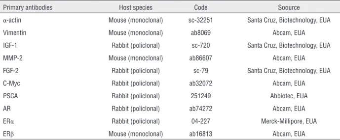
Table 1 - Characteristic of primary antibodies for immuno-staining.
Primary antibodies Host species Code Soource
α-actin Mouse (monoclonal) sc-32251 Santa Cruz, Biotechnology, EUA
Vimentin Mouse (monoclonal) ab8069 Abcam, EUA
IGF-1 Rabbit (policlonal) sc-720 Santa Cruz, Biotechnology, EUA
MMP-2 Mouse (monoclonal) ab86607 Abcam, EUA
FGF-2 Rabbit (policlonal) sc-79 Santa Cruz, Biotechnology, EUA
C-Myc Rabbit (policlonal) ab32072 Abcam, EUA
PSCA Rabbit (policlonal) 251249 Abbiotec, EUA
AR Rabbit (policlonal) ab74272 Abcam, EUA
ERα Rabbit (policlonal) 04-227 Merck-Millipore, EUA
scale and expressed as 0 (no immunoreactivity), 0% of positive epithelial and/or stromal cells, 1 (weak immunoreactivity), 1-35% of positive epi-thelial and/or stromal cells, 2 (moderate immuno-reactivity), 36-70% of positive epithelial and/or stromal cells, 3 (intense immunoreactivity), >70% of positive epithelial and/or stromal cells.
Statistical analysis
The histopathological and immunohisto-chemistry analysis for different antigens were eva-luated with the proportion test. For these analyses, an error type-I of 5% was considered statistically significant.
RESULTS
Histopathological Analysis of Reactive Stroma
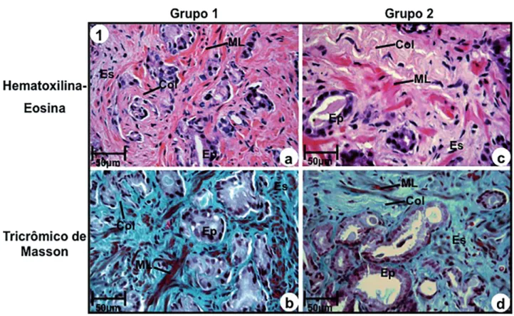
Stroma without desmoplastic reaction (Group-1) was characterized by the presence of a great amount of smooth muscular fibers, above 50% of adjacent ducts with collagen fibers inters-persed among the smooth muscular fibers (Figures 1a and 1b).
In relation to stroma with intense desmo-plastic reaction (Group-2), it was characterized by an outstanding increase and thickening of colla-gen fibers, associated to an intense reduction (be-low 14%) of smooth muscular fibers (Figures 1c
Figure 1 - Photomicrography of prostatic peripheral zone of groups 1 (a, b) and 2 (c, d). (a) and (b) Stroma without desmoplastic reaction consisting of excess of smooth muscular fibers (ML) and thin collagen fibers (Col) adjacent to prostatic ducts. Stains: Hematoxilin-Eosin (a) and Masson Trichromic (b). (c) and (d) Stroma with intense desmoplastic reactivity consisting of excess of collagen fibers (Col) and rare smooth muscular fibers (ML), stains: Hematoxillin-Eosin (c) and Masson Trichromic (d).
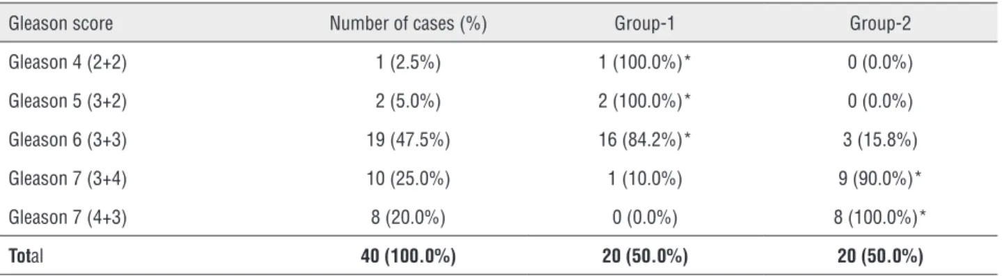
and 1d). Stroma without desmoplastic reaction (Group-1) was significantly more frequent in Gle-ason 4 (2+2), 5 (3+2) and 6 (3+3) (Table-2), and absent in high grade tumors (Gleason 7, 4+3) (Ta-ble-2).
Stroma with intense desmoplastic reactivi-ty was significantly more frequent in intermediate (Gleason 7, 3+4), and high grade tumors (Gleason 7, 4+3) and in low grade tumors it was observed in only 3 cases with Gleason 6 (3+3) (Table-2).
Immunohistochemistry of antigens:
α-actin, Vimentin, IGF-1, MMP-2, FGF-2, C--Myc, PSCA, AR, ERα and ERβ
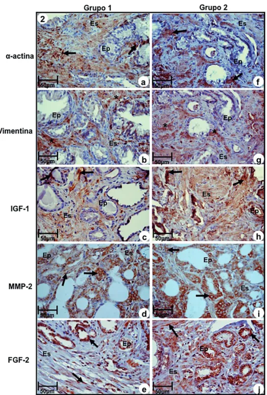
Immunoreactivity to α-actin, a marker of smooth muscle, was significantly more intense in Group-1 in relation to Group-2. The last one sho-wed moderate immune staining (Figures 2a and 2f, Table-3). On the contrary, immunoreactivity of vi-mentin, a fibroblast and myofibroblast marker, was significantly more intense in Group-2 in relation to Group-1, that showed moderate immunoreactivity (Figures 2b and 2g, Table-3).
Immunoreaction to IGF-1, MMP-2 and FGF-2 were significantly more intense in epithe-lium and stroma of samples of Group-2, compared to Group-1, that showed moderate reactivity (Fi-gures 2c, 2d, 2e, 2h, 2i, and 2, Table-3).
Immunoreactivity to C-Myc was signifi-cantly more intense in both epithelium and stroma in samples of Group-2, and moderate in Group-1
Table 2 - Distribution of Gleason score and stromal reactivity in prostatic adenocarcinoma without stromal reactivity (Group-1) and with intense stromal reactivity (Group-2).
Gleason score Number of cases (%) Group-1 Group-2
Gleason 4 (2+2) 1 (2.5%) 1 (100.0%)* 0 (0.0%)
Gleason 5 (3+2) 2 (5.0%) 2 (100.0%)* 0 (0.0%)
Gleason 6 (3+3) 19 (47.5%) 16 (84.2%)* 3 (15.8%)
Gleason 7 (3+4) 10 (25.0%) 1 (10.0%) 9 (90.0%)*
Gleason 7 (4+3) 8 (20.0%) 0 (0.0%) 8 (100.0%)*
Total 40 (100.0%) 20 (50.0%) 20 (50.0%)
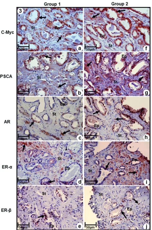
(Figures 3a. and 3f, Table-3). Likewise, immuno-reactivity to prostatic stem cell antigen (PSCA) was observed both in epithelial and stromal com-partments, being significantly more intense in samples of Group-2, compared to Group-1, that showed moderate immunoreactivity (Figures 3b and 3g, Table-3).
Immunoreactivity for AR was modera-te in both epithelium and stroma of samples of Group-2, while in Group-1 it was observed low
immunoreactivity (Figures 3c and 3h, Table-3). Immunoreactivity to ERα was predominant in stromal compartment in both groups, being inten-se in Group-2 and moderate in Group-1 (Figures 3d and 3i, Table-3).
In contrast, immunoreactivity for Erβ was predominant in epithelial compartment of both groups, and immune staining of stroma was pre-sent only in Group-2 (Figures 3e and 3j). Immuno-reactivity of this marker was moderate in Group-1 and weak in Group-2 (Figures 3e and 3, Table-3).
DISCUSSION AND CONCLUSIONS
Figure 2 - Immuno-staining of antigens α-actin, Vimentin, IGF-1, MMP-2 and FGF-2 at prostatic peripheral zone of Groups-1 (a, b, c, d, e) and 2 (f, g, h, i, j). (a) and (f) Immunoreactivity to α-actin (arrows). (b) and (g) Immunoreactivity to Vimentin (asterisks) in myofibroblasts. (c) and (h) Imunoreactivity to IGF-1 (arrows) in epithelium and stromal compartments. (d) and (i) Immunoreactivity to MMP-2 (arrows) in epithelial and stromal compartments. (e) and (j) Immunoreactivity to FGF-2 (arrows) in cells of secretory epithelium and fibroblasts of stromal compartment.
migration processes, angiogenesis, apoptosis and tumor metastasis (28).
In the present study RStr was morphologi-cally characterized by the significant reduction of smooth muscular fibers and excess of collagen fi-bers in stroma adjacent to neoplastic ducts. Inten-se stromal reactivity was obInten-served in intermediate (Gleason 7, 3+4) and high grade tumors (Gleason 7, 4+3) and in low grade tumors it was observed in only 3 cases with Gleason 6 (3+3), pointing out that RStr may be considered a predictive marker of tumor progression.
In relation to molecular characterization of RStr, the results showed increased reactivity to vimentin, IGF-1, MMP-2, FGF-2 and C-Myc in samples with intense stromal reactivity when compared to samples without reactivity. Such ma-rkers were fundamental to activation of RSTr and made the prostatic microenvironment favorable to tumor progression due to increase of imbalance of epithelium-stroma interaction.
Several studies have demonstrated that RStr is associated with lower survival free of di-sease. Yanagisawa (29) analyzed prostatic biopsies of 205 patients and demonstrated a significant di-fference between high and low reactive RSTr and
concluded that the intensity of RStr may be consi-dered a prognostic factor independent of bioche-mical recurrence. Also, Ayala (15), after analyzing samples from radical prostatectomy and Billis (30), that analyzed 266 needle prostatic biopsies showed that RStr could only be considered an prognostic factor independent of biochemical re-currence when it showed intense stromal reactivi-ty. Still, RStr with intense stromal reactivity was observed in Ayala (15), Yanagisawa (29) and Billis (30) studies in 9.0%, 6.7% and 5.3% of samples respectively, with very similar frequencies among the studies. However, RStr with low stromal reac-tivity frequencies were very distinct among these three papers: 6.25% (Ayala, 15), 0.5% (Yanagisa-wa, 29) and 53.8% (Billis, 30). That reflects that lack of uniform morphological criteria to charac-terize RSTr.
In conclusion, the present study shows a new approach to Pca diagnosis and the results de-monstrated RStr may be considered a predictive marker of PCa progression, since increase of vi-mentin, IGF-1, MMP-2, FGF-2 and C-MTC are evi-dences of worse tumor prognosis and the occur-rence of prostatic stem cells (elevation of PSCA) and the balance of AR and Erα with concurrent
Table 3 - Intensity of immuno-staining of different antigens in epithelial and stromal cells of prostatic adenocarcinomas without stromal reactivity (Group-1) and with intense stromal reactivity (Group-2).
Groups
Antigens Group 1 (n=20) Group 2 (n=20)
α-actin 3 (80.3%)* 2 (69.0%)
Vimentin 2 (61.4%) 3 (92.4%)*
IGF-1 2 (57.6%) 3 (96.8%)*
MMP-2 2 (67.3%) 3 (89.7%)*
FGF-2 2 (62.7%) 3 (91.5%)*
C-Myc 2 (65.5%) 3 (93.3%)*
PSCA 2 (56.5%) 3 (85.8%)*
AR 1 (30.9%) 2 (59.5%)*
Erα 2 (39.7%) 3 (77.9%)*
Erβ 2 (38.6%)* 1 (26.5%)
Figure 3 - Immuno-staining of antigens C-Myc, PSCA, AR, ERα and ERβ in prostatic peripheral zone of Groups 1 (a, b, c, d, e) and 2 (f, g, h, i, j). (a) and (f) Immunoreactivity to C-MyC (arrows) in epithelial and stromal compartments (b) and (g) Immunoreactivity to PSCA (arrows ) ) in epithelial and stromal compartments (c) and (h) Immunoreactivity to AR (arrows) ) in epithelial and stromal compartments (d) and (i) Immunoreactivity to ERα (arrows) ) in epithelial and stromal compartments (e) and (j) Immunoreactivity to ERβ (arrows) ) in epithelial and stromal compartments.
inhibitory action of ERβ at RStr point to a grea-ter malignancy of these tumors as well as an in-dication of recurrence. However, new studies are necessary to better understanding of this microen-vironment and upgrading of available treatments of prostate cancer, and the development of new modalities that assure better clinical results and quality of life of patients.
ABBREVIATIONS
AR = androgen receptor
EstR = Reactive stroma
IGF = Growth factor Homologous Insulin
FGF = Fibroblast Growth Factors
VEGF = Vascular Endothelial Growth Factor
MMP = Matrix metalloproteinase
CaP = Prostate cancer
PSCA = Antigen for Prostate Stem Cell
DHT = dihydrotestosterone
ERα= Estrogen Receptor Alpha
ERβ = Estrogen Receptor Beta
ACKNOWLEDGE
Academic and financil support by: Funda-ção de Amparo À Pesquisa do Estado de São Paulo (FAPESP), process number 2009/50397-9.
CONFLICT OF INTEREST
None declared.
REFERENCES
1. Berry PA, Maitland NJ, Collins AT. Androgen receptor signalling in prostate: effects of stromal factors on normal and cancer stem cells. Mol Cell Endocrinol. 2008; 288:30-7. 2. De Marzo AM, Marchi VL, Epstein JI, Nelson WG. Proliferative
inflammatory atrophy of the prostate: implications for prostatic carcinogenesis. Am J Pathol. 1999; 155:1985-92. 3. Mimeault M, Batra SK. Characterization of nonmalignant and
malignant prostatic stem/progenitor cells by Hoechst side population method. Methods Mol Biol.2009;568:139-49. 4. Zenzmaier C, Untergasser G, Berger P. Aging of the prostate
epitelial stem/progenitor cell. Exp Gerontol. 2008;43:981-5. 5. Taylor RA, Risbridger GP. The path toward identifying
prostatic stem cells. Differentiation. 2008;76:671-81.
6. Collins AT, Maitland NJ. Prostate cancer stem cells. Eur J Cancer. 2006;42:1213-8.
7. McNeal JE. Normal histology of the prostate. Am J Surg Pathol. 1988;12:619-33.
8. Bonkhoff H, Wernert N, Dhom G, Remberger K. Relation of endocrine-paracrine cells to cell proliferation in normal, hyperplastic, and neoplastic human prostate. Prostate. 1991;19:91-8.
9. Abrahamsson PA, Dizeyi N, Alm P, di Sant’Agnese PA, Deftos LJ, Aumüller G. Calcitonin and calcitonin gene-related peptide in the human prostate gland. Prostate. 2000;44:181-6. 10. Nelson EC, Cambio AJ, Yang JC, Ok JH, Lara PN Jr, Evans
CP. Clinical implications of neuroendocrine differentiation in prostate cancer. Prostate Cancer Prostatic Dis. 2007;10:6-14. 11. Ruscica M, Dozio E, Motta M, Magni P. Role of neuropeptide
Y and its receptors in the progression of endocrine-related cancer. Peptides. 2007;28:426-34.
12. Dvorak HF. Tumors: wounds that do not heal. Similarities between tumor stroma generation and wound healing. N Engl J Med. 1986;315:1650-9.
13. Tuxhorn JA, Ayala GE, Rowley DR. Reactive stroma in prostate câncer progression. J Urol. 2001;166:2472-83. 14. Ayala G, Tuxhorn JA, Wheeler TM, Frolov A, Scardino
PT, Ohori M, et al. Reactive stroma as a predictor of biochemical-free recurrence in prostate cancer. Clin Cancer Res. 2003;9:4792-801.
15. Rowley DR. What might a stromal response mean to prostate cancer progression? Cancer Metastasis Rev. 1998-1999;17:411-9.
16. Rizzo S, Attard G, Hudson DL. Prostate epithelial stem cells. Cell Prolif. 2005;38:363-74.
17. Djavan B, Waldert M, Seitz C, Marberger M. Insulin-like growth factors and prostate cancer. World J Urol. 2001;19:225-33.
18. Marszalek M, Wachter J, Ponholzer A, Leitha T, Rauchenwald M, Madersbacher S. Insulin-like growth factor 1, chromogranin A and prostate specific antigen serum levels in prostate cancer patients and controls. Eur Urol. 2005;48:34-9.
19. Cornell RJ, Rowley D, Wheeler T, Ali N, Ayala G. Neuroepithelial interactions in prostate cancer are enhanced in the presence ofprostatic stroma. Urology. 2003; 61:870-5. 20. Lakshman M, Huang X, Ananthanarayanan V, Jovanovic B,
Liu Y, Craft CS, et al. Endoglin suppresses human prostate cancer metastasis. Clin Exp Metastasis. 2011;28:39-53. 21. Gu Z, Thomas G, Yamashiro J, Shintaku IP, Dorey F, Raitano
A, et al. Prostate stem cell antigen (PSCA) expression increases with high gleason score, advanced stage and bone metastasis in prostate cancer. Oncogene. 2000; 19:1288-96. 22. Ross S, Spencer SD, Holcomb I, Tan C, Hongo J, Devaux
23. Griffiths K, Morton MS, Nicholson RI. Androgens, androgen receptors, antiandrogens and the treatment of prostate cancer. Eur Urol. 1997;32(Suppl 3):24-40.
24. Habib FK, Chen C: Pathogenesis of benign prostatic hyperplasia. In Chisholm G. Handbook on benign prostatic hyperplasia. Nova Jersey. Whitehouse Station. 1994. 25. Gardner MJ, Hall AJ, Downes S, Terrell JD. Follow up
study of children born elsewhere but attending schools in Seascale, West Cumbria (schools cohort). Br Med J (Clin Res Ed). 1987;295:819-22.
26. Cohen DW, Simak R, Fair WR, Melamed J, Scher HI, Cordon-Cardo C. Expression of transforming growth factor-alpha and the epidermal growth factor receptor in human prostate tissues. J Urol. 1994;152:2120-4.
27. Lee C, Sensibar JA, Dudek SM, Hiipakka RA, Liao ST. Prostatic ductal system in rats: regional variation in morphological and functional activities. Biol Reprod. 1990;43:1079-86. 28. Frank SR, Schroeder M, Fernandez P, Taubert S, Amati B.
Binding of c-Myc to chromatin mediates mitogen-induced acetylation of histone H4 and gene activation. Genes Dev. 2001;15:2069-82.
29. Yanagisawa N, Li R, Rowley D, Liu H, Kadmon D, Miles BJ, et al. Stromogenic prostatic carcinoma pattern (carcinomas with reactive stromal grade 3) in needle biopsies predicts biochemical recurrence-free survival in patients after radical prostatectomy. Hum Pathol. 2007;38:1611-20.
30. Billis A, Meirelles L, Freitas LL, Polidoro AS, Fernandes HA, Padilha MM, et al. Adenocarcinoma on needle prostatic biopsies: does reactive stroma predicts Biochemical recurrence in patients following radical prostatectomy? Int Braz J Urol. 2013;39:320-7.