Acurácia do lavado gástrico realizado em ambiente hospitalar e ambulatorial no diagnóstico da tuberculose pulmonar em crianças
Ethel Leonor Noia Maciel1, Reynaldo Dietze2, Renata Peres Lyrio3, Solange Alves Vinhas3,
Moises Palaci4, Rodrigo Ribeiro Rodrigues5, Claudio Jose Struchiner6
Abstract
Objective: To compare gastric lavage (GL) performed in inpatients with GL performed in outpatients in terms of its accuracy in diagnosing pulmonary tuberculosis (TB) in children. Methods: A prospective study was carried out in the state of Espírito Santo, Brazil, from 1999 to 2003. A total of 230 children suspected of having TB (103 inpatients and 127 outpatients) were selected to undergo GL. Those thus diagnosed with TB (n = 53) were divided into two groups: inpatient GL (n = 30) and outpatient GL (n = 23). All 53 children were monitored for 6 months in order to evaluate the accuracy of the diagnosis. Accuracy was determined based on any change in diagnosis, cure rate, and the percentage of positive cultures in the two groups studied. Results: The cure rate was 100% in both groups, and there was no change in diagnosis in the 53 children studied. No significant difference was found between the two groups studied in terms of detection of Mycobacterium tuberculosis (RR = 1.47; 95% CI: 0.95-2.27; p = 0.095), although the outpatient group presented a greater rate of positive cultures. Conclusions: Our results show that the accuracy of GL performed in an inpatient setting is similar to that of GL performed in an outpatient setting. This indicates that hospitalization is required only in more severe cases in which GL cannot be performed as an outpatient procedure.
Keywords: Gastric lavage; Tuberculosis; Diagnosis.
Resumo
Objetivo: Comparar a acurácia do lavado gástrico (LG) realizado em ambiente hospitalar e ambulatorial no diagnóstico da tuberculose (TB) pulmonar em crianças. Métodos: Estudo retrospectivo realizado no Estado do Espírito Santo, Brasil, de 1999 a 2003. Um total de 230 crianças com suspeita de TB foi selecionado para realizar exame de LG em ambiente hospitalar (n = 103) ou em ambiente ambulatorial (n = 27). Desse total, 53 foram diagnosticadas como casos de TB e divididas em dois grupos: LG hospitalar (n = 30) e LG ambulatorial (n = 23). Todas as 53 crianças foram monitoradas por 6 meses para avaliação da acurácia do diagnóstico. A acurácia foi determinada com base na mudança do diagnóstico, na taxa de cura e no percentual de culturas positivas nos dois grupos estudados. Resultados: A taxa de cura foi de 100% nos dois grupos, e não houve mudança de diagnóstico nas 53 crianças estudadas. Nenhuma diferença significativa foi encontrada entre os dois grupos estudados em relação ao achado do Mycobacterium tuberculosis (RR = 1,47; IC95%: 0,95-2,27; p = 0,095), apesar de o grupo LG ambulatorial ter apresentado o maior índice de cultura positivas. Conclusões: Nossos resultados mostram que a acurácia do LG realizado em ambiente hospitalar é semelhante à do realizado em ambiente ambulatorial, o que indica que a internação é necessária apenas em casos mais graves nos quais não se pode realizar o procedimento em ambiente ambulatorial.
Descritores: Lavagem gástrica; Tuberculose; Diagnóstico.
* Study carried out in Unit 2 of the Infectious Diseases Center. Fundação Oswaldo Cruz – Fiocruz, Oswaldo Cruz Foundation – Rio de Janeiro, Brazil. 1. Adjunct Professor of Epidemiology. Universidade Federal do Espírito Santo – UFES, Federal University of Espírito Santo – Vitória, Brazil. 2. Adjunct Professor of Medicine. Universidade Federal do Espírito Santo – UFES, Federal University of Espírito Santo – Vitória, Brazil.
3. Biologist in the Infectious Diseases Center. Universidade Federal do Espírito Santo – UFES, Federal University of Espírito Santo – Vitória, Brazil. 4. Adjunct Professor of Microbiology. Universidade Federal do Espírito Santo – UFES, Federal University of Espírito Santo – Vitória, Brazil. 5. Adjunct Professor of Immunology. Universidade Federal do Espírito Santo – UFES, Federal University of Espírito Santo – Vitória, Brazil. 6. Head Researcher. Fundação Oswaldo Cruz – Fiocruz, Oswaldo Cruz Foundation – Rio de Janeiro, Brazil.
Correspondence to: Dra. Ethel Leonor Noia Maciel. Núcleo de Doenças Infecciosas, Centro de Ciências da Saúde/UFES, Av. Marechal Campos, 1468, CEP 29040-090, Maruípe,Vitória, ES, Brazil.
Tel 55 27 3335-7210. E-mail: [email protected]
Financial support: Rede Brasileira de Pesquisa em Tuberculose (Rede-TB, Brazilian Tuberculosis Research Network). Submitted: 6 June 2007. Accepted, after review: 10 September 2007.
**A versão completa em português deste artigo está disponível em www.jornaldepneumologia.com.br
Introduction
Worldwide, tuberculosis (TB) is the second leading cause of death from communicable disease, killing nearly 2 million people each year. It is estimated that approximately 2 billion
A positive smear for acid-fast bacilli can be a criterion for treatment initiation. However, the sample collection procedure, the concentration of bacilli in a given sample (paucibacillary sample), and the need to hospitalize patients in order to collect samples have become a problem, especially in devel-oping countries, where hospital beds are scarce.
In a retrospective study, Mtb culture from gastric aspirates collected from children in an outpatient setting was shown to have a yield comparable to that of aspirates collected from hospitalized chil-dren, suggesting that hospitalization might not be necessary for the diagnosis of childhood TB.(11)
The objective of the present study was to compare, prospectively, inpatient and outpatient pediatric GL in terms of the yields of gastric aspi-rates collected for Mtb culture, as well as in terms of accuracy in the diagnosis of pulmonary TB. In addition, we attempted to identify factors associ-ated with a positive culture.
Methods
This was a four-year prospective study of all suspected TB cases treated at two children’s hospi-tals in the state of Espírito Santo, Brazil.
The study was conducted from July of 1999 to August of 2003. During this period, a total of 230 children under 15 years of age were referred to the two hospitals for investigation of TB. Since 1999, in the state of Espírito Santo, there has been a centralized system of allocating hospital beds known as “Central de Vagas”, which is responsible for referring children to various hospitals. We used this system to randomly select children based on the hospital to which they were referred.
The only exclusion criterion was being HIV-positive. The inclusion criteria were history of contact with an index case, signs and symp-toms consistent with TB, a positive TST result, and X-ray findings suggestive of TB. Clinical data were obtained through physical examinations performed by trained physicians.
Information related to disease history (cough, fever, contact with a TB index case, bacillus Calmette-Guérin-BCG-vaccination, and malnutrition) was obtained from parents.
The purified protein derivative RT-23 was used. The execution of TSTs and the interpretation of TST results were conducted according to the Brazilian Among children in developing countries, TB is
responsible for 1.3 million new cases and 450,000 deaths every year.(2) In Brazil, 15% of all reported
TB cases occur in pediatric patients under the age of 15.(3,4)
Programs to control TB focus almost exclusively on adults with sputum smear-positive TB, since they are most infectious individuals. However, children account for a significant proportion of the global TB caseload and experience considerable TB-related effects (morbidity and mortality).(5)
In addition, children have been shown to play an important role in the chain of TB transmission, not as an active source of disease to their household contacts but as major Mtb reservoirs. The Mtb bacilli can survive in a latent state for many years in chil-dren, who, in some cases, later progress to active TB. Therefore, childhood TB is considered an important indicator of the success of public health programs in interrupting and preventing TB transmission.(6)
Signs and symptoms of TB in children are non-specific, which makes clinical diagnosis difficult, delaying treatment and prevention. Childhood TB presents a broad clinical spectrum, ranging from asymptomatic to more severe disseminated forms, typically accompanied by wasting and frequently leading to death.(7)
It is difficult to confirm a diagnosis of TB using the methods currently available. Even in industri-alized countries, the triad of obtaining a positive tuberculin skin test (TST) result, identifying radio-graphic or clinical manifestations consistent with TB, and establishing a recent link to a known infectious TB case is the ‘gold standard’ for diagnosis. Even when these criteria have been met, problems related to the bacteriological confirmation often arise.(5)
Of the first 230 children enrolled in the study, 54 were clinically diagnosed with TB. However, one patient scheduled to undergo inpatient GL was excluded for being HIV-infected. The remaining 53 children were divided into two groups: those undergoing GL at the HINSG (inpatient group; n = 30) and those undergoing GL at the HUCAM (outpatient group; n = 23). All 53 children were monitored for 6 months in order to evaluate the accuracy of the diagnosis. Accuracy was estimated based on any change in the diagnosis, on the cure rate, and on the percentage of positive cultures in the two groups.
National Ministry of Health guidelines for the diag-nosis of childhood TB (Chart 1).(12)
Chest X-ray findings were considered suspicious when at least one of the following patterns was found: right hilar lymph node enlargement, infiltra-tion, cavitainfiltra-tion, atelectasis, and miliary pattern.(13)
Of the 230 children suspected of having TB, 103 were referred to the Hospital Infantil Nossa Senhora da Glória (HINSG, Nossa Senhora da Glória Hospital for Children) to undergo GL as inpatients, and 127 were referred to the Hospital Universitário Cassiano Antônio Moraes (HUCAM, Cassiano Antônio Moraes University Hospital) to undergo GL as outpatients.
Chart 1 - Diagnosis of pulmonary tuberculosis in children and teenagers with negative sputum smear microscopy results. TST and BCG vaccination:
If vaccinated more than 2 years ago: 5-9 mm: add 5 points. 10-14 mm: add 10 points.
≥15 mm: add 15 points. If vaccinated less than 2 years ago:
10-14 mm: add 5 points.
≥15 mm: add 15 points. Not vaccinated:
5-9 mm: add 5 points.
≥10 mm: add 15 points. Clinical and radiographic overview:
Chest X-ray showing condensation or infiltrate for more than 2 weeks: add 15 points. Chest X-ray showing condensation or infiltrate for less than 2 weeks: add 5 points. Normal chest X-ray: subtract 5 points.
Fever or symptoms such as cough, adynamia, expectoration, weight loss, or sweating for more than 2 weeks: add 15 points.
Asymptomatic or symptomatic for less than 2 weeks: 0 points.
Improvement in respiratory infection with the use of antibiotics for common germs or without the use of antibiotics: subtract 5 points.
Nutritional status:
Severe malnutrition or weight-for-age below the 10th percentile (SISVAN): add 5 points.
Weight-for-age at or above the 10th percentile (SISVAN): 0 points.
Contact with an adult with tuberculosis:
Close contact in the last 2 years: add 10 points. Occasional or no contact: 0 points.
Interpretation:
≥40 points: very likely diagnosis. 30-35 points: possible diagnosis.
≤25 points: unlikely diagnosis.
in the inpatient setting were compared with those for GL samples collected in the outpatient setting.
Although the confirmatory method is a labora-tory test, it has low sensitivity and can take as long as two months to yield results. The final diagnosis of TB in this study sample was based on clinical findings, laboratory findings, or both, at the discre-tion of the physician in charge.
In order to evaluate the association between the diagnosis of TB and the setting in which the GL sampling was performed, relative risk was calculated for categorical variables, with a confidence interval of 95%. Continuous variables were analyzed using a t-test for independent samples, with a significance level of 0.05. Statistical data were analyzed using STATA software, version 7.0 (STATA Corp., College Station, TX, USA).
The study protocol was explained to all pedi-atric patients suspected of having TB, prior to their enrollment in the study. Written informed consent was obtained from the parents or legal guardians of the participants.
The study design was reviewed and approved by the Institutional Review Board of the Federal University of Espírito Santo Biomedical Center (Grant nº. 023/99).
Results
No significant difference was found between the inpatient and outpatient groups in terms of demo-graphic variables (Table 1). In addition, there was Sampling was performed at the two study sites (the
HUCAM and the HINSG). Children were required to fast for 8 h prior to GL. Children undergoing inpatient GL were admitted to the pediatric ward. A nasopha-ryngeal catheter was introduced at midnight, and GL was performed at 7:00 a.m. by a registered nurse. The procedure was repeated on 3 consecutive days. Outpatient GL was also performed on 3 consecutive days following a protocol identical to that described above. Patients were required to fast for 8 h prior to GL and to report to the HUCAM outpatient clinic at 7:00 a.m. Parents were provided with transportation vouchers. All outpatient procedures were performed by two registered nurses.
In laboratory tests, staining of smears and solid medium culture were used in order to identify Mtb. Smear samples were processed according to the standard protocol for Ziehl-Neelsen acid-fast bacilli staining.
Sample pellets obtained from the GL samples were inoculated on Löwenstein-Jensen medium, incubated at 37 °C for 6 weeks, and evaluated weekly for bacilli growth. Growth and identification of Mtb on Löwenstein-Jensen medium was used as a confirmatory method. Three GL samples of at least 10 mL were collected from each patient on different days. The quantity to be collected was defined through a pilot laboratory test, since this quantity ranges from 5 to 50 mL in the literature.(6,7,14,15)
Positive GL samples were defined as those in which Mtb grew in one or more cultures of lavage specimens. Culture results for GL samples collected
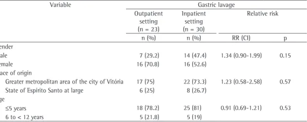
Table 1 - Distribution of demographic variables by the setting in which gastric lavage was performed.
Variable Gastric lavage
Outpatient setting (n = 23)
Inpatient setting (n = 30)
Relative risk
n (%) n (%) RR (CI) p Gender
Male 7 (29.2) 14 (47.4) 1.34 (0.90-1.99) 0.15
Female 16 (70.8) 16 (52.6)
Place of origin
Greater metropolitan area of the city of Vitória 17 (75) 22 (73.3) 1.23 (0.58-2.58) 0.57 State of Espírito Santo at large 6 (25) 8 (26.7)
Age
≤5 years 18 (78.2) 25 (81) 0.91 (0.69-1.21) 0.53 6 to < 12 years 5 (21.8) 5 (19)
Contact with an adult index case occurred in almost all cases studied, and the rates were similar in both groups.
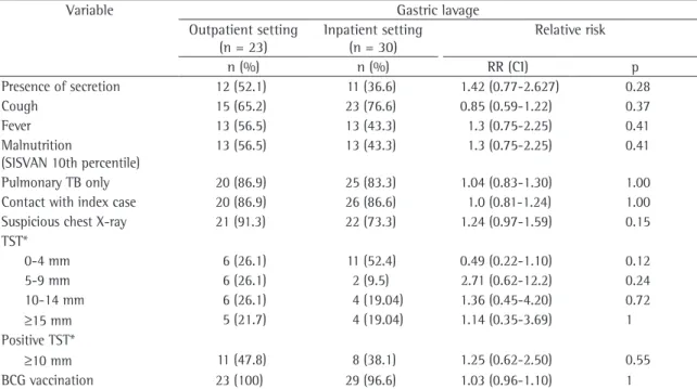
Although the rate of chest X-ray findings consistent with TB was higher in the outpatient group, the difference between the two groups was not significant. In the outpatient group, 72% of the cases presented condensation or infiltra-tion consistent with a diagnosis of TB, and 43% presented hilar lymph node enlargement. In the inpatient group, 86% of the patients whose chest X-ray findings were consistent with TB presented condensation or infiltration, and 38% presented hilar lymph node enlargement.
In our study, the TST response observed was described as pronounced in 11 (47.8%) of 23 chil-dren in the outpatient group (Table 2).
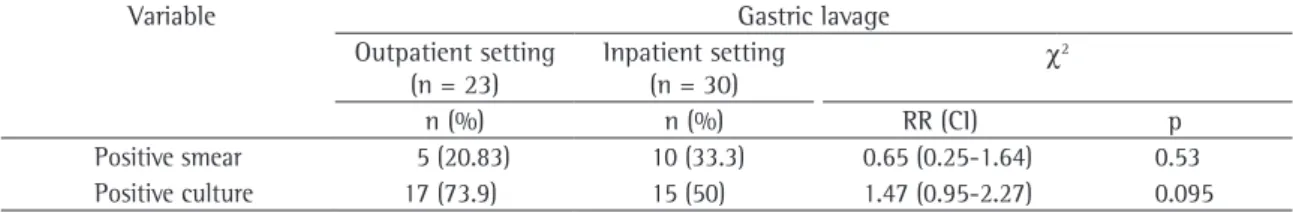
Interestingly, 2 of the 5 smear-positive patients in the outpatient group were culture negative. In parallel, 3 of the 10 smear-positive patients in the inpatient group were culture negative (Table 3). Although there was no significant difference no significant difference between the two groups
in terms of age, which ranged from 8 months to 14 years in the outpatient group and from 1 month to 12 years in the inpatient group.
Of the 23 children in the outpatient group, 17 (75%) were from the greater metropolitan area of the city of Vitória, as were 22 (73.3%) of the 30 children in the inpatient group.
Among the variables associated with clinical history, cough and malnutrition occurred at signif-icant rates in both groups (Table 2). In terms of the presence of secretion, no difference was found between the outpatient group (52.1%) and the inpatient group (36.6%). Although secretion was present in some of the children under 5 years of age, it is difficult to collect sputum from children that young.
The two groups did not differ in terms of the frequency of pulmonary TB: 12.5% of those in the outpatient group and 21.1% of those in the inpatient group presented pulmonary and extrapul-monary TB concomitantly.
Table 2 - Distribution of variables related to clinical and radiographic status of HIV-negative children by place where gastric lavage was performed.
Variable Gastric lavage
Outpatient setting (n = 23)
Inpatient setting (n = 30)
Relative risk
n (%) n (%) RR (CI) p
Presence of secretion 12 (52.1) 11 (36.6) 1.42 (0.77-2.627) 0.28 Cough 15 (65.2) 23 (76.6) 0.85 (0.59-1.22) 0.37 Fever 13 (56.5) 13 (43.3) 1.3 (0.75-2.25) 0.41 Malnutrition
(SISVAN 10th percentile)
13 (56.5) 13 (43.3) 1.3 (0.75-2.25) 0.41
Pulmonary TB only 20 (86.9) 25 (83.3) 1.04 (0.83-1.30) 1.00 Contact with index case 20 (86.9) 26 (86.6) 1.0 (0.81-1.24) 1.00 Suspicious chest X-ray 21 (91.3) 22 (73.3) 1.24 (0.97-1.59) 0.15 TST*
0-4 mm 6 (26.1) 11 (52.4) 0.49 (0.22-1.10) 0.12 5-9 mm 6 (26.1) 2 (9.5) 2.71 (0.62-12.2) 0.24 10-14 mm 6 (26.1) 4 (19.04) 1.36 (0.45-4.20) 0.72
≥15 mm 5 (21.7) 4 (19.04) 1.14 (0.35-3.69) 1 Positive TST*
≥10 mm 11 (47.8) 8 (38.1) 1.25 (0.62-2.50) 0.55 BCG vaccination 23 (100) 29 (96.6) 1.03 (0.96-1.10) 1
the age of the child: 43% in children under 1 year of age, 24% in children from 1 to 5, and 15% in the children from 10 to 15.(17) Our data corroborates the
epidemiological significance of household contacts in the chain of TB transmission.
Another important issue is that children infected prior to age 4 are highly likely to develop immediate clinical manifestations, radiographic manifestations, or both, but are unlikely to present reactivation of the disease in adulthood. In contrast, children infected in pre-adolescence or adolescence are more likely to develop the more severe, adult-type pulmo-nary TB soon after infection or in adulthood.(19)
The clinical symptoms reported in our study concur with the data from another study performed in Brazil, and, among all clinical symptoms, persistent cough was the symptom most frequently reported in both studies (75% of all patients enrolled).(20)
Analysis of the chest X-ray findings helped confirm the diagnosis of TB in both groups in our study, as in another study showing that X-ray data increases the chance of a positive diagnosis of TB by 25.4 times in infants.(13)
The rate of positive TST results obtained in our study (47.8% in the inpatient group and 38.1% in the outpatient group) differs from data published previously. In fact, TST results vary considerably, being dependent on several factors, such as age, nutritional status, BCG vaccination status, and HIV infection.(12) One group of authors evaluated 49
chil-dren with pulmonary TB and found that 45 (91.9%) presented a pronounced TST reaction.(20) In another
study, it was reported that only 7 (15.5%) of the 45 children enrolled presented a pronounced TST reaction.(16) Although presenting significant
vari-ability in pediatric TB patients, when compared to TB-negative individuals, TST results are an impor-tant surrogate marker for TB. One of the limitations of our study was the TST. When the study began, between the two groups in terms of culture or
smear results, the proportion of positive cultures was greater (73.9%) in the outpatient group than in the inpatient group (50%).
The cure rate was 100% in both groups. In the follow-up period, there was no change in the diagnostic status of the two groups. No adverse effects, other than light nasal bleeding (in 10%), were reported, which is especially surprising if we consider that each of the children enrolled was submitted to 3 separate GL procedures.
Another important result was that, in the first samples, there were 17 positive cultures in the outpatient group (76.5%) and 15 positive cultures in the inpatient group (73.3%). In the third samples, less than 8% of the cultures tested positive.
Discussion
According to published data, younger patients are the most likely to develop TB. In the present study, the great majority of the children diagnosed with TB were under 5 years of age.(2,13) In both
groups, girls predominated, which is in agreement with published data.(17)
Reports from other authors suggest that, among individuals having had contact with an index case, children are more likely to develop TB than are adults.(18-21)
One study demonstrated that 73.1% of the household contacts under 15 years of age were infected with Mtb and had a 10.67-fold greater chance of developing TB, 33% actually developing the disease.(13)
Another study revealed that 5-10% of treatment-naive immunocompetent adult patients infected with Mtb will develop TB in their lifetime; however, when pediatric patients were considered, the chance of developing TB varied significantly according to
Table 3 - Distribution of variables related to laboratory tests in HIV-negative children by the setting in which gastric lavage was performed.
Variable Gastric lavage
Outpatient setting (n = 23)
Inpatient setting (n = 30)
χ2
n (%) n (%) RR (CI) p
Positive smear 5 (20.83) 10 (33.3) 0.65 (0.25-1.64) 0.53 Positive culture 17 (73.9) 15 (50) 1.47 (0.95-2.27) 0.095
it was not standard Ministry of Health practice to perform TST in children under 2 years of age, due to the cross-reaction with BCG vaccination. In 2002, this was changed based on the modification of the cut-off point to 15 mm.(12) Therefore, only 44 of
the 53 children were submitted to TSTs, as shown in Table 2.
Mycobacteriology findings are quite important for the diagnosis of TB. However, it is extremely difficult to obtain either a positive smear or a posi-tive culture result in pediatric patients. In our study, GL sample cultures produced better diagnostic results.
A study conducted in Cape Town, South Africa, obtained promising results in children between 1 month and 5 years of age, in whom sputum induction was found to be safe and useful for microbiological confirmation of TB. Although this technique is preferable to GL for the diagnosis of pulmonary TB, to date, it has not been used in younger children.(20) A limitation of these findings is
the difficulty in implementing induced sputum on a large scale, since special rooms with negative air pressure are required.
Despite the recommendations that GL be performed in an inpatient setting,(12,21) the data
presented here suggest that there is no significant difference between samples collected in an inpa-tient setting and those collected in an outpainpa-tient setting in terms of positive culture for Mtb, which is in accordance with the findings of Lobato et al.(11)
It has also been reported that GL can provoke vagal stimulation.(23) However, that was not observed in
the present study. Another limitation is that the study sample size was small, although this limita-tion could be mitigated by the fact that the measure of association was in the direction of the inpatient group.
The rates of successful cure and correct diag-nosis were the same in both groups, which seems to show that the setting in which the GL is performed is less important than are the standards for defining a case of childhood TB. In general, there is some skepticism regarding the potential value of symp-tom-based diagnostic approaches, since current clinical diagnostic approaches are often poorly validated. However, it has been demonstrated that pulmonary TB can be diagnosed with a reasonable degree of accuracy in HIV-uninfected children using a simple symptom-based approach.(5,16)
The scoring system proposed by the Brazilian National Ministry of Health for the diagnosis of childhood TB is, in our opinion, the best system to standardize the diagnosis of TB cases in our settings.(24,25) One of the obstacles to discussing
childhood TB is the lack of standard descriptive terminology to classify the diverse disease spectrum. Accurate disease classification is important, since the correct identification of the specific disease entity has definite prognostic value. Accurate classi-fication will also improve study outcome definitions and facilitate scientific communication.
Our study corroborates the hypothesis of performing GL sampling without the need for patient hospitalization – a fact that is of great relevance, especially in developing countries, where the number of hospital beds available is extremely limited. The procedure can be performed in the outpatient setting without jeopardizing the accu-racy of the culture results. Although the yield from gastric aspirates is relatively low, important infor-mation, including drug susceptibility patterns, can be obtained.(11)
Although restricting hospitalization to more complex cases, reducing costs, and reducing patient stress are important issues, we still need to use a scoring system before performing GL. Once we establish this routine, we will reduce the number of patients who are submitted to the procedure unnec-essarily, and we will increase the number of patients in whom samples need to be collected. Based on our results, it seems possible to consider only the first two samples rather than all three samples as recommended in TB guidelines. This will reduce patient stress and discomfort related to repeating the procedure.
Despite causing considerable mortality and morbidity, childhood TB is a neglected aspect of national TB control programs.(26) This is true in
Brazil, where the official guidelines pay too little attention to this issue. Through our research, we hope to encourage the rethinking of basic TB control strategies in order to addressg problems specific to childhood TB.
References
1. Frieden TR, Sterling TR, Munsiff SS, Watt CJ, Dye C. Tuberculosis. Lancet. 2003;362(9387):887-99.
3. Sant’Anna CC, Mourgues LV, Ferrero F, Balanzat AM. Diagnóstico e Terapêutica da Tuberculose Infantil - uma visão atualizada de um antigo problema. J Pediatr (Rio J). 2002;78(Supl 2): 205-14.
4. Maciel ELN, Marinato CA , Bandeira CFR, Tonini MS, Dietze R, Ramos MC. O perfil epidemiológico da tuberculose em crianças e adolescentes menores de 15 anos na Grande Vitória, Brasil, no período de 1990-2001. Cad Saúde Colet. 2006;14(1):81-94.
5. Marais BJ, Gie RP, Hesseling AC, Schaaf HS, Lombard C, Enarson DA, et al. A refined symptom-based approach to diagnose pulmonary tuberculosis in children. Pediatrics. 2006;118(5):e1350-9.
6. Feigin, RD, Cherry, JD. Textbook of Pediatric Infectious Diseases. 3rd ed. Philadelphia: W. B. Saunders Company; 1992. p.1321-1362.
7. Houwert KA, Borggreven PA, Schaaf HS, Nel E, Donald PR, Stolk J. Prospective evaluation of World Health Organization criteria to assist diagnosis of tuberculosis in children. Eur Respir J. 1998;11(5):1116-20.
8. Pomputius WF 3rd, Rost J, Dennehy PH, Carter EJ. Standardization of gastric aspirate technique improves yield in the diagnosis of tuberculosis in children. Pediatr Infect Dis J. 1997;16(2):222-6.
9. Gómez Pastrana Durán D, Torronteras Santiago R, Caro Mateo P, López Barrio AM, Macías Mardones P, Andrés Martín A, et al. [Effectiveness of smears and cultures in gastric aspirate samples in the diagnosis of tuberculosis] [Article in Spanish]. An Esp Pediatr. 2000;53(5):405-11. 10. Laven GT. Diagnosis of tuberculosis in children using
fluorescence microscopic examination of gastric washings. Am Rev Respir Dis. 1977;115(5):743-9.
11. Lobato MN, Loeffler AM, Furst K, Cole B, Hopewell PC. Detection of Mycobacterium tuberculosis in gastric aspirates collected from children: hospitalization is not necessary. Pediatrics. 1998;102(4):E40.
12. Brasil. Ministério da Saúde. Fundação Nacional de Saúde- Centro de Referência Prof. Hélio Fraga. Sociedade Brasileira de Pneumologia e Tisiologia. Controle de Tuberculose: Uma Proposta de Integração Ensino - Serviço. 5th ed. Rio de Janeiro: FUNASA/CRPHF/SBPT; 2002.
13. Sant´Anna CC. Tuberculose na Infância e na Adolescência. São Paulo: Atheneu; 2002.
14. Migliori GB, Borghesi A, Rossanigo P, Adriko C, Neri M, Santini S, et al. Proposal of an improved score method for the diagnosis of pulmonary tuberculosis in childhood in developing countries. Tuber Lung Dis. 1992;73(3):145-9. 15. Somu N, Swaminathan S, Paramasivan CN, Vijayasekaran
D, Chandrabhooshanam A, Vijayan VK, et al. Value of bronchoalveolar lavage and gastric lavage in the diagnosis of pulmonary tuberculosis in children. Tuber Lung Dis. 1995;76(4):295-9.
16. Sant’Anna CC, Orfaliais CT, March Mde F. A retrospective evaluation of a score system adopted by the Ministry of Health, Brazil in the diagnosis of pulmonary tuberculosis in childhood: a case control study. Rev Inst Med Trop Sao Paulo. 2003;45(2):103-5.
17. Behrman RE, Jeson HB, Kliegman RM. Nelson: Tratado de Pediatria. 16 ed. Rio de Janeiro: Guanabara Koogan; 1997. 18. Miller FJ, Seale RM, Taylor MD. Tuberculosis in children.
Boston: Little Brown; 1963.
19. Starke JR. Tuberculosis in children. Semin Respir Crit Care Med. 2004;25(3):353-64.
20. Picon PD, Rizzon CF, Ott WP. Tuberculose: epidemiologia, diagnóstico e tratamento em clínica e saúde pública. Rio de Janeiro: Médica e Científica; 1993.
21. Zar HJ, Hanslo D, Apolles P, Swingler G, Hussey G. Induced sputum versus gastric lavage for microbiological confirmation of pulmonary tuberculosis in infants and young children: a prospective study. Lancet. 2005;365(9454):130-4. Erratum in: Lancet. 2005;365(9475):1926.
22. Kritski AL, Conde MB, Souza GR. Tuberculose: do ambulatório á enfermaria. São Paulo: Atheneu; 1999.
23. Castan Vidal ML, Vidal Lopez ML, Cerro Marin MJ, Rey Duran R, Ortega Calderon A, Garcia-Hortelano J. (1991). Contactos infantiles de enfermos tuberculosos. An Esp Pediat. 34(2):129-31.
24. Maciel EL, Dietze R, Struchiner CJ. Avaliação de sistemas de pontuação para o diagnóstico da tuberculose na infância. Cad Saúde Colet. 2006;14(4):655-4.
25. Maciel EL. Avaliação do diagnóstico da tuberculose na infância. [thesis]. Rio de Janeiro: Universidade do Estado do Rio de Janeiro; 2004.