Case reports
Case 1
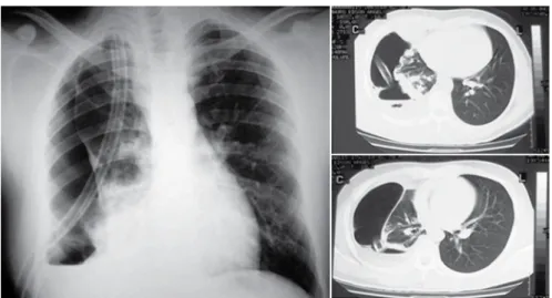
A 26-year-old Caucasian male with a history of local trauma had been experiencing an increase in left leg volume, accompanied by pain, for eight months. An X-ray of the leg revealed an osteolytic tumor in the tibial diaphysis. We performed tumor resection with histologically tumor-free margins. Seven years later, the patient presented local recurrence and underwent below-knee amputation of the affected limb. Three years later, he presented dyspnea upon moderate exertion, and, at that time, right pneumothorax was diagnosed, and nodular images, some of which were cystic, were seen in both lungs (Figure 1). Those images subsequently proved to be lung metastases of
adamanti-Introduction
Adamantinoma is a rare tumor that affects long bones, and it is estimated to account for 0.1 to 0.5% of all primary
bone tumors.(1) It is named for its histological similarity to
the tumor of the mandible (ameloblastoma).
Adamantinoma is a low-grade neoplasm that presents an indolent course, is locally aggressive, and rarely metasta-sizes. The secondary sites most frequently affected are the lungs, lymph nodes, and bones. When it is present in the lungs, hemoptysis can occur.
Although cases of pneumothorax secondary to other metastatic tumors have been described, there are, in the literature, only three references to metastatic adaman-tinoma of the lung as the etiology of spontaneous
pneumothorax.(2,3,4) Here, we report two cases followed by
the authors.
A rare case of pneumothorax: metastatic adamantinoma*
Caso raro de pneumotórax: adamantinoma metastático
Roberto Gonçalves1, Roberto Saad Júnior2, Vicente Dorgan Neto3, Marcio Botter4
Abstract
Here, we describe two cases of lung metastasis of adamantinoma of long bones, a low-grade bone neoplasm that rarely metastasizes. In both cases, the clinical presentation of the metastases was characterized by spontaneous pneumothorax secondary to tumor cavitation, a phenomenon described in only three of the studies reviewed in the literature. Clinical, radiological, and anatomopathological findings, as well as the procedures adopted in the two cases, are described.
Keywords: Adamantinoma; Pneumothorax; Neoplasm metastasis; Medical records.
Resumo
Descrevem-se dois casos de metástases pulmonares de adamantinoma de ossos longos, o qual é uma neoplasia óssea de baixo grau que raramente metastatiza. Nos dois casos a apresentação clínica das metástases se deu por pneumotórax espontâneo secundário a escavação tumoral, fenômeno descrito em apenas três dos trabalhos consultados na literatura. São descritos os achados clínicos, radiológicos e anato-mopatológicos, bem como os procedimentos adotados nos dois casos.
Descritores: Adamantinoma; Pneumotórax; Metástase neoplásica; Registros médicos.
* Study carried out in the Thoracic Surgery Section of the Department of Surgery, Santa Casa School of Medical Sciences in São Paulo, São Paulo, Brazil. 1. Masters student in Medicine in the Thoracic Surgery Section of the Department of Surgery. Santa Casa School of Medical Sciences in São Paulo, São Paulo, Brazil. 2. Full Professor in the Thoracic Surgery Section of the Department of Surgery. Santa Casa School of Medical Sciences in São Paulo, São Paulo, Brazil. 3. Adjunct Professor in the Thoracic Surgery Section of the Department of Surgery. Santa Casa School of Medical Sciences in São Paulo, São Paulo, Brazil. 4. Instructor in the Thoracic Surgery Section of the Department of Surgery. Santa Casa School of Medical Sciences in São Paulo, São Paulo, Brazil. Correspondence to: Roberto Gonçalves. Rua Doutor Cesário Mota Jr., 112, Unidade de Pulmão e Coração (UPCOR), CEP 01221-020, São Paulo, SP, Brasil. Tel 55 11 3862-6362. E-mail: [email protected]
426 Gonçalves R, Saad Junior R, Dorgan Neto V, Botter M
of 37% of predicted and forced vital capacity at 50% of predicted). Twenty days after tube removal, the patient presented another episode of right pneu-mothorax (Figure 2). It would have been impossible to resect all of the metastases, since that would have involved extensive lung parenchyma resection in a patient with compromised respiratory function. Therefore, we opted for pleural drainage followed by extensive pleurodesis. Eight months later, the patient presented bloody sputum, culminating in moderate hemoptysis. Bronchoscopy and an arte-riogram revealed that the source of the bleeding was a cavitated metastasis in the left upper lobe bronchus. Attempts to staunch the bleeding were unsuccessful. We opted for left upper lobe lobec-tomy together with resection of two metastases located in the left lower lobe (Figure 3). The patient was discharged six days later and remained under outpatient follow-up treatment.
Discussion
Fisher,(5) based on the microscopic similarity
between the neoplasm described here and adaman-tinoma of the mandible (ameloblastoma), designated this neoplasm “adamantinoma of long bones”, which preferentially affects the tibia (70%), fibula, femur, and ulna but can also affect the humerus and ribs. It typically presents as a single tumor located in the bone diaphysis.
noma, and we opted to perform pleural drainage followed by metastasectomy through a sequential double thoracotomy. Five nodules were excised from the right lung, and three nodules were excised from the left lung. The patient was discharged three days later and remained under outpatient follow-up treatment.
Case 2
A 55-year-old Caucasian male, a former smoker (25 pack-years) with a history of local trauma, had been experiencing a progressive increase in the region of the right tibia, accompanied by pain, for two years. An X-ray revealed an image, similar to that seen in Case 1, in the distal region of the right tibia. The diagnosis of adamantinoma was confirmed through frozen section biopsy, and the patient was submitted to local resection with histo-logically tumor-free margins. Two years later, the patient experienced local recurrence and a below-knee amputation was performed.
Two years after the amputation, the patient was referred to our outpatient clinic after imaging studies revealed cystic alterations, nodules, and moderate right pneumothorax. We opted for pleural drainage with complete lung expansion and tube removal. In addition, the patient underwent pulmonary function testing, which revealed a severe obstructive ventila-tory defect (forced expiraventila-tory volume in one second
reveals a dense, pinkish tumor with fibrous areas interspersed with foci that are of bony consistency. Cystic cavities of varying sizes can be observed. Histologically, it presents as a biphasic neoplasm— there is an epithelioid component consisting of stellate cells, which are strongly positive for
keratin on immunohistochemistry,(13) and a stromal
component consisting of fibrous proliferation with irregularly mineralized osteoid trabeculae and cells characteristic of myofibroblasts, with positivity for
smooth muscle markers, such as actin(14) (Figure 3).
Lung metastases of adamantinoma are extremely similar to those of primary tumors, and the differen-tial diagnosis is made with osteofibrous dysplasia.
Metastatic disease is rare (seen in only 10-15% of cases) and, when present, is a delayed event, occurring up to ten years after detection of the
primary tumor.(15,16) In the cases described above, the
patients developed lung metastases, which occurred in the form of spontaneous pneumothorax, two years after treatment of the primary tumor. During the surgical procedures, the metastases were found to affect the visceral pleura, with the formation of cystic lesions, which were the most likely etiology of
the pneumothoraces.(1-3)
The treatment of the primary tumor consists of wide local resection with tumor-free margins. It is a highly radioresistant tumor, and, to date, there is no chemotherapy that is efficacious in control-ling tumor growth. Pulmonary metastasectomy should be performed whenever the primary disease It most often occurs after skeletal maturity, in
individuals from 20 to 60 years of age, and
predom-inates in men, in whom it is more aggressive.(7)
In our patients, this was demonstrated by the occurrence of local recurrence and lung metastases.
The 10-year local recurrence rate is 18.6%.(8)
The clinical presentation is progressive volu-metric increase accompanied by local pain, and
history of trauma is common.(9) In our two cases,
the symptoms were benign and were associated with the history of local trauma, which made the patients postpone seeking treatment.
On conventional X-rays, the bone tumor is single and elongated (its longest axis parallel to the bone affected), with irregular contours, ill-defined borders, and osteolytic foci intermingled with reac-tive sclerosis (without periosteal reaction), and often
presents cysts.(10-12) In our cases, the X-rays revealed
inflated and osteolytic tumors located in the tibial diaphysis. In the second case, a magnetic resonance imaging scan of the leg was requested due to the suspicion of soft tissue invasion, which was not confirmed. The tumor showed low signal intensity on T1-weighted images and high signal intensity on T2-weighted images.
As part of the staging procedure, whole-body bone scintigraphy should be performed in search of intense radiotracer detection in order to identify possible metastases.
Regarding pathological anatomy, macroscopic examination of adamantinoma of long bones
428 Gonçalves R, Saad Junior R, Dorgan Neto V, Botter M
a disease-free interval (between the primary tumor and the metastatic tumor) of less than a year, and presenting local recurrence are factors associated
with a worse prognosis.(7)
We conclude that adamantinoma is a rare, slow-growing neoplasm that exhibits a low metastatic potential—with tropism for the lungs in the cases presented here—and can manifest as pneumothorax. Its treatment should be exclusively surgical.
References
1. Gebhardt MC, Lord FC, Rosenberg AE, Mankin HJ. The treatment of adamantinoma of the tibia by wide resection and allograft bone transplantation. J Bone Joint Surg Am. 1987;69(8):1177-88.
2. Naji AF, Murphy JA, Stasney RJ, Neville WE, Chrenka P. So-called adamantinoma of long bones. J Bone Joint Surg Am. 1964;46:151-8.
is controlled, the performance status of the patient is good, and the pulmonary function test results are
satisfactory.(17,18) Resection of all metastases should
be attempted at all costs, and wedge resection should be performed.
We believe that, since it is a slow-growing tumor, there is a potential beneficial effect in the palliative use of partial metastasectomy, especially in cases of metastases that are more central. This procedure can delay complications such as obstructive atelec-tasis or hemoptysis, and, although it should only be used in exceptional cases, we recommend its use for the treatment of cases in which there is no therapeutic alternative to surgery, such as the cases reported here.(7,8)
Regarding prognosis, the literature indicates that the mean survival of patients with metastatic disease is twelve years, and that being male, having
Figure 3 - Results of the left upper lobe lobectomy and left lower lobe nodulectomies: dense tumors of bony consistency (a); cavitation and a bleeding focus in the upper lobe tumor, causing hemoptysis (b); histology revealing a metastatic neoplasm peripheral to a bronchiole, H&E ×100 (c); and a biphasic neoplasm with epithelioid cells and a stromal osteoid component—metastatic adamantinoma, H&E ×200 (d).
a
b
c
12. Plump D, Haponik EF, Katz RS, Tipton-Donovan A. Primary adamantinoma of rib: thoracic manifestations of a rare bone tumor. South Med J. 1986;79(3):352-5.
13. Maki M, Saitoh K, Kaneko Y, Fukayama M, Morohoshi T. Expression of cytokeratin 1, 5, 14, 19 and transforming growth factors-beta1, beta2, beta3 in osteofibrous dysplasia and adamantinoma: A possible association of transforming growth factor-beta with basal cell phenotype promotion. Pathol Int. 2000;50(10):801-7.
14. Maki M, Athanasou N. Osteofibrous dysplasia and adamantinoma: correlation of proto-oncogene product and matrix protein expression. Hum Pathol. 2004;35(1):69-74. 15. Van Schoor JX, Vallaeys JH, Joos GF, Roels HJ, Pauwels
RA, Van Der Straeten ME. Adamantinoma of the tibia with pulmonary metastases and hypercalcemia. Chest. 1991;100(1):279-81.
16. De Keyser F, Vansteenkiste J, Van Den Brande P, Demedts M, Van de Woestijne KP. Pulmonary metastases of a tibia adamantinoma. Case report and review of the literature. Acta Clin Belg. 1990;45(1):31-3.
17. Rusch VW. Metastatic Neoplasms to the Lung: Introduction. Seminars in Thoracic and Cardiovascular Surgery. 2002;14:2-3.
18. Pastorino U. History of the surgical management of pulmonary metastases and development of the International Registry. Semin Thorac Cardiovasc Surg. 2002;14(1):18-28. 3. Winter WG Jr. Spontaneous pneumothorax heralding
metastasis of adamantinoma of the tibia. Report of two cases. J Bone Joint Surg Am. 1976;58(3):416-7.
4. Altmannsberger M, Poppe H, Schauer A. An unusual case of adamantinoma of long bones. J Cancer Res Clin Oncol. 1982;104(3):315-20.
5. Fischer B. Uber ein primares Adamantinom der Tibia. Frankfurt Zeitschr f Path. 1913;12: 422-41.
6. Surana SS, Mogra NK, Dube MK, Dhruva AK. Adamantinoma of tibia (a case report). J Postgrad Med. 1985;31(1):57-8. 7. Filippou DK, Papadopoulos V, Kiparidou E, Demertzis NT.
Adamantinoma of tibia: a case of late local recurrence along with lung metastases. J Postgrad Med. 2003;49(1):75-7. 8. Qureshi AA, Shott S, Mallin BA, Gitelis S. Current trends
in the management of adamantinoma of long bones. An international study. J Bone Joint Surg Am. 2000;82-A(8):1122-31.
9. Próspero JD, Bartolomei B, Cabello CJM, Ferreira FS, Pedroso RB. Adamantinoma da Tíbia. Rev. Paulista Méd. 1969;76:383.- não achei...
10. Donner R, Dikland R. Adamantinoma of the tibia. A long-standing case with unusual histological features. J Bone Joint Surg Br. 1966;48(1):138-44.