557
Eggshell Calcification of Kidney in UPJO Case Report
International Braz J Urol Vol. 32 (5): 557-559, September - October, 2006
Eggshell Calcification of Kidney in Ureteropelvic Junction
Obstruction
Narmada P. Gupta, Rajiv Yadav
Department of Urology, All India Institute of Medical Sciences, New Delhi, India
ABSTRACT
Eggshell calcification of kidney in case of ureteropelvic junction obstruction (UPJO) is an uncommon finding with only a few cases reported in literature. We report a thirty-year-old symptomatic man with curvilinear calcification in hydronephrotic right kidney. Thorough investigations to rule out genitourinary tuberculosis and hydatid disease of kidney were performed prior to the definitive management by laparoscopic approach.
Key words: ureter; hydronephrosis; kidney pelvis; calcinosis Int Braz J Urol. 2006; 32: 557-9
INTRODUCTION
Eggshell calcification of kidney in case of ureteropelvic junction obstruction (UPJO) is an uncommon finding. Such kind of curvilinear calcification is more commonly found in benign cysts, renal cell carcinoma and hydatid cyst. We report a thirty-year-old symptomatic man with eggshell calcification in hydronephrotic right kidney. Progressive deposition of calcium salts in a chronically dilated renal parenchyma may have caused calcification in the hyalinized fibrous renal tissue.
CASE REPORT
A thirty-year-old man was investigated for abdominal pain and a palpable mass in the right lumbar region. He had no associated urinary symptoms or bowel dysfunction. The only significant
558
Eggshell Calcification of Kidney in UPJO
cystoscopy, the right orifice was normal and no urine flow was detected. Right retrograde pyelogram revealed a thin ureter, which was obstructed at the ureteropelvic junction. Laparoscopic nephrectomy was done. Multiple sections examined showed a fibrocollagenous cystic wall with hyalinization, calcification and ossification. Certain areas with intact epithelial lining and thyroidization of renal tubules were also observed. No granulomatous inflammation or parasite was present.
COMMENTS
Calcification of kidney associated to ureteropelvic junction obstruction (UPJO) is rare and only a few cases have been reported in literature (1). Although such eggshell calcification is usually observed in benign condition, surgical intervention may still be indicated as the risk of malignancy in such lesion is 20% (2). Interestingly, Gold et al have
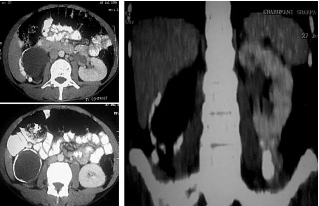
Figure 2 – Computerized tomography showing calcified right hydronephrotic kidney.
Figure 1 – Kidneys, ureters and bladder X-ray showing eggshell calcification in the region of right kidney.
559
Eggshell Calcification of Kidney in UPJO
Calcification in this situation occurred despite normal serum levels of calcium probably due to progressive deposition of calcium salts in damaged and hyalinized tissue in the wall of chronic hydronephrotic kidney.
CONFLICT OF INTEREST
None declared.
REFERENCES
1. Tsujimura A, Imazu T, Nishimura K, Sugao H, Oka T, Takaha M, et al.: Ureteropelvic junction obstruction with renal pelvic calcification: a case report. J Urol. 1993; 150: 1889-90.
2. Daniel WW Jr, Hartman GW, Witten DM, Farrow GM, Kelalis PP: Calcified renal masses. A review of ten years experience at the Mayo Clinic. Radiology. 1972; 103: 503-8.
3. Gold RP, Saitas V, Pellman C: Congenital ureteropelvic junction obstruction with calcified renal pelvis and superimposed spindle cell urothelial carcinoma. Urol Radiol. 1990; 12: 15-7.
Accepted after revision: March 26, 2006
Correspondence address:
Dr. Narmada P Gupta Head, Department of Urology
All India Institute of Medical Sciences Ansari Nagar, New Delhi 110029, INDIA Fax: + 91 11 686-2663