Retroperitoneoscopic Nephrectomy in Benign Pathology
Rodrigo S. Quintela, Leonardo R. Cotta, Marcelo F. Neves, David L. Abelha Jr, Jose E.
Tavora
Section of Urology, Hospital da Previdencia dos Servidores do Estado, Belo Horizonte, Minas
Gerais, Brazil
ABSTRACT
Introduction: We report our experience with 43 retroperitoneal laparoscopic nephrectomy for benign kidney disease.
Materials and Methods: All patients had a poor function from obstructive uropathology and renal atrophy. None of these patients had a previous lumbotomy. Retroperitoneoscopy was performed with 4 trocar port technique in a lateral position. The retroperitoneal space is created by using a Gaur’s balloon made of sterile glove. The approach to vascular pedicle was done posteriorly and vessels were clipped by metal and Hem-o-lock (Weck Closure Systems, North Carolina, USA) clips. The sample was intact extracted in an Endo-Bag prolonging one trocar incision.
Results: Median operative time was 160 minutes and median blood loss was 200 mL. Four cases (9%) were converted to open surgery: one case due to bleeding and 3 cases due to technical difficulties regarding perirenal adherences. Most patients (39) checked out from the Hospital in day two. Four of them were left over 3 days due to wound complications.
Conclusions: Retroperitoneoscopy offers a safe, effective and reproductive access to nephrectomy for benign pathologies.
Key words: kidney; nephrectomy; laparoscopy; retroperitoneal space Int Braz J Urol. 2006; 32: 521-8
INTRODUCTION
Laparoscopic nephrectomy was first success-fully performed by Clayman in 1990 using the transperitoneal route (1). The pure retroperitoneal access for nephrectomy was described by Gaur three years later, using a balloon to create the surgical work-ing space (2,3). Despite the preference for retroperi-toneal approach in open urologic surgery worldwide the transperitoneal approach is the preferred technique for laparoscopic urologic surgery. Furthermore direct retroperitoneal access (retroperitoneoscopy) is attract-ing more interest and application in urology, becom-ing the preferential approach for nephrectomy in many expertise laparoscopic centers (4-9). This data
de-scribes our experience with retroperitoneoscopic simple nephrectomy.
MATERIALS AND METHODS
pyelonephritis in 16 cases (37%) and three cases (7%) with renovascular hypertension. The work up was done in all cases following ultrasound, intravenous pyelogram (IVP) or axial computerized tomography and scint scan. Thirty-three patients (76%) presented hydronephrosis with no renal function and the other 10 patients (24%) presented renal atrophy. The etiol-ogy of the renal damage is showed in the Table-1. Three of the patients presented horseshoe kidneys with UPJ (ureteral pelvic junction) syndrome. Two patients had previous lumbar urologic interventions (percutaneous nephrolithotripsy and a percutaneous nephrostomy).
Surgical Technique
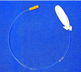
The patient is placed in lateral position as in an open surgery (classical lumbotomy position) with general anesthesia. In an attempt to get more room to face up the kidney, we flexed the table. The camera assistant stays at the surgeon’s side at a cephalic po-sition and the second assistant stays at the opposite side. A transverse incision (15 mm) is made just be-low the 12th costal arc and the muscles are dissected until the toracolumbar fascia is opened achieving the retroperitoneal space. A digital dissection is made in the retroperitoneal space pulling the peritoneum an-teriorly and displacing the fat from this body wall. A Gaur balloon (Figure-1) is placed in the space and filled with 800 mL of saline solution. The Hasson trocar is placed and fixed at the body wall to avoid air linkage. A CO2 insufflation is performed at a pres-sure of 12 mm. A 0º laparoscopic lent is introduced
by the Hasson port and the other trocars are placed under direct vision: a 10 mm trocar is placed 2 cm above the iliac crest and two 5 mm trocars are placed in anterior axillary line (one next to the costal arc and the other by the iliac crest) (Figure-2).
The psoas muscle is the main anatomic land-mark. This muscle is dissected and the ureter and the
Figure 1 – Gaur’s balloon made of a Nelaton 16F catheter and a finger glove.
Figure 2 – Superior view of trocar port incisions for a left side retroperitoneoscopic nephrectomy. The Hasson trocar is fixed with sutures. The lens port (O), the surgeon ports (S) and the assistant port (A) are signaled.
Table 1 – Etiology of the lost renal function.
Pathology Patients (%)
Ureteropelvic junction stricture 18 (42%)
Ureteral calculi 12 (27.7%)
Ureteral trauma 03 (7%)
Renal tuberculosis 03 (7%) Congenital renal atrophy 03 (7%) Renovascular hypertension 03 (7%)
Renal trauma 01 (2.3%)
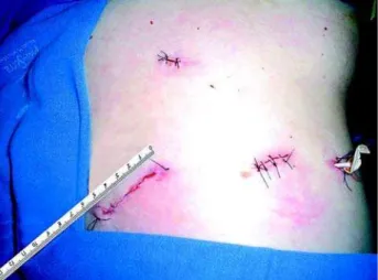
gonad vein in the left or the inferior vena cava in the right are identified. The renal vein is dissected in the right side following the vena cava and the renal ar-tery is dissected over the renal vein. In the left side, the renal vein is identified following the gonad vein and the artery is found just above the renal vein. Metal clips are used to occlude the artery and a Hem-o-lock ® (Weck Closure Systems, North Caroline, USA) clip is used at the renal vein. After the vascular control, the dissection is initiated inside the Gerota’s fascia by the renal upper pole preserving the adrenal gland in most of the cases. The lower pole then is dissected and the ureter is clip occluded with a metal clip. In cases of horseshoe kidney, the renal isthmus is co-agulated with an electrocautery and laparoscopically sutured with absorbed suture if necessary. In volumi-nous hydronephrotic kidneys the fluid in the collect-ing system is aspirated to facilitate perirenal dissec-tion. The specimen is entrapped in a handmade bag. In cases where the samples are small, we utilize the first 15 mm lumbar incision to remove the specimen and with larger samples we prolong the inguinal port incision making a small Gibson’s incision (Figure-3). A Penrose drain is placed in cases of infected kid-ney.
RESULTS
All of the surgeries were performed by the same surgeon or under his supervision. Table-2 re-sumes the results. Median operative time was 160 minutes (120 to 240 minutes) with average estimated blood loss of 200 mL. Blood transfusion was not nec-essary in any of the cases. Three patients presented intra surgical bleeding during pedicle dissection. Two patients with renal vein bleeding were laparoscopic controlled with a Hem-o-lock clip close to the vena cava. Another bleeding from an injury at the right adrenal vein required conversion to open surgery.
There were no major complications. Six pa-tients developed abdominal body wall complications: three patients developed subcutaneous emphysema with no clinical repercussion and a spontaneous reso-lution; two patients developed surgical wound he-matoma in the first trocar incision and one patient
developed surgical wound infection. All of those pa-tients were treated clinically.
Conversion was necessary in 4 cases (9%). One patient due to hemorrhage and three patients due to technical difficulties. All of them presented chronic or tuberculous pyelonephritis and perirenal adherence. In none of the cases surgical reintervention was needed.
Diet was initiated 6 hours after surgery. Most of the patients (39) were released from the hospital the second day after surgery. Four patients were left in the hospital for more than 48 hours due to surgical wound complications.
Histopathological diagnosis confirmed three cases of renal tuberculosis and 40 cases of renal atro-phy with or without chronic inflammatory process.
Figure 3 – Final aspect of the incisions. The specimen was re-moved by the 5 cm inguinal incision. A Penrose drain is left in the dorsal surgeon working port.
Table 2 – Surgical results.
Patients 43
Operative time (median) 160 minutes
Blood lost 200 ml
Conversion (%) 004 (9%)
Major complication (%) 000 Minor complication (%) 006 (13%)
COMMENTS
Laparoscopic nephrectomy is widely per-formed by the transperitoneal approach. After retro-peritoneal technique description by Gaur these has become the preferential approach in outstanding cen-ters for the surgical treatment of most kidney patholo-gies even renal tumors (4-9).
The advantage of retroperitoneoscopy are the preservation of the peritoneal cavity and the poste-rior access to the renal pedicle making possible a straight dissection and the control of the vessels in the first step of the surgery. This approach can be done without any difficulties even in hard cases (10,11).
The main disadvantage of retroperitoneosco-py is the reduced working space requiring a better syn-chronized surgical team to avoid instrument collision. The use of Gaur’s balloon creates a large space, which allow the surgeon to get enough room reaching all the kidney limits.
The main contraindication of retroperito-neoscopy is the presence of previous lumbotomy. Rel-ative contraindication because of technical aspects should be attempt in patients with chronic inflamma-tory pathologies such as renal tuberculosis or xan-togranulomatous pyelonephrosis (10-12). In those cases, the possibility of conversion is higher because of the adherences. In 3 cases of this series, patients with a chronic inflammatory process (one with renal tuberculosis) were submitted to conversion to open surgery due to technical difficulties. Furthermore, in two patients with tuberculosis the surgery was per-formed without any difficulties or complications. Some reported series show a significant number of cases of renal tuberculosis successfully treated by retroperitoneoscopy (10,11). Although it presents a better chance of conversion, the presence of a chron-ic inflammatory process is not an absolute contrain-dication for laparoscopic nephrectomy. In such cas-es, transperitoneal or hand-assisted laparoscopy should be a better option.
Previous minimally invasive lumbar proce-dures such as nephrostomy and percutaneous neph-rolithotripsy do not exclude retroperitoneoscopy (6,7,13). In two patients of this series with previous
history of nephrostomy and percutaneous nephroli-thotripsy, the procedure was performed without any difficulties.
In patients with horseshoe kidney the pres-ence of anomalous vessels require an extra careful vascular approach (3). In three patients of this series with horseshoe kidney retroperitoneal laparoscopic nephrectomy was performed without difficulties in reaching the renal pedicle but a longer operative time was needed.
The incidence of vascular lesions in transperitoneal urological laparoscopic surgery vary between 1.5% to 2% in large series (14,15). Although there is not much data for retroperitoneal approach its reported incidence is similar (16). In this data we had three cases of vascular bleeding during pedicle dissection. In one of the cases a left adrenal vein bleed-ing required conversion to open surgery. Conversion was not required in two other cases of renal pedicle bleeding. Conversion to open surgery depends on the extension of the vascular lesion and of the surgeon skills to control bleeding. A temporary increase of inflation pressure up to 15 or 20 mmHg reduces venous hemorrhage in the retroperitoneal space and a vascular control maneuver must be considered. In one case it was necessary the placement of another trocar port for a better exposition. We experienced no bleeding problems using metal clips for renal ar-tery control and Hem-o-lock (Weck Closure Systems, North Caroline, USA) clips for renal vein control.
The CO2 inflation can cause subcutaneous emphysema, especially pneumothorax and pneumomediastine if the retroperitoneal pressure reach levels over 15 mmHg (18). Usually no impor-tant clinical repercussion is seen on those patients (19). A pressure of 12 mmHg gives the surgeon a good working space. Three cases of subcutaneous emphy-sema with no clinical repercussion were observed in this data.
Total surgical conversion frequency was 9%. Most of those conversions occurred due to hemor-rhage or technique difficulties caused by perirenal adherence. The data literature shows up to 10% of conversion for simple laparoscopic nephrectomy (6,9). The majority of conversions occur due to in-flammatory processes or previous surgeries (11). When surgical conversion becomes necessary it can be rapidly performed by lumbotomy, prolonging the first trocar incision that allows the surgeon to con-tinue with the conventional lumbar incision with good cosmetic results.
The sample extraction can be performed by a new incision, amplifying the trocar incision or with morcellation. Pfannenstiel incision, often propagated for transperitoneal laparoscopic nephrectomy, can be an alternative for sample extraction opening the peri-toneum close to the upper abdominal wall at the end of the procedure. Morcellation avoids incision aug-mentation but promotes longer operative time and less accuracy for the histopathology (20). We usually ex-tract all samples by the initial trocar incision if pos-sible or by making a small 5 cm inguinal incision (Gibson’s incision) with low morbidity and good cos-metic results.
The retroperitoneal approach offers a lower morbidity when compared to the transperitoneal ap-proach because it does not violate the peritoneal cav-ity. This benefit occurs not only due to less intraperi-toneal manipulation and less organ lesions but also due to the reduction of the peritoneal contact with urine and blood (5,7,13). Infected urine elimination during the surgery was observed in many patients in this series without any further complication such as infection or postoperative ileums.
Urology departments worldwide start their laparoscopic nephrectomy experience through
transperitoneal access, which is considered the saf-est one and also the one that facilitates laparoscopic dissection especially in early experiences. This se-ries shows the initial experience using retroperito-neal approach in a public Brazilian medical center without any previous familiarity with urological laparoscopic surgery. This experience demonstrates that the retroperitoneal approach is a safe, reproduc-tive and effecreproduc-tive access for simple nephrectomy.
CONCLUSION
The laparoscopic retroperitoneal approach for simple nephrectomy is a challenge for the surgeon due to a smaller surgical working space but on the other hand it is benefic to patients because it does not violate the peritoneal cavity. Retroperitoneal and transperitoneal laparoscopic approaches have the same surgical indications but previous lumbotomy is a relative contraindication for retroperitoneal access. Minimally invasive lumbar procedures such as nephrostomy and percutaneous nephrolithotripsy are not contraindications.
An advantage to retroperitoneoscopy is the straight access to the kidney and its renal pedicle making possible an easier and faster approach as well as vascular control. Disadvantages include the need to create a retroperitoneal space and a small working area to dissect, proceed reconstructive maneuvers and bag the sample to extract it out of the body.
Retroperitoneoscopy must be considered a safe, effective and reproductive surgical technique for nephrectomy in benign renal diseases.
CONFLICT OF INTEREST
None declared.
REFERENCES
2. Gaur DD, Agarwal DK, Purohit KC: Retroperitoneal laparoscopic nephrectomy: initial case report. J Urol. 1993; 149: 103-5.
3. Gaur DD: Laparoscopic operative retroperitoneosco-py: use of a new device. J Urol. 1992; 148: 1137-9. 4. Abbou CC, Cicco A, Gasman D, Hoznek A, Antiphon
P, Chopin DK, et al.: Retroperitoneal laparoscopic versus open radical nephrectomy. J Urol. 1999; 161: 1776-80.
5. Desai MM, Strzempkowski B, Matin SF, Steinberg AP, Ng C, Meraney AM, et al.: Prospective randomized comparison of transperitoneal versus retroperitoneal laparoscopic radical nephrectomy. J Urol. 2005; 173: 38-41.
6. Gupta NP, Goel R, Hemal AK, Dogra PN, Seth A, Aron M, et al.: Should retroperitoneoscopic nephrectomy be the standard of care for benign nonfunctioning kid-neys? An outcome analysis based on experience with 449 cases in a 5-year period. J Urol. 2004; 172: 1411-3.
7. Hoznek A, Salomon L, Gettman M, Stolzenburg JU, Abbou CC: Justification of extraperitoneal laparoscopic access for surgery of the upper urinary tract. Curr Urol Rep. 2004; 5: 93-9.
8. Rassweiler JJ, Seemann O, Frede T, Henkel TO, Alken P: Retroperitoneoscopy: experience with 200 cases. J Urol. 1998; 160: 1265-9.
9. Sebe P, de la Taille A, Hoznek A, Chopin D, Abbou CC, Salomon L: Simple nephrectomy with retroperi-toneal laparoscopy. Prog Urol. 2003; 13: 577-80. 10. Hemal AK, Gupta NP, Kumar R: Comparison of
retroperitoneoscopic nephrectomy with open surgery for tuberculous nonfunctioning kidneys. J Urol. 2000; 164: 32-5.
11. Zhang X, Zheng T, Ma X, Li HZ, Li LC, Wang SG, et al.: Comparison of retroperitoneoscopic nephrectomy
versus open approaches to nonfunctioning tuberculous kidneys: a report of 44 cases. J Urol. 2005; 173: 1586-9. 12. Merrot T, Ordorica-Flores R, Steyeart H, Ginier C, Valla JS: Is diffuse xanthogranulomatous pyelonephri-tis a contraindication to retroperitoneoscopic nephroureterectomy? A case report. Surg Laparosc Endosc. 1998; 8: 366-9.
13. McDougall EM, Clayman RV: Laparoscopic nephrec-tomy for benign disease: comparison of the transperitoneal and retroperitoneal approaches. J Endourol. 1996; 10: 45-9.
14. Gill IS, Kavoussi LR, Clayman RV, Ehrlich R, Evans R, Fuchs G, et al.: Complications of laparoscopic ne-phrectomy in 185 patients: a multi-institutional review. J Urol. 1995; 154: 479-83.
15. Vallancien G, Cathelineau X, Baumert H, Doublet JD, Guillonneau B: Complications of transperitoneal laparoscopic surgery in urology: review of 1,311 pro-cedures at a single center. J Urol. 2002; 168: 23-6. 16. Meraney AM, Samee AA, Gill IS: Vascular and bowel
complications during retroperitoneal laparoscopic sur-gery. J Urol. 2002; 168: 1941-4.
17. Gill IS, Clayman RV, Albala DM, Aso Y, Chiu AW, Das S, et al.: Retroperitoneal and pelvic extraperitoneal laparoscopy: an international perspective. Urology. 1998; 52: 566-71.
18. Streich B, Decailliot F, Perney C, Duvaldestin P: In-creased carbon dioxide absorption during retroperito-neal laparoscopy. Br J Anaesth. 2003; 91: 793-6. 19. Ng CS, Gill IS, Sung GT, Whalley DG, Graham R,
Schweizer D: Retroperitoneoscopic surgery is not as-sociated with increased carbon dioxide absorption. J Urol. 1999; 162: 1268-72.
20. Savage SJ, Gill IS: Intact specimen extraction during renal laparoscopy: muscle-splitting versus muscle-cut-ting incision. J Endourol. 2001; 15: 165-9.
Accepted after revision: May 25, 2006
Correspondence address:
Dr. Rodrigo Silva Quintela Rua Correias 281 / 201
EDITORIAL COMMENT
It is a paper describing a retroperitoneal ap-proach for nephrectomy in benign disease. Endo-scopic nephrectomy is usually performed by transperitoneal rout and the merit of the paper is that it underlies the advantages of using a retroperi-toneal approach and shows that it is feasible. One important shortcoming is the absence of a control group.
I believe that vessels should be ligated with a transfixational technique since it reduces the risk of intra and postoperative bleeding.
Conversion rate was fairly high. Manual dis-section of the peritoneum gives a large working space. A hand-assisted technique also makes the operation easier, quicker and safer.
It could be argued that the total length of all incisions is longer than a small anterior extraperitoneal incision or dorsal incision for open surgery. The ad-vantage with an endoscopic approach would be an ex-traction sit with low morbidity and good cosmetic re-sults. A Pfannenstiel incision is therefore often propa-gated for the extraction (can also be used for a hand assisted procedure).
Dr. Jonas Wadström
Department of Surgical Sciences Uppsala University Hospital Uppsala, Sweden E-mail: [email protected]
EDITORIAL COMMENT
Nowadays many authors in many countries including Japan have already described retroperito-neal nephrectomy for renal disease as a standard care. From this standpoint of view, the impact and
origi-nality of this manuscript is low internationally. How-ever, as this may be a progressive approach in devel-oping countries, this manuscript will be informative for many urologists working in these countries.
Dr. K. Mita
Hiroshima University, Dept of Urology Graduate School of Medical Sciences Hiroshima City, Japan E-mail: [email protected]
EDITORIAL COMMENT
The authors are to be congratulated on their excellent clinical results with retroperitoneoscopic nephrectomy.
system to facilitate perirenal dissection in the retro-peritoneal space.
In cases with horseshoe kidney, additional renal arteries may arise from the aorta or the iliac vessels. Using the retroperitoneal approach, it might be difficult to control hemorrhage or dividing the re-nal isthmus, especially when it is thick. Therefore, I prefer to use the transperitoneal approach in such cases.
Aside from early and straightforward access to the renal vessels, the retroperitoneal approach is not an easy operation because of a narrow working space and the relative paucity of anatomical landmarks. Al-though the authors successfully performed retroperi-toneal nephrectomy despite of no previous experience with urologic laparoscopy, I think it is exceptional and inapplicable to general urologists. Educational training of laparoscopic surgery is desirable.
Dr. A. Terai
Department of Urology Kurashiki Central Hospital Kurashiki, Japan E-mail: [email protected]
EDITORIAL COMMENT
This is a retrospective review of a clinical experience with retroperitoneal laparoscopy for be-nign kidneys at a public hospital. The authors ad-equately describe their technique. The results are in general favorable, but there were several open
con-versions. Mostly for failure to progress from adhe-sions but there was at least one case of bleeding re-quired open conversion. Complications were reported and discussed. I agree with the author’s contention that the retroperitoneal technique is underutilized.