Head and Neck Lymphomas in HIV Patients: a
Clinical Perspective
Natsuki Oishi
1,2José Vicente Bagán
3Karla Javier
4Enrique Zapater
11ENT Department, Consorci Hospital General Universitari de Valencia, Valencia, Spain
2ENT Department, Universitat de Valencia Facultat de Medicina i Odontologia, Valencia, Comunitat Valenciana, Spain
3Department of Stomatology, Consorci Hospital General Universitari de Valencia, Valencia, Comunitat Valenciana, Spain
4Department of Haematology, Consorci Hospital General Universitari de Valencia, Valencia, Comunitat Valenciana, Spain
Int Arch Otorhinolaryngol 2017;21:399–407.
Address for correspondence Natsuki Oishi, ENT Department, Consorci Hospital General Universitari de Valencia, Avda. Tres Cruces s/n Valencia 46014, Spain (e-mail: [email protected]).
Introduction
T and B lymphocytes are the main cells of the immune system. In healthy people, they are mainly located in the lymphoid organs (lymphatic nodes and spleen) and in mucosa-associated lymphoid tissue (MALT). The head and neck region is rich in lymphoid tissue, especially Waldeyer's ring, the thyroid, the salivary glands, and the oral cavity. There are also several chains of regional lymph nodes. Therefore, the head and neck are fertile anatomic sites for the development of lymphoproliferative diseases, in which malignant neoplastic mutations occur in normal lymphoid tissue and give rise to a lymphoma. In the head and neck, malignant lymphomas account for 5% of all malignant neoplasms.1
Virus-linked cancers are responsible for 15% of the total incidence of malignant neoplasms.2Patients infected with human immunodeficiency virus (HIV) are at a higher risk than the uninfected population of developing a malignancy. The most frequent HIV-associated malignant neoplasm of the head and neck is Kaposi’s sarcoma, followed by lymphoma. Thus, lymphoma is regarded as an opportunistic neoplasm due to its similarities with the opportunistic infections that occur in immunosuppressed patients.3This feature is among the criteria used to establish the AIDS stage also CD4þT cell count<200/μL.4 What is clear is that specific lymphoma subtypes are caused by one or both herpesviruses of the
Keywords
►
HIV
►
lymphoma
►
head and neck
neoplasms
Abstract
Introduction
Because of the many HIV-related malignancies, the diagnosis and
treatment of lymphoma in patients infected with human immunode
fi
ciency virus are
challenging.
Objective
Here, we review current knowledge of the pathogenesis, epidemiology,
symptomatology, diagnosis, and treatment of head and neck lymphomas in HIV
patients from a clinical perspective.
Data Synthesis
Although Hodgkin
’
s lymphoma is not an AIDS-de
fi
ning neoplasm, its
prevalence is ten times higher in HIV patients than in the general population. NHL is the
second most common malignancy in HIV patients, after Kaposi
’
s sarcoma. In this group
of patients, NHL is characterized by rapid progression, frequent extranodal involvement,
and a poor outcome. HIV-related salivary gland disease is a benign condition that shares
some features with lymphomas and is considered in their differential diagnosis.
Conclusion
The otolaryngologist may be the
fi
rst clinician to diagnose head and neck
lymphomas. The increasing survival of HIV patients implies clinical and epidemiological
changes in the behavior of this disease. Early diagnosis is important to improve the
prognosis and avoid the propagation of HIV infection.
received
July 16, 2016
accepted
October 31, 2016
published online
February 1, 2017
DOI https://doi.org/ 10.1055/s-0036-1597825.
ISSN 1809-9777.
Copyright © 2017 by Thieme Revinter Publicações Ltda, Rio de Janeiro, Brazil
gamma subfamily: Epstein Barr virus (EBV/HHV-4) and Ka-posi’s sarcoma herpesvirus (KSHV/HHV-8). The EVB infection occurs in 40% of Large Cell Lymphomas (LCL) and in 90% of Immunoblastic Lymphomas (IBL) cases.5Plasmablastic lym-phoma (PBL) nearly always occur in HIV positive patients, in whom they preferentially localize in the oral mucosa; and are strongly associated with infection by HHV8 and EBV.6
Because of the many HIV-related malignancies, the diag-nosis and treatment of lymphoma in HIV patients are chal-lenging. In this article, we summarize current knowledge of HIV-related head and neck lymphomas, focusing on a clinical perspective.
Literature Review
Pathology
In 2008, the World Health Organization proposed a classifi -cation system for lymphomas that distinguished six groups: precursor lymphoid neoplasms, mature B cell neoplasms, mature T- and natural killer (NK)-cell neoplasms, Hodgkińs lymphoma, Immunodeficiency-associated lymphoprolifera-tive disorders, and histiocytic and dendritic cell neoplasms.7 Non-Hodgkin’s lymphoma (NHL) is the most frequent tumor of the head and neck, accounting for75% of lympho-mas in this anatomic region.1
In HIV patients, most lymphomas are derived from B-cells, with the most common NHLs being immunoblastic large-cell lymphomas and diffuse large B cell lymphomas (DLBCLs). Both tumors generally occur in the context of moderate to severe immunosuppression (CD4þcell count
<100 mm3/ml) and they are often associated with EBV.8 Together with Burkitt’s lymphomas and Burkitt’s-like lymphomas, they serve as indicators in the staging of AIDS.9
Epidemiology
Epidemiological studies have shown that Hodgkin’s lympho-mas and NHLs are the most common tumors in HIV patients. Both are usually diagnosed at an early stage of HIV infection, when CD4þcell counts are relatively high.10HIV patients are
at a 60- to 200-fold greater risk than the general population of developing NHL11,12.
Immunosuppression related to HIV facilitates virus-induced carcinogenesis. Type 8 human herpes virus (HHV-8), EBV, and human papilloma virus have all been related to the develop-ment of Kaposi’s sarcoma.13Nearly half of all cases of lympho-ma in HIV patients are associated with concomitant infection with gamma herpes virus, EBV, or HHV-8.8Recentfindings are consistent with a role of HIV as a critical microenvironmental factor promoting lymphoma development. These data are changing the current paradigm, which assumes that HIV is only indirectly related to lymphomagenesis.14
Antiretroviral treatment has led to a decline in the inci-dence of opportunistic infections and, thus, to an increase in the survival of HIV patients. Within this group, there has been a decrease in the incidence of Kaposís sarcoma, whereas similar declines in Hodgkin’s lymphoma and NHL have yet to be confirmed.15,16
Symptomatology and Examination
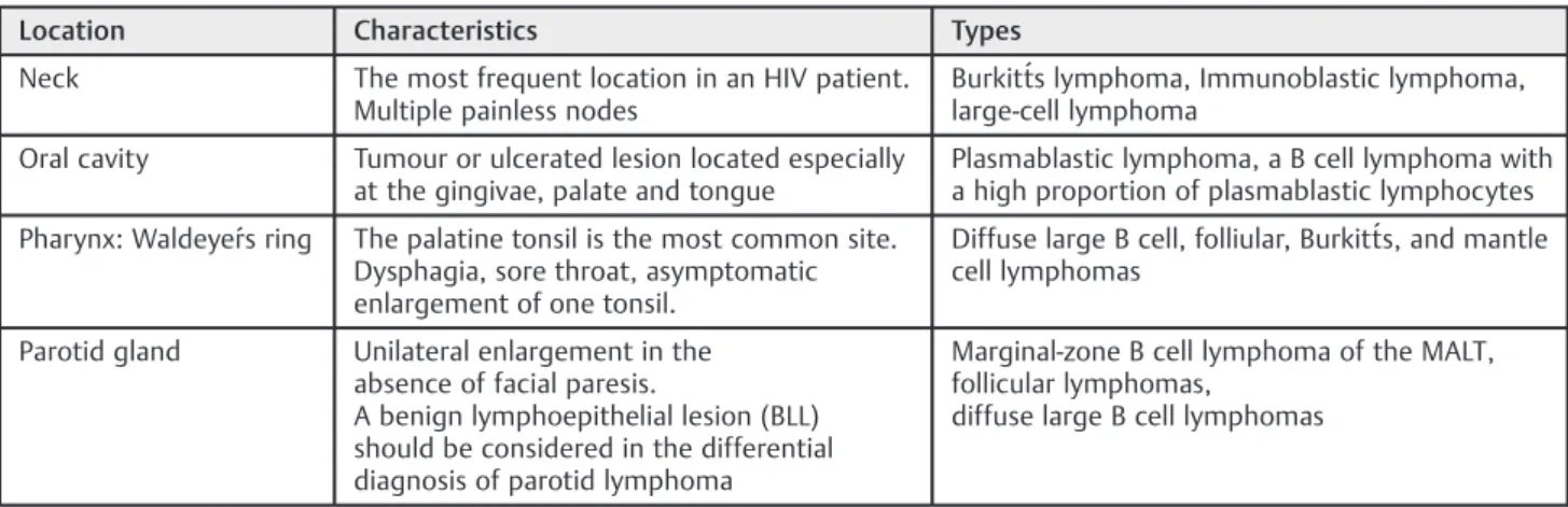
It is important to carefully examine every neck node suggest-ing malignancy, as it can be thefirst manifestation of a cancer located in the head and neck. Using endoscopic techniques, the otolaryngologist is able to locate tumors in diffi cult-to-exam-ine areas such as the nasopharynx, larynx, and hypopharynx. The most frequent location of a lymphoma in an HIV patient is the neck (50%), followed by the central nervous system, oral cavity, pharynx, parotid gland, mandible, para-nasal sinuses, larynx, and orbit17,18(►Table 1). Lymphomas in the HIV-infected population develop in relatively young patients and may be thefirst disease to awaken a suspicion of HIV infection. Indeed, the diagnosis of head and neck lym-phoma should always include the exclusion of HIV.17 Con-versely, the mean time from the diagnosis of HIV to lymphoma development is 1.5 years.18
Lymphomas in HIV patients generally manifest with atypical symptoms. The characteristic aggressiveness of these tumors and their typically advanced stage at diagnosis reflect the deterioration of immunity, increased viral load, and low CD4þ lymphocyte counts.18,19In addition, as noted above, HIV-related lymphoma generally occurs together with opportunistic infec-tions such as Pneumocystis carinii pneumonia, candidiasis, cytomegalovirus retinitis, and herpes zoster. Lymphomas can
Table 1 Table of the most common location, characteristics, and types of head and neck lymphoma in HIV patients
Location Characteristics Types
Neck The most frequent location in an HIV patient. Multiple painless nodes
Burkitt́s lymphoma, Immunoblastic lymphoma, large-cell lymphoma
Oral cavity Tumour or ulcerated lesion located especially at the gingivae, palate and tongue
Plasmablastic lymphoma, a B cell lymphoma with a high proportion of plasmablastic lymphocytes
Pharynx: Waldeyeŕs ring The palatine tonsil is the most common site. Dysphagia, sore throat, asymptomatic enlargement of one tonsil.
Diffuse large B cell, folliular, Burkitt́s, and mantle cell lymphomas
Parotid gland Unilateral enlargement in the absence of facial paresis.
A benign lymphoepithelial lesion (BLL) should be considered in the differential diagnosis of parotid lymphoma
Marginal-zone B cell lymphoma of the MALT, follicular lymphomas,
also coexist with Kaposís sarcoma, but, although the latter may regress when the immune system recovers, this is not the case with lymphoma.18
Given the relative infrequency in the general population of Hodgkin’s lymphoma in the head and neck, systemic symp-toms, apart from those deriving from HIV infection, are uncommon but may consist of fever, night sweats, or pruritus. Involved neck nodes in lymphoma are usually multiple and painless. They are not as hard on palpation as the metastatic nodules of squamous cell carcinoma and do not adhere to either the skin or deep tissue planes. The most common location of affected nodes is the lower region of the neck, but the axilla and groin may also be involved.20It is important to emphasize that nodal-based lymphomas may be difficult to identify in patients with the persistent generalized lymph-adenopathy that characterizes HIV.
Extranodal manifestations are a common clinical presenta-tion in HIV-associated lymphomas. In the general populapresenta-tion with NHL, extranodal involvement occurs in 24–48% of cases; 10% of these involve the head and neck region.21,22Primary extranodal Hodgkińs disease is rare. NHL in the head and neck, at the beginning, is usually submucosal rather than ulcerative, as seen in squamous cell carcinoma. Disease evolution can lead to ulceration.23Half of the extranodal lymphomas of the head and neck are located in Waldeyeŕs ring. The involvement of this location is associated with an increased incidence of disease spread to the gastrointestinal tract. The palatine tonsil is the most common site of lymphoma development; it accounts for the typical symptoms of dysphagia and sore throat. Because the asymptomatic enlargement of one tonsil is common, some authors consider routine excision of abnormally large asym-metrical tonsils to be advisable24(►Fig. 1).
Cavum lymphoma against a background of HIV is usually diagnosed from an enlarged neck node. Thus, prior to a surgical procedure, the detection of any neck adenopathy should be followed by an endoscopic examination of the upper cavities.20 Oral lymphomas occur more frequently in patients with HIV infection. Typical symptoms include oral swelling, pain, and ulcers. These lymphomas may present as a tumor or ulcerated lesion located anywhere in the mouth, but especially the gingivae, palate,
and tongue.25,26 Plasmablastic lymphoma, a B cell lymphoma with a high proportion of plasmablastic lymphocytes, is the most common HIV-associated subtype in this location.15,27 Constitu-tional symptoms such as fever and night sweats are frequent in PBL. Its typical appearance is a nonspecific ulceration that spreads rapidly and may eventually lead to hemorrhage, necrosis, and lytic bone destruction. A possible explanation for the high frequency of PBL in lymphoid areas of Waldeyer’s ring and in epithelial cells of the cavum and oropharynx is the predilection of EBV to accumu-late at these sites28(►Fig. 2).
Laryngeal lymphoma in HIV patients is usually located in the supraglottic region and therefore typicallyfirst manifests as dysphagia, although with disease evolution hoarseness and dyspnea may also develop. HIV patients with pharyngo-laryn-geal symptoms should always be examined by an otolaryngol-ogist, who will be able to detect lesions in difficult-to-examine locations. This is particularly important because an early diagnosis is essential to improve the prognosis.29
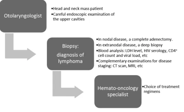
Diagnosis
The diagnosis of lymphoma is best made based on the pathology results (►Fig. 3). In nodal disease, a complete adenectomy is required. If the neck node is located at superficial planes, which is the most frequently involved site, the procedure can be performed under local anesthesia with or without sedation. It is important to resect the node completely and to preserve it as undistorted as possible. In extranodal disease, a deep biopsy should be taken. The biopsy site should be close to healthy mucosa/skin to avoid necrotic tissue. Local anesthesia is possible in patients with lesions in the oral cavity, oropharynx or nasopharynx and with some supraglottic and hypopharyngeal lesions. In salivary gland tumors as well as thyroid and some laryngeal and hypophar-yngeal lesions, general anesthesia is required. Biopsy material should be processed fresh to allow immunohistochemical staining, which is necessary for an accurate diagnosis.
The results of a blood analysis will not be specific for lymphoma but will instead detect neutrophilia, eosinophilia, lymphopaenia, thrombocytosis, thrombocytopaenia, or ane-mia. Neutrophilia, lymphopaenia and anemia are considered
Fig. 1 Unilateral tonsillar hypertrophy in diffuse large B cell lymphoma.
signs of a poor prognosis in lymphoma patients. Increases in serum alkaline phosphatase and calcium levels may indicate bone infiltration. Liver and kidney analytical indicators should also be measured.
Bone marrow involvement is more common in patients with immunodeficiency and in patients with advanced stage disease, less favorable histologic types, or constitutional symptoms. A bone marrow biopsy is recommended for all patients with B symptoms, advanced disease stages, bulky disease, or peripheral blood cytopaenias.
Complementary examinations are indicated only for pur-poses of disease staging (►Table 2).
Radiological studies are useful to assess tumor extension; whereas thorax radiography and CT are necessary. Thorax, abdominal, and pelvic CT scans are compulsory; however, abdominal scans will not accurately detect retroperitoneal, liver, or spleen involvement. Orthopantomography and CT
scanning of the cranial base, paranasal sinuses, and maxilla are useful to detect bone invasion in the proximity of an extranodal lymphoma (►Fig. 4).
Magnetic resonance imaging (MRI) are used to study soft tissues, including the parapharyngeal space, tongue, bone marrow, and brain.20A typicalfinding is homogeneous lymph nodes that are increased in size, well-defined, rounded, ovoid, or lobed.30 Nodal involvement in Hodgkin’s lymphoma is continuous and usually affects the internal jugular chain. Necrosis of the neck nodes or extranodal extension is often seen in patients with aggressive lymphoma.31Node calcifi ca-tions are often apparent after chemotherapy or irradiation.30 67Ga scintigraphy is used to evaluate disease extension and
to monitor the effectiveness of treatment. However, there may be positives if there is active infection and false-negatives after recent chemotherapy treatment (minimum 3 weeks) in necrotic areas or in lesions<2 cm in diameter. PET Fig. 3 Algorithm of a recommended diagnostic workup.
Table 2 Ann Arbor staging system for lymphomas
Stage I Involvement of a single lymph node region (I) or localized involvement of a single extralymphatic organ site (IE).
Stage II Involvement of two or more lymph node regions on the same side of the diaphragm (II) or localized involvement of an extralymphatic organ or site and one or more lymph node regions on the same side of the diaphragm (IIE)
Stage III Involvement of lymph node regions on both sides of the diaphragm (III), which may also be a accompanied by localized involvement of an associated extralymphatic organ or site (IIIE), by involvement of the spleen (IIIS), or both (IIISþE)
is the standard for staging in aggressive lymphomas and HL. PL is performed as part of staging in ARL and is used to identify lymphoma cells and to perform a PCR for EBV.
Treatment
The introduction of highly active antiretroviral therapy (ART) in HIV-infected patients, together with the administration of prophylactic antibiotics to prevent infections and of hemato-poietic factors, has changed the prognosis of patients with HIV-related lymphoma. Thus, today the outcome of these patients is comparable to that of the population with non-HIV-related lymphoma. The optimal therapy for lymphomas in the setting of HIV has yet to be defined. ART is administered initially and may be later modified to control HIV infection and allow chemotherapy and/or radiation therapy. The choice of therapy is determined by the histopathologic subtype and stage of the disease; however, in HIV patients, modifications should be made based upon the degree of virus-linked immunosuppression, as measured by the CD4þcell count.32 The therapeutic approach will depend on the subtype of HIV-associated lymphoma, as described below.
DLBCL: HIV-positive patients with DLBCL usually present with the disease in advanced stages. For most of these patients, combination chemotherapy with CHOP (cyclophosphamide, doxorubicin, vincristine, and prednisone) is recommended; the addition of immunotherapy with rituximab in those with CD20-positive disease and in whom the CD4þ cell count is>50 cells/µl, has been suggested.33,34 In patients with CD20-positive disease and a CD4þ cell count
<50 cells/µl, the decision to add rituximab must be made on a patient-specific basis. For patients with disease characterized by a high tumor-cell growth fraction (Ki 67>80%) or plasmablastic histology, dose-adjusted (DA) EPOCH (etoposide, vincristine, doxorubicin, oral prednisone, and an intravenous bolus of cyclophosphamide) plus rituximab is recommended.35,36 This choice of treatment requires prophylaxis forPneumocystis jirovecii pneumonia and antibiotic prophylaxis for enteric
organisms. Antiviral and antifungal prophylaxis can also be instituted. When the paranasal sinuses, epidural space, or bone marrow are involved, or if disease involvement includes more than two extranodal sites, lumbar puncture should be per-formed as part of the diagnostic workup.
Burkitt’s lymphoma: For most patients with AIDS-related Burkitt’s lymphoma, CDE (cyclophosphamide, doxorubicin, and etoposide) plus rituximab is the recommended treat-ment.37–39 Hyper CVAD (cyclophosphamide, vincristine,
doxorubicin, and dexamethasone, alternating with high-dose methotrexate and cytarabine) plus rituximab40–42and
modified CODOX-M/IVAC (cyclophosphamide, doxorubicin, vincristine, methotrexate, intrathecal cytarabine and metho-trexate/ifosfamide, etoposide, cytarabine and intrathecal methotrexate) are the suggested chemotherapeutic regi-mens.43–45 For patients who may not tolerate these highly
toxic regimens, alternative treatment with DA EPOCH plus rituximab may be considered. For all patients with a CD4þcell count<100 cells/µl, tumor elimination using rituximab should be considered. During treatment, ART should be continued along with P. jirovecii antifungal prophylaxis as well as antivirals. All patients should receive granulocyte colony stimulating factor and undergo lumbar puncture. They should also be administered intrathecal chemotherapy and high doses of antineoplastic agents that cross the blood-brain barrier. Patients treated with short, intensive chemo-therapeutic regimens such as CODOX-M/IVAC can delay ART until after they have completed chemotherapy.
PBL: The same regimens recommended for Burkitt’s lym-phoma are recommended for PBL. In this group of patients, intrathecal chemotherapy is an option given the high rates of proliferation and extranodal involvement, the strong associ-ation of PBL with HIV infection, and the presence of MYC translocations. The role of radiotherapy is not clear, as there are only 20–30 reported cases in which PBL patients under-went radiotherapy. In patients with chemotherapy-sensitive PBL, autologous stem cell transplantation during the first
remission is a treatment option that may increase overall survival.46Because of the plasmablastic differentiation of PBL cells, patients have been administered antimyeloma agents, including bortezomib in combination with chemotherapy, in those with disease relapse; however, this approach may be of limited efficacy.47
Primary effusion lymphoma: The prognosis of these patients is extremely poor, and combination ART has shown little impact on this lymphoma subtype. The most common therapeutic regimens used in patients with primary effusion lymphoma are CHOP-like therapies in association with ritux-imab. DA EPOCH and CDE are usually well tolerated and effective. Bortezomib and lenalidomide have also been tested in patients with primary effusion lymphoma, but favorable results have yet to be reported.48 This subtype has been related to HHV-8 infection, which is why complete remission has been achieved in patients administered the antiviral nucleotide analogue cidofovir, which is effective in the lytic phase of HHV-8/Kaposi’s sarcoma-associated herpesvirus replication.49–51
Relapsed lymphoma is associated with a poor prognosis; the median survival of these patients is usually<1 year. For patients with relapsed NHL of B cell origin, a good second-line treatment is ESHAP (etoposide, methylprednisolone, cytar-abine, and cisplatin). In addition, given the significant im-provements in HIV control and immune function, autologous stem cell transplantation can be a therapeutic option in select patients with relapsed lymphoma, as is the case in their HIV-negative counterparts.
Hodgkin’s lymphoma: All patients with Hodgkin’s lymphoma should be treated with potent ART and a standard chemothera-peutic regimen such as ABVD (adriamycin, bleomycin, vinblas-tine and dacarbazine) or BEACOPP (bleomycin, etoposide, doxorubicin, cyclophosphamide, vincristine, procarbazine, and prednisone) as the initial treatment, followed byfield radiation therapy.52
Early relapsed (<1 year) disease must be treated with drugs different than those used in thefirst-line treatment. One option is MOPP (nitrogen mustard, vincristine, procarba-zine, and prednisone). If relapse occurs later, the same regimen can be used. However, a better strategy is to achieve a second complete remission followed by consolidation treatment with autologous stem cell transplantation.
NK-/T cell lymphoma: For localized disease, the currently preferred standard approach is radiotherapy together with chemotherapy because systemic relapse rates are as high as 25–40%, which suggests that apparently localized nasal NK-/T cell lymphomas can indeed become disseminated. Plane ra-diotherapy with MRI or PET/CT should be used to ensure coverage of all involved areas. The delivered dose of radiation should not be<50 Gy; lower doses will result in high in-field failure rates.53,54 CHOP-based chemotherapy is not recom-mended for patients with NK-/T cell lymphoma. The regimens most often used with radiotherapy with a very good overall response, complete response, and survival rates are those based on alkylating agents such as⅔DeVIC (dexamethasone,
etoposide, ifosfamide, and carboplatin) or VIPD (etoposide, ifsofamide, cisplatin, and dexamethasone) or onL
-asparagi-nase, including LVP (L-asparaginase, vincristine, and predni-sone), GELOX (gemcitabine, Escherichia coli L-asparaginase, and oxaliplatin) or SMILE (dexamethasone, methotrexate, ifosfamide,E. coliL-asparaginase, and etoposide).55–58
Prognostic Factors
Lymphomas in HIV patients have frequent extranodal involvement, in which case the prognosis is poor.12Indolent lymphomas are often diagnosed in HIV patients, and the incidence of these tumors is expected to increase. Indolent lymphomas arising in infected patients are different from intermediate- and high-grade lymphomas in terms of the features of the underlying HIV disease, as they develop in patients with relatively preserved CD4þ cell counts. Prolonged survival and a propensity for bone marrow in-volvement are common features of low-grade lymphomas, just as in uninfected patients.59
The International Prognostic Index (IPI) is a widely accepted measure to assess the prognosis of patients with aggressive lymphomas, and it is also relevant for those with HIV-associated lymphomas. The IPI categorises patients into low-, intermediate-and high-risk groups according to the baseline characteristics of age, Eastern Cooperative Oncology Group (ECOG) performance status (PS), lactate dehydrogenase (LDH) level, Ann Arbor stage, and extranodal involvement.60 The age-adjusted IPI (aaIPI) stratifies prognosis based on age (<60 versus60 years) and relies solely on stage, PS, and LDH level.61
Discussion
For a minority of cases, the otolaryngologist is thefirst physi-cian to see an HIV patient due to the frequent head and neck manifestations of the disease.62It is estimated that>40% of HIV patients have symptoms related to the ear, nose, and throat region, including unusual recurrent infections or malig-nancies. The correct diagnosis of HIV will be facilitated by a recognition of its relationship to these conditions.63
determined to guide appropriate use of HIV post-exposure prophylaxis.69
Lymphoma of the salivary glands accounts for 2–5% of salivary gland neoplasms. The parotid gland is the most frequently affected; patients show unilateral enlargement in the absence of facial paresis on examination.20A benign lymphoepithelial lesion (BLL) should be considered in the differential diagnosis of parotid lymphoma. In fact, BLL is the most common parotid tumor in HIV patients, in whom the prevalence is 10%; by contrast, it is sporadic in the general population (<1%).70Thus, a diagnosis of BLL should trigger a suspicion of AIDS.71 Malignant lymphomas of the parotid gland are thought to arise in BLL lesions; thus, this benign condition may progress to malignant lymphoma. BLL can therefore be considered a transitional lesion.72 The most frequent histopathological forms are marginal-zone B cell lymphoma of the MALT type, follicular lymphomas, and diffuse large B cell lymphomas.30
Barta et al73 created the HIV-Related Lymphoma IPI (ARL-IPI), a new prognostic index to assess the risk of death in patients with AIDS-related lymphomas. The ARL-IPI was based on 487 patients newly diagnosed with HIV-related lymphomas who were evaluated for pretreatment features. Survival following treatment with rituximab-containing che-moimmunotherapy was then predicted. The index considers age, sex, ECOG performance status, CD4þcell count, and viral load at diagnosis, history of any AIDS-defining illness prior to NHL diagnosis, type of lymphoma, Ann Arbor stage, LDH level, number of involved extranodal sites, and type of chemother-apy. The authors found that the ARL-IPI performed signifi -cantly better than the age-adjusted IPI in predicting the risk of death and better discriminated the risk of death between each risk category.
Final Comments
The otolaryngologist may be the first clinician to diagnose head and neck lymphomas. Detecting a neck node requires an endoscopic examination of the whole upper aero-digestive tract to rule out a primary tumor. The diagnosis of extranodal NHL should also exclude HIV infection. Lymphomas in the HIV-positive population develop at early ages, are generally very aggressive, and are diagnosed at an advanced stage. Patients with HIV-associated lymphoma will often also have opportunistic infections such as P. jirovecii pneumonia, candidiasis, cytomegalovirus retinitis, and herpes zoster. The increasing survival of HIV patients implies clinical and epidemiological changes in the behavior of this disease. Early diagnosis is important to improve the prognosis and avoid the propagation of HIV infection.
References
1 Boring CC, Squires TS, Tong T. Cancer statistics, 1993. CA Cancer J Clin 1993;43(1):7–26
2 zur Hausen H. Viruses in human cancers. Science 1991;254(5035) 1167–1173
3 Ioachim HL. Immunopathogenesis of human immunodeficiency virus infection. Cancer Res 1990;50(17, Suppl)5612S–5617S 4 Centers for Disease Control. Revisions of the CDC surveillance case
definition for adquired immunodeficiency syndrome. MMWR 1987;36:1–15
5 Carbone A, Tirelli U, Gloghini A, Volpe R, Boiocchi M. Human immunodeficiency virus-associated systemic lymphomas may be subdivided into two main groups according to Epstein-Barr viral latent gene expression. J Clin Oncol 1993;11(9):1674–1681 6 Cioc AM, Allen C, Kalmar JR, Suster S, Baiocchi R, Nuovo GJ. Oral
plasmablastic lymphomas in AIDS patients are associated with human herpesvirus 8. Am J Surg Pathol 2004;28(1):41–46 7 Jaffe ES, Harris NL, Stein H, Isaacson PG. Classification of lymphoid
neoplasms: the microscope as a tool for disease discovery. Blood 2008;112(12):4384–4399
8 Carbone A. AIDS-related non-Hodgkin’s lymphomas: from pathol-ogy and molecular pathogenesis to treatment. Hum Pathol 2002; 33(4):392–404
9 Goedert JJ. The epidemiology of acquired immunodeficiency syndrome malignancies. Semin Oncol 2000;27(4):390–401 10 Tirelli U, Errante D, Dolcetti R, et al. Hodgkin’s disease and human
immunodeficiency virus infection: clinicopathologic and virologic features of 114 patients from the Italian Cooperative Group on AIDS and Tumors. J Clin Oncol 1995;13(7):1758–1767
11 Beral V, Peterman T, Berkelman R, Jaffe H. AIDS-associated non-Hodgkin lymphoma. Lancet 1991;337(8745):805–809
12 Tirelli U, Spina M, Gaidano G, Vaccher E, Franceschi S, Carbone A. Epidemiological, biological and clinical features of HIV-related lymphomas in the era of highly active antiretroviral therapy. AIDS 2000;14(12):1675–1688
13 Bower M, Stebbing J. AIDS-associated malignancies. Cancer Che-mother Biol Response Modif 2005;22:687–706
14 Dolcetti R, Gloghini A, Caruso A, Carbone A. A lymphomagenic role for HIV beyond immune suppression? Blood 2016;127(11): 1403–1409
15 Corti M, Villafañe M, Bistmans A, Narbaitz M, Gilardi L. Primary extranodal non-hodgkin lymphoma of the head and neck in patients with acquired immunodeficiency syndrome: a clinico-pathologic study of 24 patients in a single hospital of infectious diseases in Argentina. Int Arch Otorhinolaryngol 2014;18(3): 260–265
16 Purgina B, Pantanowitz L, Seethala RR. A Review of Carcinomas Arising in the Head and Neck Region in HIV-Positive Patients. Pathol Res Int 2011;2011:469150
17 Zapater E, Bagan JV, Campos A, Armengot M, Abril V, Basterra J. Non-Hodgkin’s lymphoma of the head and neck in association with HIV infection. Ann Otolaryngol Chir Cervicofac 1996;113(2): 69–72
18 Finn DG. Lymphoma of the head and neck and acquired immuno-deficiency syndrome: clinical investigation and immunohistolog-ical study. Laryngoscope 1995; 105(4 Pt 2, Suppl 68)1–18 19 Wood C, Harrington W Jr. AIDS and associated malignancies. Cell
Res 2005;15(11-12)947–952
20 Zapater E, Bagán JV, Carbonell F, Basterra J. Malignant lymphoma of the head and neck. Oral Dis 2010;16(2):119–128
21 Weisenburger DD. Epidemiology of non-Hodgkin’s lymphoma: recentfindings regarding an emerging epidemic. Ann Oncol 1994; 5(Suppl 1)19–24
22 Laskar S, Mohindra P, Gupta S, Shet T, Muckaden MA. Non-Hodgkin lymphoma of the Waldeyer’s ring: clinicopathologic and thera-peutic issues. Leuk Lymphoma 2008;49(12):2263–2271 23 Nathu RM, Mendenhall NP, Almasri NM, Lynch JW. Non-Hodgkin’s
lymphoma of the head and neck: a 30-year experience at the University of Florida. Head Neck 1999;21(3):247–254
25 Kemp S, Gallagher G, Kabani S, Noonan V, O’Hara C. Oral non-Hodgkin’s lymphoma: review of the literature and World Health Organization classification with reference to 40 cases. Oral Surg Oral Med Oral Pathol Oral Radiol Endod 2008;105(2):194–201 26 Bagan JV, Carbonell F, Gómez MJ, et al. Extra-nodal B-cell
non-Hodgkin’s lymphomas of the head and neck: a study of 68 cases. Am J Otolaryngol 2015;36(1):57–62
27 Levine AM. Acquired immunodeficiency syndrome-related lym-phoma. Blood 1992;80(1):8–20
28 Ferrazzo KL, Mesquita RA, Aburad AT, Nunes FD, de Sousa SO. EBV detection in HIV-related oral plasmablastic lymphoma. Oral Dis 2007;13(6):564–569
29 Simo R, Hartley C, Malik T, Wilson GE, Taylor PH, Mandal BK. Primary non-Hodgkin’s lymphoma of the larynx in an AIDS patient. J Laryngol Otol 1998;112(1):77–80
30 Weber AL, Rahemtullah A, Ferry JA. Hodgkin and non-Hodgkin lymphoma of the head and neck: clinical, pathologic, and imaging evaluation. Neuroimaging Clin N Am 2003;13(3):371–392 31 Aiken AH, Glastonbury C. Imaging Hodgkin and non-Hodgkin
lymphoma in the head and neck. Radiol Clin North Am 2008; 46(2):363–378, ix–x
32 Swedlow SH, Campo E, Harris NL, et al. 2008. Eds. WHO Classifi ca-tion of Tumours of Haematopoietic and Lymphoid Tissues, 4th ed. WHO⁄International Agency for Research on Cancer: Lyon 33 Boué F, Gabarre J, Gisselbrecht C, et al. Phase II trial of CHOP plus
rituximab in patients with HIV-associated non-Hodgkin’s lym-phoma. J Clin Oncol 2006;24(25):4123–4128
34 Ribera JM, Oriol A, Morgades M, et al; PETHEMA, GELTAMO, GELCAB and GESIDA Groups. Safety and efficacy of cyclophospha-mide, adriamycin, vincristine, prednisone and rituximab in patients with human immunodeficiency virus-associated diffuse large B-cell lymphoma: results of a phase II trial. Br J Haematol 2008;140(4):411–419
35 Little RF, Pittaluga S, Grant N, et al. Highly effective treatment of acquired immunodeficiency syndrome-related lymphoma with dose-adjusted EPOCH: impact of antiretroviral therapy suspension and tumor biology. Blood 2003;101(12):4653–4659
36 Barta SK, Lee JY, Kaplan LD, Noy A, Sparano JA. Pooled analysis of AIDS malignancy consortium trials evaluating rituximab plus CHOP or infusional EPOCH chemotherapy in HIV-associated non-Hodgkin lymphoma. Cancer 2012;118(16):3977–3983 37 Sparano JA, Lee S, Chen MG, et al. Phase II trial of infusional
cyclophosphamide, doxorubicin, and etoposide in patients with HIV-associated non-Hodgkin’s lymphoma: an Eastern Cooperative Oncology Group Trial (E1494). J Clin Oncol 2004;22(8):1491–1500 38 Spina M, Jaeger U, Sparano JA, et al. Rituximab plus infusional cyclophosphamide, doxorubicin, and etoposide in HIV-associated non-Hodgkin lymphoma: pooled results from 3 phase 2 trials. Blood 2005;105(5):1891–1897
39 Spina M, Simonelli C, Vaccher E, et al. Long-term follow-up of rituximab and infusional cyclophosphamide, doxorubicin, and etoposide (CDE) in combination with HAART in HIV related Non-Hodgkin Lymphomas (NHL) [abstract]. Blood 2008; 1467:112available at: http://www.bloodjournal.org/content/ 112/11/1467
40 Cortes J, Thomas D, Rios A, et al. Hyperfractionated cyclophospha-mide, vincristine, doxorubicin, and dexamethasone and highly active antiretroviral therapy for patients with acquired immuno-deficiency syndrome-related Burkitt lymphoma/leukemia. Cancer 2002;94(5):1492–1499
41 Thomas DA, Faderl S, O’Brien S, et al. Chemoimmunotherapy with hyper-CVAD plus rituximab for the treatment of adult Burkitt and Burkitt-type lymphoma or acute lymphoblastic leukemia. Cancer 2006;106(7):1569–1580
42 Thomas DA, Kantarjian HM, Cortes J, et al. Long-term outcome after hyper-CVAD and rituximab chemoimmunotherapy for Bur-kitt (BL) or BurBur-kitt-like (BLL) leukemia/lymphoma and mature B-cell acute lymphocytic luekemia (ALL) [abstract]. Blood 2008;
1929:112available at: http://www.bloodjournal.org/content/ 112/11/1929
43 Wang ES, Straus DJ, Teruya-Feldstein J, et al. Intensive chemother-apy with cyclophosphamide, doxorubicin, high-dose methotrex-ate/ifosfamide, etoposide, and high-dose cytarabine (CODOX-M/ IVAC) for human immunodeficiency virus-associated Burkitt lym-phoma. Cancer 2003;98(6):1196–1205
44 Barnes JA, Lacasce AS, Feng Y, et al. Evaluation of the addition of rituximab to CODOX-M/IVAC for Burkitt’s lymphoma: a retrospec-tive analysis. Ann Oncol 2011;22(8):1859–1864
45 Noy A, Kaplan L, Lee J, et al. Modified dose intensive R-CODOX-M/ IVAC for HIV-associated Burkitt (BL) (AMC 048) shows efficacy and tolerability, and predictive potential of IRF4/MUM1 expression. Infect Agent Cancer 2012;7(Suppl 1):14
46 Castillo JJ, Bibas M, Miranda RN. The biology and treatment of plasmablastic lymphoma. Blood 2015;125(15):2323–2330 47 Dunleavy K, Pittaluga S, Czuczman MS, et al. Differential efficacy of
bortezomib plus chemotherapy within molecular subtypes of diffuse large B-cell lymphoma. Blood 2009;113(24):6069–6076 48 Okada S, Goto H, Yotsumoto M. Current status of treatment for
primary effusion lymphoma. Intractable Rare Dis Res 2014;3(3): 65–74
49 Hocqueloux L, Agbalika F, Oksenhendler E, Molina JM. Long-term remission of an AIDS-related primary effusion lymphoma with antiviral therapy. AIDS 2001;15(2):280–282
50 Luppi M, Trovato R, Barozzi P, et al. Treatment of herpesvirus associated primary effusion lymphoma with intracavity cidofovir. Leukemia 2005;19(3):473–476
51 Halfdanarson TR, Markovic SN, Kalokhe U, Luppi M. A non-chemotherapy treatment of a primary effusion lymphoma: dura-ble remission after intracavitary cidofovir in HIV negative PEL refractory to chemotherapy. Ann Oncol 2006;17(12):1849–1850 52 Hentrich M, Berger M, Wyen C, et al. Stage-adapted treatment of
HIV-associated Hodgkin lymphoma: results of a prospective mul-ticenter study. J Clin Oncol 2012;30(33):4117–4123
53 Kim SJ, Kim WS. Treatment of localized extranodal NK/T cell lymphoma, nasal type. Int J Hematol 2010;92(5):690–696 54 Wang ZY, Li YX, Wang WH, et al. Primary radiotherapy showed
favorable outcome in treating extranodal nasal-type NK/T-cell lymphoma in children and adolescents. Blood 2009;114(23): 4771–4776
55 Kim WS, Song SY, Ahn YC, et al. CHOP followed by involvedfield radiation: is it optimal for localized nasal natural killer/T-cell lymphoma? Ann Oncol 2001;12(3):349–352
56 Kim SJ, Kim K, Kim BS, et al. Phase II trial of concurrent radiation and weekly cisplatin followed by VIPD chemotherapy in newly diagnosed, stage IE to IIE, nasal, extranodal NK/T-Cell Lymphoma: Consortium for Improving Survival of Lymphoma study. J Clin Oncol 2009;27(35):6027–6032
57 Yamaguchi M, Tobinai K, Oguchi M, et al. Concurrent chemo-radiotherapy for localized nasal natural killer/T-cell lymphoma: an updated analysis of the Japan clinical oncology group study JCOG0211. J Clin Oncol 2012;30(32):4044–4046
58 Jiang M, Zhang H, Jiang Y, et al. Phase 2 trial of“sandwich” L-asparaginase, vincristine, and prednisone chemotherapy with radiotherapy in newly diagnosed, stage IE to IIE, nasal type, extranodal natural killer/T-cell lymphoma. Cancer 2012; 118(13):3294–3301
59 Levine AM, Sadeghi S, Espina B, Tulpule A, Nathwani B. Characteristics of indolent non-Hodgkin lymphoma in patients with type 1 human immunodeficiency virus infection. Cancer 2002;94(5):1500–1506 60 Hermans J, Krol AD, van Groningen K, et al. International
Prognos-tic Index for aggressive non-Hodgkin’s lymphoma is valid for all malignancy grades. Blood 1995;86(4):1460–1463
62 Corey JP, Seligman I. Otolaryngology problems in the immune compromised patient–an evolving natural history. Otolaryngol Head Neck Surg 1991;104(2):196–203
63 Leess FR, Kessler DJ, Mickel RA. Non-Hodgkin’s lymphoma of the head and neck in patients with AIDS. Arch Otolaryngol Head Neck Surg 1987;113(10):1104–1106
64 Bell DM. Occupational risk of human immunodeficiency virus infection in healthcare workers: an overview. Am J Med 1997; 102(5B):9–15
65 Ippolito G, Puro V, De Carli G. The risk of occupational human immunodeficiency virus infection in health care workers. Italian Multicenter Study. The Italian Study Group on Occupational Risk of HIV infection. Arch Intern Med 1993;153(12):1451–1458 66 Haines T, Stringer B, Herring J, Thoma A, Harris KA. Surgeons’and
residents’ double-gloving practices at 2 teaching hospitals in Ontario. Can J Surg 2011;54(2):95–100
67 Mischke C, Verbeek JH, Saarto A, Lavoie MC, Pahwa M, Ijaz S. Gloves, extra gloves or special types of gloves for preventing percutaneous exposure injuries in healthcare personnel. Cochrane Database Syst Rev 2014;3(3):CD009573
68 Parantainen A, Verbeek JH, Lavoie MC, Pahwa M. Blunt versus sharp suture needles for preventing percutaneous exposure incidents in surgical staff. Cochrane Database Syst Rev 2011;(11):CD009170 69 Kuhar DT, Henderson DK, Struble KA, et al. US Public Health
Service Working Group.Updated US Public Health Service guide-lines for the management of occupational exposures to human immunodeficiency virus and recommendations for postexposure prophylaxis. Infect Control Hosp Epidemiol 2013;34(9):875–892 70 Ihrler S, Zietz C, Riederer A, Diebold J, Löhrs U. HIV-related parotid lymphoepithelial cysts. Immunohistochemistry and 3-D recon-struction of surgical and autopsy material with special reference to formal pathogenesis. Virchows Arch 1996;429(2-3)139–147 71 Greaves WO, Wang SA. Selected topics on lymphoid lesions in the
head and neck regions. Head Neck Pathol 2011;5(1):41–50 72 Fishleder A, Tubbs R, Hesse B, Levine H. Uniform detection of
immunoglobulin-gene rearrangement in benign lymphoepithelial lesions. N Engl J Med 1987;316(18):1118–1121