Antioxidant enzyme profile and lipid peroxidation products
in semen samples of testicular germ cell tumor patients
submitted to orchiectomy
_______________________________________________
Camila Sposito
1, Mariana Camargo
1, Danielle Spinola Tibaldi
1, Valéria Barradas
1, Agnaldo Pereira
Cedenho
1, Marcílio Nichi
2, Ricardo Pimenta Bertolla
1, Deborah Montagnini Spaine
11 Departamento de Cirurgia, Divisão de Urologia, Setor de Reprodução Humana, Universidade Federal
de São Paulo, SP, Brasil; 2 Departamento de Reprodução Animal, Faculdade de Medicina Veterinária,
Universidade de São Paulo, SP, Brasil
ABSTRACT
ARTICLE
INFO
______________________________________________________________ ______________________
Purpose: To determine enzymatic antioxidant and lipid peroxidation levels in seminal plasma of patients orchiectomized for testicular tumors.
Materials and Methods: The study included 52 patients: 26 control men and 26 orchi-ectomized patients for testicular tumor, of which 12 men had seminoma tumor and 14 men non-seminoma tumor. After semen analysis performed according to the WHO guidelines, an aliquot of semen was centrifuged and the seminal plasma was collected. Lipid peroxidation was performed by thiobarbituric acid reactive substances (TBARS) assay and antioxidant profile was assessed by analyzing catalase, glutathione per-oxidase (GPx) and superoxide anion (SOD) activities using colorimetric assays with a standard spectrophotometer. Data were tested for normality and compared using one-way ANOVA (p<0.05).
Results: Seminoma and non-seminoma groups presented lower sperm concentration and morphology when compared to control group (p=0.0001). Both study groups (sem-inoma and non-sem(sem-inoma) presented higher TBARS levels when compared to con-trol group (p=0.0000013). No differences were observed for SOD (p=0.646) andGPx (p=0.328). It was not possible to access the enzymatic activity of catalase in any group.
Conclusion: Patients with testicular tumor present increased semen oxidative stress, but no differences were observed in antioxidant levels, even after orchiectomy. This indicates that most likely an increased generation of oxidative products takes place in these patients.
Keywords:
Antioxidants; Oxidative Stress; Testicular Germ Cell Tumor; Orchiectomy; Semen
Int Braz J Urol. 2017; 43: 644-51
_____________________
Submitted for publication: September 21, 2016
_____________________
Accepted after revision: December 18, 2016
_____________________
Published as Ahead of Print: February 06, 2017
INTRODUCTION
Testicular tumors account for about 1 to 2% of all kinds of tumors that affect men, cau-sing approximately 0.1% of deaths from cancers in men (1-3). In Brazil, in 2013, the National Ins-titute of Cancer (INCA) estimated approximately 343 deaths caused by testicular cancer (4).
The treatment for patients with a testi-cular tumor is orchiectomy of the affected testis via inguinal surgery, followed, in most cases, by adjuvant treatments such as chemotherapy, and/ or radiotherapy, and/or retroperitoneal lymphade-nectomy (10, 11). Although unilateral orchiectomy does not result directly in infertility, because the remaining testicle may present normal sperma-togenesis, potential side effects are disruption of retroperitoneal sympathetic nerves, which may re-sult in retrograde ejaculation (12), and a high risk of hypogonadotropic hypogonadismdue to the re-duction in testosterone prore-duction (13).
Regardless of their classification, studies have reported the association of testicular tumors with testicular oxidative imbalance (14, 15) whi-ch can compromise sperm motility, concentration, and morphology, as well as interfere with sperm capacitation and fertilization (16-18). The semi-nal plasma, however, represents the main protec-tion system, which includes a chain of enzymatic antioxidants, composed by superoxide dismutase (SOD), catalase (CAT) and glutathione peroxidase (GPx), which are capable of neutralizing ROS, thus preventing damage caused by oxidative stress (19).
Although it is well known that testicular tumors are harmful to the sperm membrane (20), it still remains to be clarified if orchiectomized pa-tients return to a normal state after the surgery, or if oxidative stress is still present. Therefore, this study set out to investigate the enzymatic antio-xidant profile and lipid peroxidation products in seminal samples of patients with testicular germ cell tumors after orchiectomy.
MATERIALS AND METHODS
Study design and patients
A cross-sectional study was performed with 52 men between 18-45 years old. The stu-dy groups comprised patients referred for semen cryopreservation at the Cell and Germinative Tis-sue Bank in the Federal University of São Paulo. The study groups included 12 patients orchiecto-mized for a unilateral seminoma testicular germ cell tumor (study group S) and 14 patients orchiec-tomized for a unilateral non-seminoma testicular germ cell tumor (study group NS), which did not
present post-operative complications after the surgery. The semen collection was realized one month after surgery, before the beginning of any adjuvant therapy. The control group was com-posed of 26 healthy men without testicular germ cell tumor and semen quality within the 95% fertile range of the 2010 World Health Organiza-tion (WHO) guidelines (21), seeking the Human Reproduction Sector, Division of Urology, Sao Paulo Federal University Andrology Laboratory for semen analysis. Patients referring fever in the 90 days period prior to semen analysis, with evidence of urogenital infection, and patients with a history of cancer or endocrinophaties (and their treatments), smokers, individuals with BMI
≥30 and individuals who had not yet been sub-mitted to orchiectomy were excluded from the study. Institutional Review Board approval was obtained from the Sao Paulo Federal University Research Ethics Committee.
Semen analysis
Semen samples were obtained by mastur-bation after 2 to 5 days of ejaculatory abstinence. After liquefaction, semen analysis was conducted according to the WHO guidelines (21). Immediate-ly after the semen anaImmediate-lysis, 0.5mL was centrifuged at 16.000g for 1h at 4ºC to remove all the cellular debris. The supernatant seminal plasma was then collected and stored at -20ºC until the time of the analyses. All of the remaining semen volume was transported to the Germ Cell and Tissue Bank of the Human Reproduction Sector / Division of Uro-logy of the Sao Paulo Federal University for cryo-preservation (22).
Lipid Peroxidation assessment
sodium hydroxide in glass tubes were placed into a boiling water bath (100ºC) for 10 min, and sub-sequently cooled in an ice bath (0ºC) to stop the chemical reaction. The TBARS were then quanti-fied using a spectrophotometer (UV-vis Spectro-photometer Ultrospec 3300 Pro; BiochromLtd., Cambridge, UK) at a wave length of 532nm. The results were compared with a previously prepared standard curve with a standard solution of ma-londialdehyde (Sigma-10.838-3-St Louis, USA). Lipid peroxidation levels were described in nano-grams of TBARS/mL of seminal plasma. TBARS le-vels were then normalized to sperm concentration (TBARS/sperm).
Antioxidant seminal profile
Catalase activity was determined indi-rectly through monitoring hydrogen peroxide consumption (H2O2). The reaction solution con-tained 10µL of seminal plasma added to 90µL of Tris(hydroxymethyl)-amino-methane/EDTA bu-ffer solution (50 and 250mM, respectively) and 900µL of H2O2 (9.0mM). The reaction was allowed to take place at pH 8.0, 30ºC, for 8 min, and then the enzymatic activity was measured using a spec-trophotometer (wavelength, 230nm). The absor-bance was measured every 5 seconds, generating a curve of H2O2 consumption that was compared with a blank sample. The calculations considered the value 0.071M-1 x cm-1 as a molar extinction coefficient of H2O2. The activity of the enzyme ca-talase was calculated based on the formula [ca-talase activity=(initial absorbance-final absorban-ce)/0.071 x dilution] and the result expressed in UI/mL (24).
The enzymatic activity of the Glutathione peroxidase(GPx) was determined indirectly by me-asuring the consumption of reduced nicotinamide adenine dinucleotide phosphate (NADPH) (24). The assay mixture consisted of NADPH (0.12mM, 1mL), GSH (1mM, 100mL), GSSGr (0.25U/mL, 20mL), and sodium azide (0.25mM, 20mL). A vo-lume of 100µL of seminal plasma was used. The spectrophotometer cell was brought up to a volu-me of 1.9mL with phosphate buffer 143mM, EDTA 6.3mM, (pH 7.5), that was also used to dissolve the NADPH. The GSH was dissolved in 5% metaphos-phoric acid. Sodium azide was used to inhibit the
action of catalase. This reaction was initiated with the addition of 1.2mM of tert-butyl hydro pero-xide (TBHP, 100mL), and the consumption of NA-DPH was detected at a wavelength of 340nm, for 10 min at 37oC (measurements performed every 5 seconds). The results of GSH-Px were expressed as units of GSH-Px/mL of semen, and calculations used 6.22mM-1 x cm-1 as the extinction coeffi-cient of NADPH (24).
The SOD activity was measured indirectly by means of the reduction rate of cytochrome C by superoxide anion (O2-) controlled every 5 min. The xanthine-xanthine oxidase system was used as a continuous generated of O2- that, in turn, causes the reduction of cytochrome C. The total SOD in seminal sample competes with cytochro-me C and superoxide anion (O2-). The total SOD activity was measured indirectly by determining the decrease rate in the reduction of cytochrome C (Floré & Otting, 1984). The assay mixture consis-ted of 10µL of seminal plasma, 835µL of a solution containing cytochrome C (1mM) and xanthine (50mM), and 155µL of xanthine oxidase diluted in sodium phosphate/EDTA buffer (50 and 100mM, respectively, pH 7.8). The concentration of xan-thine oxidase was calculated to generate the op-timum amount of O2 with a consequent reduction of cytochrome C that was calculated as the rate of cytochrome C reduction of 0.025 units of absor-bance/min (at 550nm of wavelength); the basis of this calculation is that 1 unit of total SOD activity corresponded to 50% of this value. Therefore, SOD activity in the sample decreased the rate of cyto-chrome reduction when compared to the blank.
Statistical analysis
Significant Differences (LSD) post-hoc test was used to compare the groups. An α of 5% was con-sidered for all the analyses.
RESULTS
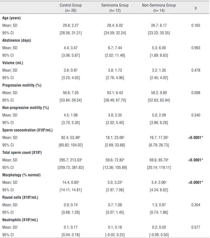
Individual characteristics and semen qua-lity results are presented in Table-1. No statisti-cally significant differences were observed in age, ejaculate volume, sperm motility, and round cells and neutrophil concentration between the three groups. The seminoma and non-seminoma pre-sented lower sperm concentration, morphology, and total sperm count when compared to the con-trol group.
The variables TBARS, GPx, SOD and TBARS/sperm of the three groups (seminoma, non-seminoma, and control) are shown in Ta-ble-2. The seminoma and non-seminoma groups presented higher TBARS levels when compared to controls. No statistically significant differen-ces were observed for SOD and GPx activities. No reading levels were achieved for catalase enzyme activity by the Beutler method in any group.
DISCUSSION
Whichever the histological origin of the testicular germ cell tumor (seminoma or non-se-minoma), orchiectomy is the initial therapeutic ap-proach, usually followed by gonadotoxic adjuvant therapies, such as chemotherapy and radiotherapy (25). However, even before any treatment for the cancer begins, patients may display reduced fertili-ty potential because of alterations to the testicular environment due to the disease itself (14, 15).
To our knowledge, there is no study in the literature that evaluates the role of oxidative stress and antioxidants in the seminal plasma of testi-cular germ cell tumor patients after orchiectomy. Thus, this study aimed to verify the activity of the main seminal antioxidant enzymes SOD, GPx, and catalase as well as the oxidative by-product ma-londialdehyde. The seminal non-enzymatic antio-xidant milieu consists of GPx, SOD and catalase to physiologically control the balance between ROS production and neutralization (26). In this study, catalase levels were undetected. However,
determining catalase activity in seminal plasma remains a matter of debate, because the presence of this antioxidant in the semen is usually due mainly to the presence of neutrophils (24).Because the semen of our patients presented low concen-tration of these cells (under 0.5million/mL in ei-ther group), this could explain why catalase was not detected in this study.
GPx protects sperm membrane from oxi-dative stress and is involved in redox regulation (27). In addition, SOD is responsible for dismuta-tion of superoxide radicals (O2-)into H2O2 and O2 (28). Regarding the seminoma and non-seminoma groups,when compared to healthy control men, no differences were observed in the SOD and GPx activity, suggesting that antioxidant mechanisms were not altered in the presence of a testicular germ cell tumor followed by orchiectomy. This may stem from the fact that most of the seminal antioxidant levels is supported by secretions from the prostate and seminal vesicles (27, 28), which are apparently unaffected by the presence of a tes-ticular cancer, at least in terms of their production of enzymatic antioxidants. It could be argued that epididymal antioxidants also contribute to the se-minal antioxidant capacity, but their contribution is generally described as much lower (29), and it is still not known whether epididymal secretion of antioxidants is affected by the tumor.
Table 1 - Semen analysis of orquiectomized men and healthy control men. Groups were compared by ANOVA followed by LSD post-hoc test.
Control Group (n= 26)
Seminoma Group (n= 12)
Non-Seminona Group
(n= 14) p
Age (years)
Mean; SD 29.8; 3.27 28.4; 6.02 26.7; 6.17 0.165 95% CI [28.56; 31.21] [24.59; 32.24] [23.22; 30.35]
Abstinence (days)
Mean; SD 4.4; 3.47 6.7; 7.44 5.3; 6.00 0.993 95% CI [3.06; 5.87] [2.02; 11.48] [1.89; 8.83]
Volume (mL)
Mean; SD 3.6; 0.97 3.8; 1.73 3.2; 1.30 0.478 95% CI [3.23; 4.02] [2.76; 4.96] [2.45; 4.02]
Progressive motility (%)
Mean; SD 56.6; 7.05 63.1; 6.43 58.2; 9.80 0.098 95% CI [53.84; 59.54] [58.49; 67.70] [52.63; 63.94]
Non-progressive motility (%)
Mean; SD 4.5; 1.98 3.8; 2.35 5.0; 2.09 0.340 95% CI [3.70; 5.30] [2.02; 5.40] [3.86; 6.28]
Sperm concentration (X106/mL)
Mean; SD 82.4; 53.48b 18.1; 23.06a 16.7; 17.26a <0.0001*
95% CI [60.82; 104.02] [2.69; 33.68] [6.79; 26.73]
Total sperm count (X106)
Mean; SD 295.7; 213.03b 59.6; 72.82a 69.6; 85.70a <0.0001*
95% CI [209.73; 381.82] [13.36; 105.89] [20.14; 119.11]
Morphology (% normal)
Mean; SD 14.4; 0.85b 5.0; 3.22a 5.4; 2.06a <0.0001*
95% CI [14.11; 14.81] [2.97; 7.06] [4.24; 6.62]
Round cells (X106/mL)
Mean; SD 0.9; 0.74 0.7; 1.08 1.3; 0.97 0.304 95% CI [0.68; 1.28] [0.07; 1.45] [0.74; 1.86]
Neutrophils (X106/mL)
Mean; SD 0.1; 0.17 0.1; 0.18 0.2; 0.50 0.577 95% CI [0.04; 0.18] [-0.02; 0.22] [-0.09; 0.50]
SD=Standard deviation
95% CI =Confidence interval of 95% of the mean * – significant difference
Table 2- TBARS, Glutathione Peroxidase (GPx) and Superoxide Dismutase (SOD) levels from orquiectomized men and
healthy control men. Groups were compared by ANOVA followed by LSD post-hoc test.
Control Group (n= 26)
Seminoma Group (n=12)
Non-seminoma Group
(n=14) p
TBARS/sperm
Mean; SD 3.2; 2.59b 53.2; 127.19a 22.7; 25.95a <0.001*
95% CI [2.16; 4.26] [-27.62; 134.01] [7.74; 37.71]
GPx (UI/mL)
Mean; SD 65.0; 20.02 59.9; 19.45 67.5; 23.05 0.646 95% CI [56.97; 73.15] [47.63; 72.35] [54.21; 80.83]
SOD (UI/mL)
Mean; SD 63.9; 59.84 34.3; 30.64 50.0; 27.49 0.328 95% CI [39.79; 88.13] [14.88; 53.83] [34.16; 65.92]
SD=Standard deviation
95% CI=Confidence interval of 95% of the mean * – significant difference
Different Letters in the same row indicate significant difference (post-hoc LSD test – p<0.05).
stress (as demonstrated by our results) would ex-plain why lipid peroxidation levels would depend on sperm concentration - the latter acting as a substrate for the former (32). Given that,in our study, testicular germ cell tumor patients collec-ted semen samples on average one month after orchiectomy,the inflammatory state would not have had time to be fully resolved, as one full cycle of spermatogenesis would not have yet oc-curred. This is further supported by the fact that the study group presented decreased sperm con-centration (quantity) and morphology (quality). The findings corroborate with Tavilani et al. (33) study, which observed antioxidant profile and oxidative stress in asthenozoospermic men and only observed a significant difference in TBARS/ sperm, indicating that the mechanism of oxida-tive stress occurs similarly in infertile men and testicular germ-cell tumor patients.
Besides, this study is limited to the fact that the patients have a short period to perform a sample collection, because after they are sub-mitted to orchiectomy, the adjuvant therapy is required. This way, it is not possible to observe antioxidant profile difference after a complete
spermatogenesis cycle. In addition, the number of patients included in the study could not be enou-gh to demonstrate difference in some studied pa-rameters. Moreover, a further path for future rese-arch would be increase the number of patients and include pre-operative samples, which would add information regarding how the antioxidants and lipid peroxidation act in a tumor milieu.
CONCLUSIONS
CONFLICT OF INTEREST
None declared.
REFERENCES
1. Gandini L, Lombardo F, Salacone P, Paoli D, Anselmo AP, Culasso F, et al. Testicular cancer and Hodgkin’s disease: evaluation of semen quality. Hum Reprod. 2003;18:796-801. 2. Jemal A, Siegel R, Ward E, Hao Y, Xu J, Thun MJ. Cancer
statistics, 2009. CA Cancer J Clin. 2009;59:225-49.
3. Valsero Herguedas ME, Pascual Samaniego M, Garcia Lagarto E, Martín Martin S, Muñoz Moreno MF, Cortiñas Gonzalez JR. Testicular cancer: our experience after 10 years. Arch Esp Urol. 2012;65:467-75.
4. INCA-CÂNCER-Tipo-Testículo [Internet]. 2016 [cited 2016 Feb 17]; Available at.<http://www2.inca.gov.br/wps/wcm/ connect/tiposdecancer/site/home/testiculo>
5. Huyghe E, Matsuda T, Thonneau P. Increasing incidence of testicular câncer worldwide: a review. J Urol. 2003;170:5-11. 6. Winter C, Albers P. Testicular germ cell tumors: pathogenesis,
diagnosis and treatment. Nat Rev Endocrinol. 2011;7:43-53. 7. Ma YT, Cullen MH, Hussain SA. Biology of germ cell tumors.
Hematol Oncol Clin North Am. 2011;25:457-71.
8. Leman ES, Magheli A, Yong KM, Netto G, Hinz S, Getzenberg RH. Identification of nuclear structural protein alterations associated with seminomas. J Cell Biochem. 2009;108:1274-9. 9. Chung P, Warde P. Testicular cancer: seminoma. BMJ Clin
Evid. 2011;2011.
10. Simon SD, Srougi M. Neoadjuvant M-VAC chemotherapy and partial cystectomy for treatment of locally invasive transitional cell carcinoma of the bladder. Prog Clin Biol Res. 1990;353:169-74.
11. Powles T. Stage I nonseminomatous germ cell tumor of the testis: more questions than answers? Hematol Oncol Clin North Am. 2011;25:517-27.
12. Feldman DR, Bosl GJ, Sheinfeld J, Motzer RJ. Medical treatment of advanced testicular cancer. JAMA. 2008;299:672-84.
13. Bandak M, Aksglaede L, Juul A, Rørth M, Daugaard G. The pituitary-Leydig cell axis before and after orchiectomy in patients with stage I testicular cancer. Eur J Cancer. 2011;47:2585-91.
14. Masotti L, Casali E, Gesmundo N, Sartor G, Galeotti T, Borrello S, et al. Lipid peroxidation in cancer cells: chemical and physical studies. Ann N Y Acad Sci. 1988;551:47-57; discussion 57-8.
15. Agarwal A, Allamaneni SS. Disruption of spermatogenesis by the cancer disease process. J Natl Cancer Inst Monogr. 2005;34:9-12.
16. Halliwell B. Free radicals, antioxidants, and human disease: curiosity, cause, or consequence? Lancet. 1994;344:721-4. 17. Sharma RK, Agarwal A. Role of reactive oxygen species in
male infertility. Urology. 1996;48:835-50.
18. Baumber J, Ball BA, Gravance CG, Medina V, Davies-Morel MC. The effect of reactive oxygen species on equine sperm motility, viability, acrosomal integrity, mitochondrial membrane potential, and membrane lipid peroxidation. J Androl. 2000;21:895-902.
19. Mancini A, Festa R, Silvestrini A, Nicolotti N, Di Donna V, La Torre G, et al. Hormonal regulation of total antioxidant capacity in seminal plasma. J Androl. 2009;30:534-40. 20. Petersen PM, Skakkebaek NE, Vistisen K, Rørth M,
Giwercman A. Semen quality and reproductive hormones before orchiectomy in men with testicular cancer. J Clin Oncol. 1999;17:941-7.
21. WHO | WHO laboratory manual for the examination and processing of human semen [Internet]. [cited 2015 Oct 6];Available at. <http://www.who.int/reproductivehealth/ publications/infertility/9789241547789/en/>
22. Esteves SC, Spaine DM, Cedenho AP. Effects of pentoxifylline treatment before freezing on motility, viability and acrosome status of poor quality human spermatozoa cryopreserved by the liquid nitrogen vapor method. Braz J Med Biol Res. 2007;40:985-92.
23. Ohkawa H, Ohishi N, Yagi K. Assay for lipid peroxides in animal tissues by thiobarbituric acid reaction. Anal Biochem. 1979;95:351-8.
24. Nichi M, Bols PE, Züge RM, Barnabe VH, Goovaerts IG, Barnabe RC, et al. Seasonal variation in semen quality in Bos indicus and Bos taurus bulls raised under tropical conditions. Theriogenology. 2006;66:822-8.
25. Fedyanin M, Tryakin A, Bulanov A, Fainshtein I, Zakharova T, Matveev V, et al. Effect of the timing of orchiectomy on survival in patients with metastatic germ cell tumors of testis. Urol Oncol. 2014;32:32.e27-33.
26. Kankofer M, Kolm G, Aurich J, Aurich C. Activity of glutathione peroxidase, superoxide dismutase and catalase and lipid peroxidation intensity in stallion semen during storage at 5 degrees C. Theriogenology. 2005;63:1354-65. 27. Ursini F, Maiorino M, Valente M, Ferri L, Gregolin C. Purification
from pig liver of a protein which protects liposomes and biomembranes from peroxidative degradation and exhibits glutathione peroxidase activity on phosphatidylcholine hydroperoxides. Biochim Biophys Acta. 1982;710:197-211. 28. Mruk DD, Silvestrini B, Mo MY, Cheng CY. Antioxidant
superoxide dismutase - a review: its function, regulation in the testis, and role in male fertility. Contraception. 2002;65:305-11.
30. Tong L, Chuang CC, Wu S, Zuo L. Reactive oxygen species in redox câncer therapy. Cancer Lett. 2015;367:18-25.
31. Waris G, Ahsan H. Reactive oxygen species: role in the development of câncer and various chronic conditions. J Carcinog. 2006;5:14.
32. Intasqui P, Antoniassi MP, Camargo M, Nichi M, Carvalho VM, Cardozo KH, et al. Differences in the seminal plasma proteome are associated with oxidative stress levels in men with normal semen parameters. Fertil Steril. 2015;104:292-301.
33. Tavilani H, Goodarzi MT, Vaisi-raygani A, Salimi S, Hassanzadeh T. Activity of antioxidant enzymes in seminal plasma and their relationship with lipid peroxidation of spermatozoa. Int Braz J Urol. 2008;34:485-91.
_______________________ Correspondence address: