Laparoscopic donor nephrectomy in unusual venous anatomy
– donor and recepient implications
_______________________________________________
Avinash Bapusaheb Patil
1,Tarun Dilip Javali
1,Harohalli K. Nagaraj
1, S. M. L. Prakash Babu
1, Arvind Nayak
11 Department of Urology, M.S. Ramaiah Hospital, Bangalore
ABSTRACT
ARTICLE
INFO
______________________________________________________________ ______________________
Objectives: Laparoscopic donor nephrectomy is now a commonly performed procedure in most of renal transplantation centers. However, the suitability of laparoscopy for donors with abnormal venous anatomy is still a subject of debate.
Materials and methods: Between August 2007 and August 2014, 243 laparoscopic donor nephrectomies were performed in our institution. All donors were evaluated with preoperative three-dimensional spiral computed tomography (CT) angiography Thirteen (5.35%) donors had a left renal vein anomaly. A retrospective analysis was performed to collect donor and recipient demographics and perioperative data.
Results: Four donors had a type I retroaortic vein, seven had type II retroaortic vein and a circumaortic vein was seen in three donors. The mean operative time was 114±11 minutes and mean warm ischemia time was 202±12 seconds. The mean blood loss was 52.7±18.4mL and no donor required blood transfusion. Mean recipient creatinine at the time of discharge was 1.15±0.18mg/dL, and creatinine at six months and one year follow-up was 1.12±0.13mg/dL and 1.2±0.14mg/dL, respectively. There were no significant differences in operative time, blood loss, warm ischemia time, donor hos-pital stay or recipient creatinine at 6 months follow-up, following laparoscopic donor nephrectomy in patients with or without left renal vein anomalies.
Conclusion: Preoperative delineation of venous anatomy using CT angiography is as important as arterial anatomy. Laparoscopic donor nephrectomy is safe and feasible in patients with retroaortic or circumaortic renal vein with good recipient outcome.
Keywords:
Laparoscopy; Veins; Kidney Transplantation
Int Braz J Urol. 2017; 43: 671-8
_____________________
Submitted for publication: May 31, 2016
_____________________
Accepted after revision: October 01, 2016
_____________________
Published as Ahead of Print: February 10, 2017
INTRODUCTION
With the advent of laparoscopic live do-nor nephrectomy, there has been an increase in number of donors for kidney transplantation. The first laparoscopic donor nephrectomy was performed by Ratner et al. in 1995 (1). Since then laparoscopic donor nephrectomy has be-come the standard of care in most transplant centers around the world. Compared with open
rative pain, shorter length of hospital stay, and faster return to work (2-6).
MATERIALS AND METHODS
Between August 2007 and August 2014, 243 laparoscopic donor nephrectomies were per-formed in our institution. A retrospective analy-sis was performed to collect donor and recipient demographics and perioperative data. All donors underwent standard preoperative evaluation in-cluding medical, surgical, psychological and im-munological evaluation, and detailed informed consent. Three-dimensional spiral CT angiography was used to define the renal vascular anatomy and a renal isotope scan was performed to determine the choice of kidney for nephrectomy.
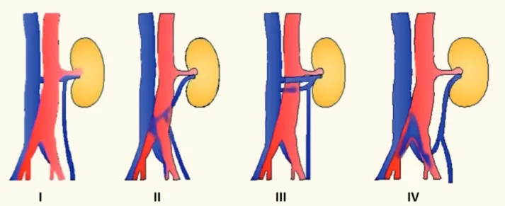
Left renal vein abnormalities are categori-zed into four types (7). Types I, II and IV are “re-troaortic” left renal veins, while type III is consi-dered as “circumaortic” vein. Type I retroaortic left renal vein typically joins the IVC in the orthotopic position, while type II retroaortic left renal vein joins the IVC at level L4-5. The type IV retroaor-tic vein joins the left common iliac vein. The cir-cumaortic or type III left renal vein anomaly has both a pre-aortic as well as a retroaortic compo-nent (Figure-1).Information on donor age, gender, body mass index (BMI), relation to the recipient
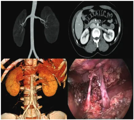
and type of renal vein anomaly was collected. Of the 243 donors, 13 (5.35%) donors had a left re-nal vein anomaly. Among these, four had a type I retroaortic vein, seven had type II retroaortic vein and three donors had a circumaortic vein. Among these 13 patients with left renal vein anomalies, 11 patients had a single left renal artery (Figure-2) while one patient with type I retroaortic vein had bilateral two renal arteries (Figure-3) and one pa-tient had bilateral two renal arteries with right two renal veins and left circumaortic vein (Figure-4). Laparoscopic right donor nephrectomy was per-formed in 38 patients hence they were excluded from the study. Remaining 192 patients without any left renal vein anomaly had undergone laroscopic left donor nephrectomy. Of these 192 pa-tients, 20 patients had multiple left renal arteries and left kidney was selected due to anomalous ri-ght renal vein/artery. Surgical data included ope-rative time, warm ischemia time, estimated blood loss, complications, and nadir serum creatinine.
The laparoscopic procedure was performed transperitoneally. All donor nephrectomies were performed by either one of two surgeons: HKN/TDJ, and all renal transplants were performed by a single recipient team. Briefly, dissection was started at the
Figure 2- Type II retroaortic left renal vein with bilateral single renal arteries.
lower pole of kidney and the ureter and gonadal vein were dissected. The gonadal vein was traced towards the left renal vein. The adrenal vein, gonadal vein and lumbar vein (if present) were identified, clipped and transected. In case of retroaortic vein, renal vein was dissected up to lateral border of aorta and cli-pped using two Hemolock clips (Weck clips) and
one metallic clip at the donor side. Renal artery was clipped using two Hemolock clips and one metallic clip. In two patients with circumaortic veins, smaller caliber retroaortic component was sacrificed. Kidney was delivered out through Pfannenstiel incision.
Operative time was defined as the time from the initial skin incision to delivery of the kidney to the recipient transplant team. Warm ischemia time was calculated as the time from renal artery ligation to immersion of the kidney in ice slush. Information was also collected on recipient allograft function, including serum cre-atinine at the time of discharge, at six months and at one year follow-up, as well as complica-tions. Delayed graft function was defined as the patient requiring dialysis postoperatively.
Statistical analysis was performed using Ins-tat® software (GraphPad Software, San Diego Cali-fornia USA). Both discrete and continuous variables were analyzed with the Student’s t-test. A P value of less than 0.05 was considered statistically significant.
RESULTS
Donor demographic data, including age, gender, BMI, type of left renal vein anomaly, opera-tive time, warm ischemia time and blood loss for 13 donors who underwent transperitoneal laparoscopic donor nephrectomy are listed in Table-1. There were two (15%) men and 11 (85%) women, with a mean age of 44.5±7.1 years. The mean operative time was 114±11 minutes and mean warm ischemia time was 202±12seconds.There were no intraoperative compli-cations. The mean blood loss was 52.7±18.4mL,and no donor required blood transfusion. Donors were discharged on the third postoperative day and no donor required readmission. Mean donor creatinine preoperatively and at discharge was 1.0±0.14mg/dL and 1.27±0.18mg/dL, respectively.
Recipient outcomes are shown in Table-2. Mean recipient age was 45.2±7.3 years. The mean cold ischemia time was 50.2±8 minutes. The mean recipient blood loss was 317±39mL and mean hos-pital stay was 11.3±2.4 days. One patient required temporary dialysis after surgery due to delayed graft function due to acute tubular necrosis. Mean recipient creatinine at the time of discharge was 1.15±0.18mg/ dL, and creatinine at six months and one year follow-up was 1.12±0.13mg/dL and 1.2±0.14mg/ dL, respectively. We routinely used ureteral stents
Table 1 - Donor characteristics.
Sl. No.
Donor Age (Years)
Sex BMI (Kg/m2)
Type of renal vein anomaly
Number of renal arteries
Operative time (minutes)
Warm ischemia time
(seconds)
Blood loss (mL)
Complications Serum Creatinine (mg/dL)
Before surgery
At discharge
1 46 F 26.2 Retroaortic type II 1 118 212 45 Nil 1.1 1.3
2 30 F 22.3 Retroaortic type II 1 112 194 60 Nil 0.9 1.2
3 41 F 24.6 Retroaortic type I 1 109 190 25 Nil 1 1.2
4 52 M 27.2 Retroaortic type II 1 103 210 40 Nil 1.1 1.4
5 41 F 24.5 Circumaortic 2 133 205 85 Nil 0.9 1
6 47 F 26.8 Retroaortic type II 1 112 188 70 Nil 0.8 1.1
7 53 F 27 Retroaortic type I 1 114 217 65 Nil 1.3 1.6
8 36 M 23.4 Retroaortic type I 1 108 196 80 Nil 0.8 1
9 43 F 24.1 Retroaortic type II 1 98 208 45 Nil 1 1.3
10 39 F 25.3 Retroaortic type II 1 115 220 55 Nil 0.9 1.3
11 46 F 27.4 Circumaortic 1 124 185 40 Nil 1.1 1.4
12 54 F 28.5 Retroaortic type I 2 136 208 45 Nil 1 1.3
13 50 F 29.2 Retroaortic type II 1 103 190 30 Nil 1.1 1.5
M= Male; F= Female; BMI= Body mass index
Table 2 - Recipient outcomes.
Sl. No. Recepient Age (years)
Sex Cold ischemia time (minutes)
Blood loss (mL)
Hospital stay (days)
Graft related complications Serum Creatinine (mg/dL)
At discharge At 1 month At 6 months
1 50 M 52 280 11 Nil 1.3 1.2 1.3
2 33 M 48 310 12 Nil 1 1.1 1.1
3 38 M 45 350 9 Nil 1.2 1.2 1.3
4 46 F 53 290 13 Nil 1.4 1.3 1.4
5 45 M 67 285 11 Nil 0.9 1 1
6 51 M 43 370 10 Nil 1 1 1.2
7 45 M 40 335 8 Nil 1.3 1.3 1.2
8 40 M 53 390 12 Nil 0.9 1 1.1
9 35 M 41 325 17 Delayed graft function 1.2 1 1
10 44 M 47 290 11 Nil 1.2 1.3 1.4
11 49 M 54 265 14 Nil 1.4 1.2 1.3
12 58 M 63 340 9 Nil 1 1 1.1
in all uretero-vesical anastomosis and these stents were removed after 21 days.
These results were compared to the outco-mes of 192 patients without left renal vein ano-maly (Table-3). Incidences of multiple left renal arteries were comparable in both the groups. There was no statistically significant difference in donor outcomes(operative time, blood loss, warm ische-mia time) and recipient outcomes (serum creatini-ne at 6 months follow-up).
DISCUSSION
Incidence of end stage renal disease and number of kidneys available for renal transplant has always been a major medical concern. With the advent of laparoscopic donor nephrectomy, there has been increase in live donor pool over last decade (8). Laparoscopic donor nephrectomy offers low morbidity, shorter length of hospitali-zation, less pain medication requirements, and
re-duced convalescence as compared to open donor nephrectomy (9).
The left kidney is favored for laparoscopic nephrectomy because it provides a graft with a longer renal vein (2,3). Traditionally, right open donor nephrectomy is chosen when the left kid-ney has multiple renal arteries or veins or other vascular anomalies. Major concernin right-sided laparoscopic donor nephrectomy is short length right renal vein which is further shortened by use of vascular clips. There is increased risk of va-sospasm and iatrogenic vascular injury during la-paroscopic right donor nephrectomy as the right renal artery is located directly posterior to short right renal vein. Some authors have reported a higher potential for vascular complications with eventual graft loss with laparoscopic right donor nephrectomy (10,11).
The most common renal venous anomaly is the occurrence of dual renal veins, accounting for 15%-30%, frequently on the right side (12-15).
Table 3 - Comparison of demographics and outcomes in patients with and without left renal vein anomaly.
Donors with left renal vein anomalies (n=13)
Donors without left renal vein anomalies (n=192)
P value
Donor age (years), mean±SD 44.5±7 48.4±8.1 >0.05
Recipient age (years), mean±SD 45.2±7.3 40.1±6.9 <0.05
Multiple left renal arteries (%) 2 (15.4) 20 (10.4)
Donor BMI (Kg/m2), mean±SD 25.9±2.0 25.5±3.7 >0.05
Operative Details Operative time (minutes), mean±SD 114±11 109±17 >0.05
Blood loss (mL), mean±SD 53±18 64±23 >0.05
Warm ischemia time (seconds), mean±SD
202±12 211±18 >0.05
Hospital stay (Donors) 60 Hours 60 Hours
Recipient outcomes
Delayed graft function (%) 1 (7.7) 11 (5.7)
Cold ischemia time (minutes), mean±SD
50.2±8 54.6±11 >0.05
Serum Creatinine at 6 months (mg/dL), mean±SD
Circumaortic and retroaortic variants constitute the most common anomalies of the left renal vein with incidence of 6.2%-14% (10,16,17).Because of the higher risk of vascular injury, the presence of a circumaortic or retroaortic renal vein has pre-viously been considered a relative contraindication for left donor nephrectomy by some surgeons (18). Some authors have reported that there was no sig-nificant difference regarding parameters such as operative time, warm ischemia time, length of allo-graft vessels, and estimated blood loss in patients with circumaortic or retroaortic renal vein when compared to control group (19,20).
In this retrospective study, we analyzed safety and feasibility of laparoscopic donor ne-phrectomy in patients with left renal vein ano-maly and we compared donor and recipient ou-tcomes with group of patients without left renal vein anomaly. The use of CT angiography allows preoperative identification of venous anomalies (21). In our hospital, 243 patients underwent la-paroscopic donor nephrectomy. On preoperative evaluation with three-dimensional spiral CT an-giography, 13 patients (5.35%) were diagnosed to have left renal vein anomaly in form of retroaortic vein (11) or circumaortic vein (2). Retroaortic vein will have an abnormal course posterior to aorta. Adrenal, gonadal, and lumbar veins may enter the renal vein at abnormal position. Hence, after me-ticulous dissection and control of these tributaries, retroaotic vein can be clipped at the level of late-ral border of aorta. In case of circumaortic vein, usually retroaortic component will have smaller caliber and it can be safely sacrificed. Preaortic component can be clipped at the opening into IVC. Careful preoperative radiological evaluation of vascular anatomy is mandatory and intraope-rative potential variation in vascular anatomy has to be kept in mind. In our experience, operative time and warm ischemia time were not prolonged. The mean warm ischemia time was 202±12secon-ds and mean operative time was 114±18 minutes. One-year graft survival was 100%.
CONCLUSIONS
Preoperative delineation of venous anatomy
anatomy. Laparoscopic donor nephrectomy is safe and feasible in patients with retroaortic or circuma-ortic renal vein with good recipient outcome.
CONFLICT OF INTEREST
None declared.
REFERENCES
1. Ratner LE, Ciseck LJ, Moore RG, Cigarroa FG, Kaufman HS, Kavoussi LR. Laparoscopic live donor nephrectomy. Transplantation. 1995;60:1047-9.
2. Jacobs SC, Cho E, Dunkin BJ, Flowers JL, Schweitzer E, Cangro C, et al. Laparoscopic live donor nephrectomy: the University of Maryland 3-year experience. J Urol. 2000;164:1494-9.
3. Ratner LE, Montgomery RA, Kavoussi LR. Laparoscopic live donor nephrectomy: the four year Johns Hopkins University experience. Nephrol Dial Transplant. 1999;14:2090-3. 4. Nicholson ML, Kaushik M, Lewis GR, Brook NR, Bagul A,
Kay MD, et al. Randomized clinical trial of laparoscopic versus open donor nephrectomy. Br J Surg. 2010;97:21-8. 5. Greco F, Hoda MR, Alcaraz A, Bachmann A, Hakenberg OW,
Fornara P. Laparoscopic living-donor nephrectomy: analysis of the existing literature. Eur Urol. 2010;58:498-509. 6. Yuan H, Liu L, Zheng S, Yang L, Pu C, Wei Q, et al. The
safety and efficacy of laparoscopic donor nephrectomy for renal transplantation: an updated meta-analysis. Transplant Proc. 2013;45:65-76.
7. Nam JK, Park SW, Lee SD, Chung MK. The clinical significance of a retroaortic left renal vein. Korean J Urol. 2010;51:276-80.
8. Romagnoli J, Salerno MP, Mamode N, Calia R, Spagnoletti G, Bianchi V, et al. Expanding the Living Donor Pool “Second Act”: Laparoscopic Donor Nephrectomy and ABO-Incompatible Kidney Transplantation Improve Donor Recruitment. Transplant Proc. 2015;47:2126-9.
9. Tooher RL, Rao MM, Scott DF, Wall DR, Francis DM, Bridgewater FH, et al. A systematic review of laparoscopic live-donor nephrectomy. Transplantation. 2004;78:404-14. 10. Mandal AK, Cohen C, Montgomery RA, Kavoussi LR, Ratner
11. Leventhal JR, Kocak B, Salvalaggio PR, Koffron AJ, Baker TB, Kaufman DB, et al. Laparoscopic donor nephrectomy 1997 to 2003: lessons learned with 500 cases at a single institution. Surgery. 2004;136:881-90.
12. Pozniak MA, Balison DJ, Lee FT Jr, Tambeaux RH, Uehling DT, Moon TD. CT angiography of potential renal transplant donors. Radiographics. 1998;18:565-87.
13. Namasivayam S, Kalra MK, Waldrop SM, Mittal PK, Small WC. Multidetector row CT angiography of living related renal donors: is there a need for venous phase imaging? Eur J Radiol. 2006;59:442-52.
14. Holden A, Smith A, Dukes P, Pilmore H, Yasutomi M. Assessment of 100 live potential renal donors for laparoscopic nephrectomy with multi-detector row helical CT. Radiology. 2005;237:973-80.
15. Smith PA, Ratner LE, Lynch FC, Corl FM, Fishman EK. Role of CT angiography in the preoperative evaluation for laparoscopic nephrectomy. Radiographics. 1998;18:589-601.
16. Schmidt GP, Loeweneck H. [Frequency of the retroaortic left renal vein in adults (author’s transl)]. Urol Int. 1975;30:332-40.
17. Trigaux JP, Vandroogenbroek S, De Wispelaere JF, Lacrosse M, Jamart J. Congenital anomalies of the inferior vena cava and left renal vein: evaluation with spiral CT. J Vasc Interv Radiol. 1998;9:339-45.
18. Walker TG, Geller SC, Delmonico FL, Waltman AC, Athanasoulis CA. Donor renal angiography: its influence on the decision to use the right or left kidney. AJR Am J Roentgenol. 1988;151:1149-51.
19. Lin CH, Steinberg AP, Ramani AP, Abreu SC, Desai MM, Kaouk J, et al. Laparoscopic live donor nephrectomy in the presence of circumaortic or retroaortic left renal vein. J Urol. 2004;171:44-6.
20. Troppmann C, Wiesmann K, McVicar JP, Wolfe BM, Perez RV. Increased transplantation of kidneys with multiple renal arteries in the laparoscopic live donor nephrectomy era: surgical technique and surgical and nonsurgical donor and recipient outcomes. Arch Surg. 2001;136:897-907.
21. El Fettouh HA, Herts BR, Nimeh T, Wirth SL, Caplin A, Sands M, et al. Prospective comparison of 3-dimensional volume rendered computerized tomography and conventional renal arteriography for surgical planning in patients undergoing laparoscopic donor nephrectomy. J Urol. 2003;170:57-60.
_______________________ Correspondence address: