1Instituto de Neurologia de Curitiba, Curitiba PR, Brasil; 2Departamento de Neurocirurgia, da Universidade Estadual de Campinas
(UNICAMP), Campinas SP, Brazil; 3Professor assistente de Neurologia, Universidade Federal do Paraná, Curitiba PR, Brasil.
Received 15 July 2004. Accepted 12 October 2004.
Dr. Ricardo Ramina - Rua Gonçalves Dias 713 - 80340-240 Curitiba PR - Brasil. E-mail: [email protected]
INTRINSIC TECTAL LOW GRADE ASTROCYTOMAS
IS SURGICAL REMOVAL AN ALTERNATIVE TREATMENT?
Long-term outcome of eight cases
Ricardo Ramina
1,2, Mauricio Coelho Neto
1, Yvens Barbosa Fernandes
2,
Guilherme Borges
2, Donizeti Cesar Honorato
2, Walter Oleschko Arruda
1,3ABSTRACT - Low-grade gliomas arising in dorsal midbrain in children and young patients usually present few neurological symptoms and findings, and patients´ management is controversial. Some authors pro-pose only clinical observation until the patient present signs of increased intracranial pressure when a shunt with or without biopsy, is inserted; others recommend radiotherapy after stereotactic or open biopsy. Micro-surgical total removal of tumor may be curative. We present a retrospective analysis of eight patients (mean age 16.6 ±11.5 years-old) with low-grade astrocytoma of the tectal region operated on using an infraten-torial/ supracerebellar approach between 1981 and 2002. All patients presented hydrocephalus and had a shunt insertion before surgical resection of the lesion. The tumour could be totally resected in seven patients. In one case radical removal was not possible due to infiltrative pattern of the lesion. Postoperative radiothe-rapy was performed in two cases, one patient at the beginning of this series and in the case with infiltrati-ve tumor. This patient presented progressiinfiltrati-ve tumor growth and died fiinfiltrati-ve years after surgery. No recur-rence occurred after total removal. Post-surgical follow-up time ranged from 2 1/2 to 22 1/2 years (mean 9.9 ± 5.9 years). Radical microsurgical removal of non invasive tumors is possible without mortality or sig-nificant morbidity. It may be curative and should remain as an alternative to be discussed with the patient.
KEY WORDS: astrocytoma, tectal tumours, glioma, hydrocephalus.
Astrocitomas tectais de baixo grau: o tratamento cirúrgico é uma alternativa? análise de oito casos com longa evolução
RESUMO - Gliomas de baixo grau originários da porção dorsal do mesencéfalo ocorrem em crianças e adul-tos jovens. Geralmente apresentam pouca sintomatologia e tardia, com hipertensão intracraniana por hidroce-falia não-comunicante. O seu tratamento é controverso. Alguns autores propõem somente observação clíni-ca até o aparecimento de sintomas decorrentes de hipertensão intracraniana, quando é realizada derivação ventrículo-peritoneal (DVP), com ou sem biópsia da lesão. Outros recomendam radioterapia após compro-vação histológica por biópsia estereotáxica. A remoção cirúrgica total pode ser curativa. Analisamos retros-pectivamente 8 pacientes com astrocitoma de baixo grau na região tectal operados entre 1981 e 2002. A idade média foi 16,6 ±11,5 anos (variando de 8 a 44 anos). As lesões foram abordadas por acesso infraten-torial / supra-cerebelar. Todos os pacientes apresentaram hidrocefalia e uma DVP foi colocada em todos antes da remoção cirúrgica da lesão. A lesão tumoral foi removida completamente em 7 dos 8 casos. Em um único caso a remoção total foi impossível devido ao caráter infiltrativo do tumor. Radioterapia pós-operatória foi indicada em 2 casos, o primeiro no início da série e o segundo caso com tumor de caráter infiltrativo. Este último paciente apresentou crescimento tumoral progressivo e veio a falecer 5 anos após a cirurgia. Nos demais 7 pacientes não houve recurrência tumoral. O tempo de acompanhamento foi 2,5 a 22,5 anos (média 9,9 ± 5,9 anos). Remoção microneurocirúrgica radical pode e deve ser sempre cogita-da em pacientes com tumores não-invasivos, pois a baixa morbi/mortalicogita-dade é possível e aceitável, além do procedimento poder ser curativo.
Surgical resection of brain stem tumours is
current-ly limited to dorsal exophytic tumours arising from
the floor of the fourth ventricle and some intrinsic
lesions of the cervicomedullary junction and
mid-brain
1. Radiotherapy and chemotherapy remain
al-ternative therapies for other infiltrative and
biologi-cally aggressive brainstem tumours. Long-term
prog-nosis is usually very poor
2. Low-grade astrocytomas
of the tectal region present characteristic
neuro-radiological findings (specially in the MRI
examina-tion) and are often slow growing lesions causing
late-onset aqueductal stenosis and hydrocephalus
3.
Due to this indolent growth and few clinical
sym-ptoms the management of these lesions remains
controversial
1,3-7. Some authors recommend only a
shunt procedure and clinical observation
3,8,9,
oth-ers indicate biopsy and radiotherapy
10-12and in
some centres radical surgery has been
perfor-med
1,5,13,14.
We reviewed our experience with eight patients
harbouring intrinsic tectal low-grade astrocytomas.
METHOD
Between 1981 and 2002, eight patients (six males and two females) with low-grade astrocytomas of the tectal plate were operated on in the Instituto de Neurologia de Curitiba (Table 1). Seven patients were younger than 20 years and only one was older than 40 years (mean age 16.6±11.5 years-old). During this period of time, 43 pineal region tumors were surgically removed. All patients com-plained of headache, nausea and vomiting for several months. Three patients had papilledema. Two patients presented Parinaud’s syndrome and one was referred with the diagnosis of communicating hydrocephalus with no tumor visible in a high definition contrast enhanced com-puterized tomography (CT) examination. One patient had a ventricle-peritoneal shunt surgery 33 years before admission due to communicating hydrocephalus. She pre-sented headache and Parinaud’s syndrome and magnet-ic resonance image (MRI) examination revealed a lesion in the tectal region. All patients presented hydrocephalus, focal calcification in the pineal region was seen in two cases and contrast enhancement in the pineal and tec-tal region was observed in two patients. The first patient of this series had only CT scan which showed a
enhanc-Table 2. Treatment and outcome.
Case Follow-up (years) Approach Tumor removal Complications Radiotherapy
1 22 1/2 SC/IT total none 5.5 Gy
2 5 SC/IT sub-total none 5.5 Gy*
3 10 1/2 SC/IT total VI CN palsy** no
4 10 1/2 SC/IT total PS** no
5 10 1/2 SC/IT total none no
6 10 1/2 SC/IT total PS** no
7 7 1/2 SC/IT total none no
8 2 1/2 SC/IT total VI CN palsy** PS** no
SC/IT, supracerebellar/infratentorial; PS, Parinaud´s Syndrome; *Tumor recurrence; **Transient. Table 1. Clinical and radiological findings.
Case Age (years) Clinical CT presentation MRI
1 14 Headache, diplopia Tectal mass,
hydrocepahlus Not done
2 8 Parinaud syndrome,
headache, diplopia Focal calcification,
hydrocephalus Tectal mass
3 11 ICPS Hydrocephalus Tectal mass
4 9 ICPS Hydrocephalus Tectal mass
5 13 ICPS Calcification
hydrocephalus Tectal mass
6 17 ICPS Hydrocephalus Tectal mass
7 17 ICPS, Parinaud
Syndrome Hydrocephalus Tectal mass
8 44 Headache, diplopia Previous shunt Tectal mass
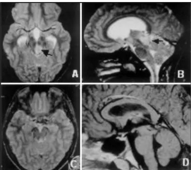
Postoperative the patient developed Parinaud’s syndro-me that completely resolved in four months after ope-ration. The histological diagnosis was low-grade astro-cytoma. The patient remained asymptomatic and no tumor recurrence was observed in the control MRI 10 years after surgery (Fig1).
Case 2 – This 17-year-old boy started to complain of headache and diplopia, 8 months before admission to our clinic. The neurological examination revealed Parinaud’s syndrome. MRI revealed hydrocephalus due to aqueduct occlusion by a tectal tumor. The lesion showed no contrast enhancement (Fig 2). A shunt was inserted and the tumor totally removed through a supracerebellar/infratentorial. The postoperative course was uneventful. The histopathological examination revealed a low-grade astrocytoma. A control MRI exam-ination 7 years after operation showed no tumor recur-rence (Fig 2).
Case 3 – This 44-year-old woman had a history of hydrocephalus treated by shunt placement 33 years before admission to our clinic. Several CT-scan examina-tions and two MRI showed normal ventricles and no tumor. Two months before admission to our clinic she pre-sented symptoms of headache and diplopia and Parinaud’s syndrome. MRI revealed a non-enhancing tumor in the tectal region (Fig 3). The lesion could be rad-ically removed through a supracerebellar/infratentorial approach. There was no postoperative complication and the Parinaud’s syndrome disappear completely 6 months after operation. The histological diagnosis was low-grade astrocytoma. The patient remained asymp-tomatic and a MRI, 2 years after surgery, showed total removal of the lesion (Fig 3).
DISCUSSION
Gliomas of the tectal plate are rare lesions. In
a review of 486 brain tumors in children, only 6
cas-es were observed
10. They may also occur in adults
but are rare and corresponded to 8% in a series of
48 brain stem tumors
15. These tumors are silent
pre-senting very often only late-onset aqueduct stenosis
and hydrocephalus
3,16,17. The diagnosis of low-grade
astrocytomas of the tectal region was difficult
until the advent of MRI
18. CT-scan may fail to reveal
these tumors, for they may be isodense and
non-enhancing. Therefore all patients with aqueduct
stenosis should be investigated with MRI. MRI
examination of these low-grade astrocytomas led
to earlier diagnosis and they can be followed
with-out invasive techniques
19. Low-grade astrocytomas
of this region typically present as isointense or
hy-pointense lesions on T1-weighted images
compa-ing tectal tumor. The other patients underwent (MRI) exa-mination with and without gadolinium administration. The patients studied with MRI presented, in the tectal region, an iso or hypointense mass on T1- weighted stu-dies and hyperintense on T2-weighted images. No enhan-cement or edema around the lesion were observed.
Hydrocephalus was treated with shunt procedure (sev-en pati(sev-ents) and (sev-endoscopic third v(sev-entriculostomy (one case). Tumor removal was performed through an infra-tentorial/ supracerebellar approach in a modified “con-corde position”. The first patient in this series was irradia-ted (6 Mv linear accelerator, 5500 cGy) and presenirradia-ted memory impairment probably related to radiotherapy. One patient with recurrent infiltrative astrocytoma re-ceived postoperative radiotherapy. Control neurologi-cal examination and MRI- examinations were performed (annually) in all cases between 2 1/2 and 22 1/2 years (mean 9.9 ± 5.9 years) after operation.
RESULTS
There were no complications related to the shunt placement or third ventriculostomy in this series. Hydro-cephalus resolved in all patients. In the postoperative period, three patients presented incomplete Parinaud’s syndrome, which disappeared completely three to six months after surgery. The two patients with preopera-tive Parinaud’s syndrome showed marked improvement of the upward gaze after the operation. Radical tumor removal was possible in seven patients. Total tumor removal of the lesion was confirmed by MRI in seven cas-es. Sub-total tumor removal, due to infiltration of the brainstem (a small portion in the cerebral peduncle re-mained), was performed in one patient. Growth of this residual tumour was observed in MRI 3 years after oper-ation. The tumor progressed and the patient died five years after surgery in spite of radiotherapy. There was no major complications in this series, three patients presented post-operative transient abducens palsy, which completely resolved within 3 and 6 months after surgery. No sign of tumor recurrence was observed in the follow-up exami-nation of the remained seven patients. Histological diag-nosis of all patients was low grade astrocytoma. A sum-mary of treatment and outcome is presented in Table 2.
ILUSTRATIVE CASES
red to the surrounding brain and hyperintense in
T2-weighted images. Tectus enlargement is
char-acteristic
20,21and gadolinium enhancement is not
always observed. The natural history of these
le-sions is not well known and the choice of the best
therapeutic approach is still controversial
3,5,6,20,22-26.
In a series of 16 patients, 4 cases demonstrated
clini-cal signs of tumor progression (Parinaud’s
syndro-me) with a median interval of 11.5 years from
on-set of initial symptoms and hydrocephalus
27. Two
cases of spontaneous tumor involution after third
ventriculostomy were reported
28Daglioglu et al.
29conclude that intrinsic tectal gliomas of childhood
which are less than 2 cm in diameter, without any
tumor extension or contrast enhancement, should
be managed conservatively.
The treatment of these patients varies from
clinical observation with control MRI examinations
to radical surgical removal. Increase of intracranial
pressure due to hydrocephalus is the most common
symptom of these patients. Shunt-procedures are
indicated for hydrocephalus due to aqueductal
stenosis
8,12. Radical removal and/or radiotherapy
for these benign lesions are usually indicated when
radiological or clinical signs of tumor progression
is observed
11,14. Total tumor resection is possible
wi-th low morbidity
5,30. Recently, Pollack et al.
recom-mended biopsy and radiotherapy for patients with
progressing symptoms
27. Biopsy may be performed
with neuroendoscopic techniques
31. A biopsy may,
however, produce sampling error and has some risks.
The proliferate index studies with Ki-67 labelling
is proving to be a valuable tool to confirm the
ag-gressive behaviour of some astrocytic lesions
32but
their routine use as a prognostic index still need to
be established.
Radical surgical removal is possible in non
infil-trative lesions with no mortality and low
morbid-Fig 2. A, B - Preoperative MRI with gadolinium showing no enhan-cement (arrows). C, D - Postoperative MRI 7 1/2 years after sur-gery demonstrating total tumor removal.
Fig 1. Preoperative T2-weighted MRI (A, B) tumor (black arrows). Postoperative MRI (C, D) 10 1/2 years after surgery showing total removal.
ity, it has two main advantages: confirm the
diag-nosis and may cure the patient. The approach used
in this series, was the
infratentorial/supracerebel-lar in a modified “concorde position”
33. There is no
clear evidence of beneficial effect of
radiothera-py (radiosurgery) for low-grade astrocytomas.
Rou-tine use of radiotherapy or chemotherapy is
ques-tionable
21. In a series of seven patients harbouring
low-grade tectal gliomas treated with stereotatic
radiosurgery, two developed severe radio-induced
edema, one with permanent deficits and one did
not respond to that treatment
34. Radiation
thera-py should be avoided in children because it may
cause anaplastic transformation of low-grade
astro-cytomas
35,36. Tarlov
37first suggested this
complica-tion in 1937 and development of radiacomplica-tion-induced
tumours in other areas has been also reported
38-40.
Other deleterious effects of radiotherapy as
psycho-motor impairment, cranial neuropathy,
leukoence-phalopathy, endocrinopathy and vasculopathy
ha-ve been also described
24, 41-47. When an infiltrative
pattern is present, a subtotal resection might be
done to reduce morbidity. The efficacy of
radiothe-rapy for incompletely resected low-grade
astrocy-tomas remains to be proven
48,49. The optimal
man-agement of high-grade astrocytomas of this region
is unclear too. Attempt of radical surgery in patients
with high-grade infiltrative lesions may produce
severe postoperative neurological deficits. If gross
total removal is, however, possible and followed
by radio and chemotherapy, the prognosis of these
patients is better
50.
In conclusion, low-grade astrocytomas of the
tec-tal region have an indolent clinical course. Most
of these lesions are truly focal neoplasms with
lit-tle tendency to infiltrate surrounding structures.
Headache, hydrocephalus and Parinaud´s syndrome
are the most common symptoms. Some tumors may
suffer transformation to higher grades.
Hydroce-phalus should be treated by shunt insertion or
en-doscopic third ventriculostomy. Radical removal of
the lesion is possible in the majority of patients, may
be curative and present very few complications.
Sur-gical removal should be presented to the patient
as a possibility of treatment, specially in growing
low-grade tumors.
REFERENCES
1. Epstein F, McClearly EL. Intrinsic brain-stem tumours of childhood: sur-gical indications. J Neurosurg 1986;64:11-15.
2. Grigsby PW, Thomas PR, Schwartz HG, Fineberg BB. Multivariant analysis of prognostic factors in pediatric and adult thalamic and bra-instem tumours. Int J Radiat Oncol 1989;16:649-655.
3. May PL, Blaser SI, Hoffman HJ, Humphreys RP, Harwood-Nash DC. Benign intrinsic tectal “tumours”in children. J Neurosurg 1991;74:867-871. 4. Albright AL, Guthkelch AN, Packer RJ, Price RA, Rourke LB. Prognostic factors in pediatric brain-stem gliomas. J Neurosurg 1986;65:751-755. 5. Lapras C, Bognar L, Turjman F, et al. Tectal plate gliomas: Part I. micro-surgery of the tectal plate gliomas. Acta Neurochir (Wien) 1994;126:76-83. 6. Parker RJ, Nicholson HS, Vezina LG, Johnson DL. Brainstem gliomas. in Berger MS (ed). Neurosurg clinic, vol.3. Philadelphia: Saunders, 1992:863-879.
7. Yeh DD, Warnick RE, Ernst RJ. Management strategy for adult patients with dorsal midbrain gliomas. Neurosurgery 2002;50:735-740. 8. Gomez-Gosalvez FA, Menor F, Morant A, et al. Tectal tumours in
pae-diatrics: a review of eight patients. Rev Neurol 2001;33:605-661. 9. Stroink AR, Hoffmann HJ, Hendrick EB, Humphreys RP. Diagnosis and
management of pediatric brainstem gliomas. J Neurosurg 1986;65:745-750. 10. Boydston WR, Sanford RA, Muhlbauer MS, et al. Gliomas of the tec-tum and periaqueductal region of the mesencephalon. Pediatr Neurosurg 1992;17:234-238.
11. Robertson PL, Muraszko KM, Brunberg JA, Axtell RA, Dauser RC, Turrisi AT. Pediatric midbrain tumours: a benign subgroup of brain-stem gliomas. Pediatr Neurosurg 1995;22:65-73.
12. Squires LA, Allen JC, Abbott R, Epstein FJ. Focal tectal tumours: man-agement and prognosis. Neurology 1994;44:953-956 .
13. Heffez DS, Zinreich SJ, Long DM. Surgical resection of intrinsic brain stem lesions: an overview. Neurosurgery 1990;27:789-798.
14. Poussaint TY, Kowal JR, Barnes PD, et al. Tectal tumours of childhood: clinical and imaging follow-up. Am J Neuroradiol 1998;19:977-983. 15. Guillamo JS, Monjour A, Taillandier L, et al. Brainstem gliomas in
adults: prognostic factors and classification. Brain 2001;124:2528-2539. 16. Panitch HS, Berg BO. Brainstem tumours of childhood and adolescence.
Am J Dis Child 1970;119:465-472.
17. Vandertop WP, Hoffman HJ, Drake JM, et al. Focal midbrain tumours in children. Neurosurgery 1992;31:186-192.
18. Yoshida Y, Kitakami A, Kikuchi Y, Hidaka T, Ogawa A. Two surgical cases of tectal glioma. No Shinkei Geka 1995;23:705-709.
19. Smith RR, Zimmerman RA, Packer RJ, et al. Pediatric brainstem glioma; post-radiation clinical and radiographic follow-up. Neuroradiology 1990;32:265-271.
20. Bowers DC, Georgiades C, Aronson LJ, et al. Tectal gliomas: natural his-tory of an indolent lesion in pediatric patients. Pediatr Neurosurg 2000;32:24-29.
21. Hamilton MG, Lauryssen C, Hagen N. Focal midbrain glioma: long term survival in a cohort of 16 patients and the implications for management. Can J Neurol Sci 1996;23:204-207.
22. Chapman PH. Indolent gliomas of the midbrain tectum, In Marlin AE (ed): Concepts in pediatric neurosurgery, vol.10. Basel: Karger, 1990;97-107. 23. Grant GA, Avellino AM, Loeser JD, Ellenbogen RG, Berger MS, Roberts
TS. Management of intrinsic gliomas of the tectal plate in children. A ten-year review. Pediatr Neurosurg 1999;31:170-176.
24. Parker RJ, Zimmerman RA, Bilaniuk LT, et al. Magnetic resonance ima-ging in the evaluation of treatment-related central nervous system da-mage. Cancer 1986;58:635-640.
25. Pollack IF, Hoffman HJ, Humphreys RP, Bwcker L. The long-term out-come after surgical treatment of dorsally exophytic brain-stem gliomas. J Neurosurg 1993;78:859-863.
26. Vertosick FT Jr, Selker RG, Arena VC Survival of patients with well-differentiated astrocytomas diagnosed in the era of computed tomog-raphy. Neurosurgery 1991;28:496-501.
27. Pollack IF, Pang D, Albright AL. The long-term outcome in children with late-onset aqueductal stenosis resulting from benign intrinsic tectal tumours. J Neurosurg 1994;80:81-84.
28. Alkahani AM, Boop FA, Rutka JT. Involution of enhancing intrinsic tec-tal after endoscopic third ventriculostomy. Report of two cases. J Neuro-surg 1999;91:863-869.
29. Daglioglu E, Cataltepe O, Akalan N. Tectal gliomas in children: the impli-cations for natural history and management strategy. Pediatr Neurosurg 2003;38:223-231.
30. Kaku Y, Yokenawa Y, Taub E. Transcollicular approach to intrinsic tec-tal lesions. Neurosurgery 1999,44:338-344.
31. Mizoguchi M, Inamura T, Hikita T, Cheng CL, Ohgami S. Neuro-endoscopic biopsy of tectal glioma: a case report. Minim Invasive Neurosurg 2000;43:53-55.
33. Stein BM Surgical treatment of pineal tumours. Clin Neurosurg 1979;26:490-510.
34. Kihlstrom L, Lindquist C, Lindquist M, Karlsson B. Stereotactic radiosur-gery for tectal low-grade gliomas. Acta Neurochir (Wien) 1994;62:55-57. 35. Dirks PB, Jay V, Becker LE, et al. Development of anaplastic changes in low-grade astrocytomas of childhood. Neurosurgery 1994;34:68-78. 36. Shapiro K, Katz M The recurrent cerebellar astrocytoma. Childs Brain
1983;10:168-176.
37. Tarlov IM. Effect of roentgenotherapy on gliomas. Arch Neurol Psychiatry 1937;38:513-536.
38. Kumar PP, Good RR, Skultety FM, Leibrock LG, Severson GS. Radiation-induced neoplasms of the brain. Cancer 1987;59:1274-1282. 39. Liwnicz BH, Berger TS, Liwnicz RG, Aron BS. Radiation-associated
glio-mas: report of four cases and analysis of postradiation tumours of the central nervous system. Neurosurgery 1985;17:436-445.
40. Ron E, Modan B, Boice JD, et al. Tumours of the brain and nervous sys-tem after radiotherapy in childhood. N Engl J Med 1988;319:1033-1039. 41. Donahue B. Short and long-term complications of radiation therapy for
pediatric brain tumours. Pediatr Neurosurg 1992;18:207-217. 42. Duffner PK, Cohen ME, Voorhess ML, et al. Long-term effects of
cra-nial irradiation on endocrine function in children with brain tumours. A prospective study. Cancer 1985;56:2189-2193.
43. Ellenberg L, McComb JG, Siegel SE, et al. Factors affecting intellectual out-come in pediatric brain tumour patients. Neurosurgery 1987;21:638-644. 44. Murros KE, Toole JF. The effect of radiation on carotid arteries. Arch
Neurol 1989;46:449-455.
45. Parsons JT, Fitzgerald CR, Hood CI, Ellingwood KE, Bova FJ, Million RR. The effects of irradiation on the eye and optic nerve. Int J Radiat Oncol Biol Phys 1983;9:609-622.
46. Scanlon PW, Taylor WF. Radiotherapy of intracranial astrocytomas: analysis of 417 cases treated from 1960 through 1969. Neurosurgery 1979;5:301-308.
47. Tomita T. Long-term effects of treatment for childhood brain tumous, In Berger MS (ed): Neurosurg Clin N Am, vol. 3. Philadelphia: Saunders, 1992:959-968.
48. Hoffman H, Soloniuk DS, Humphreys RP, et al. Management and out-come of low-grade astrocytomas of the midline in children: a retrospec-tive review. Neurosurgery 1993;33:964-970.
49. Pollack IF, Claassen D, Al-Shboul Q, Janosky JE, Deutsch M. Low-grade gliomas of the cerebral hemispheres in children: an analysis of 71 cases. J Neurosurg 1995;82:536-547.