New formula to objectively evaluate
skeletal maturation using lateral
cephalometric radiographs
Nova fórmula para avaliação objetiva da
maturação esquelética em radiografias
cefalométricas laterais
Abstract: The aim of this study was to establish two new formulas for objectively evaluat-ing skeletal maturation of cervical vertebrae in female and male Brazilian subjects usevaluat-ing lateral cephalometric radiographs. The sample included 128 girls and 110 boys, aged 7.0 to 15.9 years, from the iles of the Oral Radiology Clinic, Piracicaba Dental School, Uni-versity of Campinas (Unicamp), SP, Brazil. The cervical vertebral bodies of C3 and C4 were traced and measured and regression formulas were developed in order to determine cervical vertebral bone age. Another sample of lateral teleradiographs and hand-wrist ra-diographs of 55 girls and 54 boys (aged 7.0 to 15.9 years) was used to verify the reliability of the developed regression formulas, as compared with bone age assessed using the Tan-ner et al.15 (2001) Method (TW3) in hand-wrist radiographs. The analysis of both the
boys’ and girls’ data (ANOVA) showed no statistical difference between cervical vertebral bone age, bone age, and chronological age, indicating that these formulas can be used in this population (p = 0.5721 and p = 0.6007 for girls and boys, respectively). Female cervi-cal vertebral bodies of C3 and C4 increased in an accelerated manner from 10 to 13 years. Analysis of the male sample showed that C3 measurements increased in an accelerated manner from 12 to 15 years. The C4 measurements, however,did not increase at all. Us-ing cervical vertebral bone age it is possible to evaluate skeletal maturation objectively in cephalometric radiographs.
Descriptors: Puberty; Cervical vertebrae; Growth; Measures.
Resumo: O objetivo deste estudo foi estabelecer dois novos métodos para meninas e me-ninos brasileiros, no intuito de determinar de forma objetiva a maturação esquelética das vértebras cervicais em radiograias cefalométricas laterais. Foram selecionados 128 meni-nas e 110 meninos, com faixa etária variando entre 7 e 15,9 anos, pertencentes à Clínica de Radiologia da Faculdade de Odontologia de Piracicaba da Universidade de Campinas (Unicamp). Os corpos da terceira e quarta vértebras cervicais foram traçados e medidos e fórmulas de regressão foram criadas no intuito de se estabelecer a idade óssea das vér-tebras cervicais. Uma outra amostra composta por telerradiograias em normal lateral e radiograias carpais de 55 meninas e 54 meninos com a mesma faixa etária foi utilizada para veriicar a coniabilidade das fórmulas criadas, em comparação à idade óssea deter-minada pelo método de Tanner et al.15 (2001) (TW3) em radiograias carpais. A análise
da amostra feminina e masculina (ANOVA) revelou não haver diferença estatística signi-icativa entre idade óssea da vértebra cervical, idade esquelética e idade cronológica, in-dicando que as fórmulas desenvolvidas podem ser utilizadas nesta população (p = 0,5721 e p = 0,6007 para meninas e meninos, respectivamente). Os corpos da terceira e quarta vértebras cervicais aumentaram de forma acelerada dos 10 aos 13 anos nas meninas. A análise da amostra masculina revelou aumento acelerado de C3 dos 12 aos 15 anos. A vér-tebra C4, no entanto, não aumentou em tamanho. Utilizando a idade óssea de vérvér-tebras cervicais, é possível avaliar a maturidade esquelética de forma objetiva em radiograias cefalométricas laterais.
Descritores: Puberdade; Vértebras cervicais; Crescimento; Medidas.
Maria de Paula Caldas(a)
Gláucia Maria Bovi Ambrosano(b) Francisco Haiter Neto(c)
(a) DDS, Resident, Division of Oral Diagnosis; (b)AGR ENG, PhD, Professor, Department of Community Dentistry;(c)PhD, Professor, Division of Oral Diagnosis – School of Dentistry of Piracicaba, State University of Campinas.
Corresponding author: Francisco Haiter Neto Av. Limeira, 901
Piracicaba - São Paulo - Brazil CEP: 13414-900
E-mail: [email protected]
Introduction
Determination of maturation and subsequent evaluation of growth potential during pre-adoles-cence or adolespre-adoles-cence is extremely important. Growth stages can be identiied by chronological age, sexual maturation characteristics, dental development, body height, weight, and skeletal development.3,5,13 However, chronological age by itself cannot be used for identifying the stages of developmental progres-sion.2
Because of individual variations in timing, dura-tion and velocity of growth, skeletal age assessment is essential for designing orthodontic treatment plans.7 Hand-wrist radiographs have been used for determination of maturation and subsequent evalu-ation of growth potential. However, a more reliable and objective method that does not require radio-graphs is needed to reduce X-ray exposure as much as possible.
Cervical vertebrae appear on cephalometric ra-diographs, which are usually used by orthodontists to plan treatment. It is known that the morphol-ogy of the cervical vertebral bodies changes with growth, as seen on lateral cephalograms.12,16 Lam-parski9 (1972) published a method that simulated morphological changes in cervical vertebral bodies and found them to be as reliable and as valid as the hand-wrist area for assessing skeletal age. The effec-tiveness of the cervical vertebrae as a maturational indicator has been corroborated by Hassel, Farman7 (1995) and Garcia-Fernandes et al.4 (1998), who found a high correlation between cervical vertebral maturation and the skeletal maturation of the hand-wrist area. The limitation inherent to Lamparski’s method, though, is that it cannot be used to evalu-ate growth in an objective manner.
Mito et al.10 (2002) reported that cervical ver-tebral bone age can be calculated based on cepha-lometric radiographs. They measured cervical ver-tebral bodies and determined a formula to obtain skeletal age. However, the sample used to derive the formula consisted of Japanese people.
The purpose of this study was to establish two new formulas to objectively evaluate skeletal matu-ration in female and male Brazilian subjects using cephalometric radiographs.
Material and Methods
Lateral cephalometric and hand-wrist radio-graphs obtained from the iles of the Oral Radiology Clinic, Piracicaba Dental School, State University of Campinas, São Paulo, SP, Brazil, were examined. Group 1 was composed of 238 subjects (128 girls and 110 boys), aged 7.0 to 15.9 years. This group was used to derive two different formulas for obtain-ing cervical vertebral bone age in female and male subjects. A different sample (Group 2) consisting of 55 girls and 54 boys (aged 7.0 to 15.9 years) was used to verify the reliability of the newly developed regression formula, as compared with the bone age data assessed by the Tanner et al.15 (2001) method (TW3) in hand-wrist radiographs. Ethical approval to conduct this study was granted from the Piracica-ba Dental School Committee and the patients signed an informed consent form prior to participating.
All cephalometric radiographs were used in group 1 to calculate cervical vertebral bone age, which were traced by hand on mate acetate ilm (Converlex Ltda., Lordelo, Guimarães, Portugal) and measured with micrometer calipers (Acrimet Ind. e Com. Ltda., São Paulo, SP, Brazil) by the same operator. The measurements made were an-terior vertebral body height (AH), vertebral body height (H), posterior vertebral body height (PH), and anteroposterior vertebral body length (AP) on the third and fourth cervical vertebrae (Figure 1).
Bone age was evaluated by the TW3 method, which assessed speciic ossiication centers of the hand and wrist (radius, ulna, and selected metacar-pals and phalanges), leading to their classiications into one of several stages; scores were derived from each bone stage and calculated to compute the skele-tal age. The hand-wrist radiographs were used as gold standard to determine the reliability of the formulas developed to assess cervical vertebral bone age.
A stepwise multiple regression analysis was used for determining the formulas to obtain cervical ver-tebral bone age using bone age as dependent vari-able and the ratios (AH, H, PH, AP) as independent variables. Adequacy of the model was evaluated by Mallows Cp statistics.
Analysis of variance (ANOVA) was used to de-termine if there was a statistically signiicant differ-ence between cervical vertebral bone age, bone age, and chronological age. All analysis were performed with SAS (SAS Inc., Cary, North Carolina, USA) and with a signiicance level of 5%.
Results
Parameters were measured on the third and fourth cervical vertebrae (Graphs 1, 2, 3 and 4) of both females’ and males’ cephalometric radio-graphs. When analyzing the female sample, AH3, PH3, H3 increased in an accelerated manner from 10 to 13 years. AH4, PH4 and H4 increased in an accelerated manner from 11 to 13 years. Analysis of the male sample showed that AH3, AP3, PH3,and
H3 increased in an accelerated manner from 12 to 15 years. On the other hand, AH4, AP4, PH4 and H4 did not increase at all. A stepwise multiple regres-sion analysis was developed in order to determine the formulas to obtain cervical vertebral bone age:
Female cervical vertebral bone age = 1.3523 + 6.7691 x AH3/AP3 + 8.6408 x AH4/AP4
Male cervical vertebral bone age = 1.4892 + 11.3736 x AH3/AP3 + 4.8726 x H4/AP4
Cervical vertebral bone age, bone age, and chronological age in group 2 were calculated in or-der to determine the reliability of the formulas. The analysis of both boys’ and girls’ data showed no statistically signiicant difference between cervical vertebral bone age, bone age, and chronological age, indicating that these formulas can be used in Brazil-ians (Tables 1 and 2).
Discussion
The use of skeletal age has been shown to be more reliable and accurate than the use of chronological age in assessing an individual’s progress toward
ma-•
•
AH4
PH4
H4
AP4
turity.11 In recent years, evaluation of cervical verte-brae has been increasingly used to determine skele-tal maturation. Almost all previous evaluations with cervical vertebrae on cephalometric radiographs used the method reported by Lamparski9 (1972). This method takes into account morphological
char-acteristics of the cervical vertebrae, as concavity of the lower border and height and shape of the verte-bral bodies. However, cervical vertebrae are used to evaluate growth in a subjective manner, because the method uses only a qualitative comparison between the patient’s images and those of an atlas. Thus, the Table 1 - Mean (years) and standard deviation (SD) of
cer-vical vertebral bone age (CVBA), bone age (BA) and chron-ological age (CA) for the girls’ sample.
Group Mean SD
CVBA 10.57856 2.18149
BA 10.41636 2.93302
CA 10.40072 2.57364
p = 0.5721.
Table 2 - Mean (years) and standard deviation (SD) of cer-vical vertebral bone age (CVBA), bone age (BA) and chron-ological age (CA) for the boys’ sample.
Group Mean SD
CVBA 10.77162 1.71684
BA 10.60240 2.45301
CA 10.57129 2.61778
p = 0.6007. Graph 1 - Average change in each part of the third cervical
vertebral body in females (group 1).
Chronological age (years)
mm
14.14
6.74 7.12 7.31
8.03 9.01
10.26
12.18 12.01 13.17 12.95 12.83 13.40
14.17 13.79 14.82 14.39 14.87 15.02
9.05 9.56
10.05 10.19 11.20
12.20
13.51 13.4214.20
8.07 8.49
9.22 9.41
10.37 11.77
13.15 12.81
7 8 9 10 11 12 13 14 15
0 2 4 6 8 10 12 14 16
AH3 AP3 PH3 H3
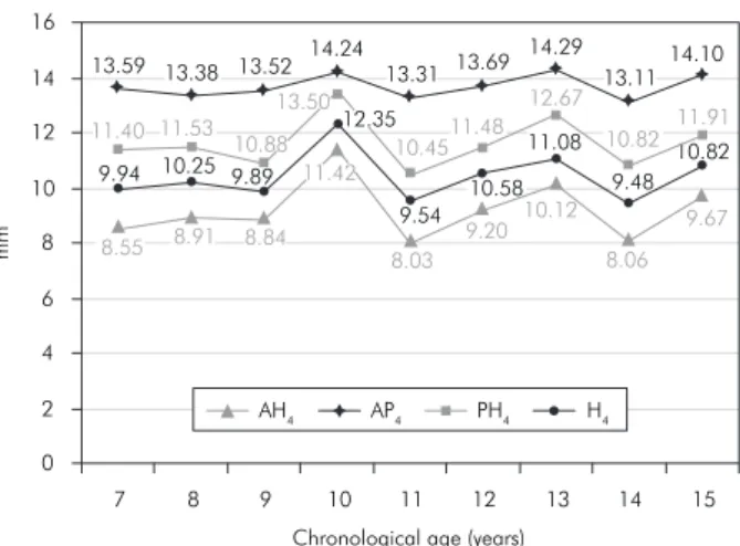
Graph 2 - Average change in each part of the fourth cervi-cal vertebral body in females (group 1).
Chronological age (years) mm 8.55 8.91 8.84
11.42
8.03
9.20 10.12 8.06
9.67 13.59 13.38 13.52 14.24 13.31 13.69
14.29 13.11 14.10 11.40 11.53 10.88 13.50 10.4511.48 12.67 10.82 11.91 9.94 10.25 9.89
12.35 9.54 10.58 11.08 9.48 10.82 0 2 4 6 8 10 12 14 16
7 8 9 10 11 12 13 14 15
AH4 AP4 PH4 H4
Graph 3 - Average change in each part of the third cervical vertebral body in males (group 1).
0 2 4 6 8 10 12 14 16 18 mm
Chronological age (years)
7 8 9 10 11 12 13 14 15
6.10 6.30 6.85
7.75 8.15 8.50
9.65
11.60 11.79 13.52 13.79 14.20 14.31
15.16 14.76 15.68 16.16 16.59
8.61 9.32
9.50
10.43 11.26 11.58
12.19 12.97 13.88
7.81 7.73 8.67
9.49 9.77 10.47 11.28
12.77 13.12
AH3 AP3 PH3 H3
Graph 4 - Average change in each part of the fourth cervi-cal vertebral body in males (group 1).
0 2 4 6 8 10 12 14 16 18 mm
Chronological age (years)
7 8 9 10 11 12 13 14 15
8.22 7.66 7.95 7.27 7.88 8.53 8.21
8.82 8.76
14.57 14.61
13.99 13.86 14.26 14.54 14.50
15.00 15.22
11.42
10.76 10.85
9.23
10.69 11.0710.23
11.94 11.51
9.69 9.41 9.47
8.70 9.81
10.14 9.98 10.74 10.34
method developed in the present study may be of great importance because it allows skeletal age to be calculated in an objective manner.
Mito et al.10 (2002) established a formula for ob-jectively evaluating skeletal maturation on cephalo-metric radiographs. They examined only Japanese girls because of sex-dependent differences with re-gard to the timing of morphological changes in cer-vical vertebral bodies.14 However, when the formula was applied to a different population of children developing under different environmental circum-stances, we found that the method developed could be applied only to Brazilian women. In the present study, the sample selected was composed by both female and male subjects in order to establish two different formulas to objectively evaluate skeletal maturation using cephalometric radiographs.
We measured cervical vertebral bodies because many investigators have suggested that the size and shape of the cervical vertebrae change from birth to full maturity at each level of skeletal development.8 We selected the third and fourth vertebral bodies because the cervical vertebrae lower than C4 cannot be observed when a thyroid protective collar is worn during radiation exposure. Baccetti et al.1 (2002) showed that only the shape change of C2, C3, and C4 was enough to show skeletal maturation. How-ever, C2 is very dificult to measure and it shows little morphological change. In the present study, we measured only C3 and C4.
Hägg, Taranger6 (1980) reported that the puber-tal growth spurt begins at the age of 10 years in girls and at the age of 12 years in boys. In both genders, the growth peak occurs two years after the spurt be-gins and then the growth goes on up to the ages of
15 and 17 years in girls and boys, respectively. In the present study, analysis of the female sample showed that AH3, PH3, H3 increased in an accelerated manner from 10 to 13 years. AH4, PH4 and H4 increased in an accelerated manner from 11 to 13 years. Analysis of the male sample showed that AH3, AP3, PH3, and H3 increased in an accelerated manner from 12 to 15 years of age. The data for AH4, AP4, PH4 and H4 did not increase at all. The lack of growth in C4 could be explained by the fact that maturational changes oc-cur in cervical vertebrae at different times. Concavity of the lower border of the vertebrae appears sequen-tially from C2 to C6. The differences in the skeletal age assessments according to time of observation be-tween female and male subjects were expected since girls reach skeletal maturity before boys do.
When determining the reliability of the formu-las, both female and male subjects showed no sta-tistically signiicant difference between cervical ver-tebral bone age, bone age, and chronological age, indicating that the formulas can be applied to Bra-zilians. The establishment of an objective method to evaluate skeletal maturation in this population us-ing cephalometric radiographs might contribute to a better orthodontic diagnosis and treatment plann for growing children.
Conclusions
The results suggest that the method established in the present study for objectively evaluating skel-etal maturation in cephalometric radiographs is re-liable and can be applied to both female and male subjects. A computer software to automatically cal-culate cervical vertebral bone age is needed to in-crease objectivity.
References
1. Baccetti T, Franchi L, McNamara Jr JA. An improved ver-sion of the cervical vertebral maturation (CVM) method for the assessment of mandibular growth. Angle Orthod. 2002;72(4):316-23.
2. Fishman LS. Chronological versus skeletal age, an evaluation of craniofacial growth. Angle Orthod. 1979;49(3):181-9. 3. Fishman LS. Maturational patterns and prediction during
adolescence. Angle Orthod. 1987;57(3):178-93.
4. Garcia-Fernandes P, Torre H, Flores L, Rea J. The cervi-cal vertebrae as maturational indicators. J Clin Orthod. 1998;32(4):221-5.
5. Hägg U, Taranger J. Maturation indicators and the pubertal growth spurt. Am J Orthod. 1982;82(4):299-309.
7. Hassel B, Farman AG. Skeletal maturation evaluation us-ing cervical vertebrae. Am J Orthod Dentofacial Orthop. 1995;107(1):58-66.
8. Kucukkeles N, Acar A, Biren S, Arun T. Comparisons between cervical vertebrae and hand-wrist maturation for the assess-ment of skeletal maturity. J Clin Pediatr Dent. 1999;24(1):47-52.
9. Lamparski DG. Skeletal age assessment utilizing cervical ver-tebrae [Dissertation]. Pittsburgh: University of Pittsburgh; 1972.
10. Mito T, Sato K, Mitani H. Cervical vertebral bone age in girls. Am J Orthod Dentofacial Orthop. 2002;122(4):380-5. 11. Moed G, Wight BW, Vandegrift HN. Studies of physical
dis-ability: reliability of measurement of skeletal age from hand films. Child Dev. 1962;33:37-41.
12. Remes VM, Heinanen MT, Kinnunen JS, Marttinen EJ. Refer-ence values for radiological evaluation of cervical vertebral body shape and spinal canal. Pediatr Radiol. 2000;30(3):190-5. 13. Singer J. Physiologic timing of orthodontic treatment. Angle
Orthod. 1980;50(4):322-33.
14. Singh GD, McNamara JA Jr, Lozanoff S. Procrustes, Euclid-ean and cephalometric analyses of the morphology of the mandible in human Class III malocclusions. Arch Oral Biol. 1998;43(7):535-43.
15. Tanner JM, Whitehouse RH, Cameron N, Marshall WA, Healy MJR, Goldstein NH. Assessment of skeletal maturity and prediction of adult height (TW3 method). 3rd ed. London:
W.B. Saunders; 2001.