Orthodontics
Mônica Costa Armond(a) Rodrigo Generoso(a)
Saulo Gabriel Moreira Falci(b) Maria Letícia Ramos-Jorge(c) Leandro Silva Marques(a)
(a)Department of Orthodontics, School of Dentistry, University of Vale do Rio Verde (UNINCOR), Três Corações, MG, Brazil. (b)Graduate Program in Dentistry, School of
Dentistry, Federal University of Vales do Jequitinhonha e Mucuri, Diamantina, MG, Brazil.
(c)Department of Pediatric Dentistry, School of Dentistry, Federal University of Vales do Jequitinhonha e Mucuri, Diamantina, MG, Brazil.
Author for correspondence: Leandro Silva Marques
E-mail: [email protected]
Received for publication on Sep 13, 2011 Accepted for publication on Nov 17, 2011
Skeletal maturation of the cervical
vertebrae: association with various
types of malocclusion
Abstract: The identiication of the skeletal maturation stage of the cer-vical vertebrae has proven an important reference for orthodontic diag-nosis. The aim of the present study was to determine the association be-tween the skeletal maturation stage of the cervical vertebrae and types of malocclusion according to the age and gender of participants. A total of 361 individuals (168 males and 193 females) between 8 and 14 years of age were selected from a convenience sample. Malocclusions were diag-nosed through study models using the Angle classiication. Maturation stages of the cervical vertebrae were determined using the method pro-posed by Hassel and Farman. Statistical analysis involved the chi-square test (p ≤ 0.05) and multiple logistic regression (forward stepwise proce-dure). Signiicant differences were observed between the stage of skel-etal maturation of the cervical vertebrae and gender at ages 11, 12 and 14 years. Males with Class II malocclusion were twice as likely to be in Stage 1 or 2 of cervical vertebra maturation than individuals with Class I malocclusion (OR = 2.1 [CI 95%, 1.33-3.18]). There were no differences between individuals with Class I and Class III malocclusions. The as-sociation between skeletal maturation of the cervical vertebrae and type of malocclusion was signiicant, suggesting a skeletal component in the determination of Class II malocclusions.
Descriptors: Malocclusion; Cervical Vertebrae; Growth and Development.
Introduction
Malocclusions are characterized as a developmental problem capable of causing alterations in the dental occlusion. Thus, the correct identii-cation of a child’s developmental stage is a decisive factor in considering the manifestation of different types of malocclusion, as well as their di-agnosis and treatment.1
Stages of development can be assessed through a number of differ-ent growth indicators, including chronological age, ddiffer-ental developmdiffer-ent, height/weight, secondary sexual characteristics and skeletal age.2 In this context, identiication of the skeletal maturation stage of the cervical ver-tebrae has proven an important benchmark for orthodontic diagnosis, as it allows distinguishing children of the same chronological age but with different skeletal ages.3 Moreover, it is considered by some authors as valid as the radiographic analysis of the hand and wrist, while offering Declaration of Interests: The authors
the advantage of reducing the radiation of growing patients.4-7
The objective of the present study was to de-termine the association between the skeletal matu-ration stage of the cervical vertebrae and types of malocclusion according to the age and gender of participants.
Methodology
The initial sample was composed of 984 lateral cephalometric radiographs, along with the respec-tive study models and clinical charts of patients who had never undergone any type of orthodontic treatment. All documentation was taken from the archives of the Orthodontic Center S/C Ltda., a pri-vate orthodontic and facial orthopedic clinic in the city of Varginha, MG, Brazil. Exposures were made using the Funk Orbital ×15 radiographic unit using the following settings: 84 kVp, 80 mA, 0.6-second exposure time, for ilm and Lanex Screens GMT. The ilms were developed in a Rubzomatic 130-EMB automatic processor, whose factors were set at 35.5 and 2.1 minutes. After inspection of the ma-terials, 361 patients were selected – 168 males and 193 females between 8 and 14 years of age.
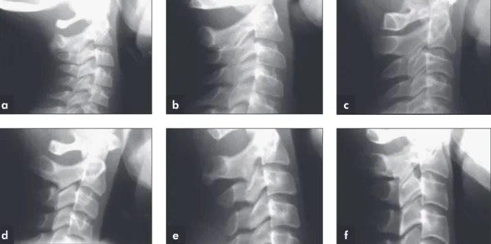
The radiographs were observed by means of an x-ray ilm viewer with standard light intensity for inspecting the morphology of the second (C2), third (C3) and fourth (C4) cervical vertebrae, following the classiication proposed by Hassel and Farman.8 Figures 1a through 1f illustrate the vertebral bod-ies with respective descriptions of each maturation stage of the cervical vertebrae according to these au-thors’ classiication. The study models and ANB an-gles were used to identify Class I (ANB = 0-4 mm), Class II (ANB ≥ 4 mm) and Class III (ANB < 0 mm) malocclusions. Visual inspection was performed on both sides of the models.
The following were considered exclusion factors:
• radiographs lack quality,
• patients with different malocclusions on each side,
• those with no irst premolars,
• history of early loss of deciduous teeth and con-sequent displacement of the permanent molars,
• permanent irst molars that failed to completely erupt and
• models exhibiting a dubious evaluation as a re-sult of movement when in occlusion.
Figure 1 - Vertebral bodies with respective descriptions of each maturation stage of cervical vertebrae according to the Hassel and Farman8 classification (1995). a – Initiation; b – Acceleration; c – Transition; d – Deceleration; e – Maturation; f – Finalization.
b
e
c
f a
Stage of skeletal maturation of cervical vertebrae
p* Phase 1
n (%) Phase 2n (%) Phase 3n (%) Phase 4n (%) Phase 5n (%)
8
ye
ars
Gender
Male 10 (43.5) 14 (51.9) - -
-0.344
Female 13 (56.5) 13 (48.1) 2 (100.0) -
-Malocclusion
Class I 4 (17.4) 4 (14.8) - -
-0.802
Class II 19 (82.6) 23 (85.2) 2 (100.0) -
-Class III - - - -
-9
ye
ars
Gender
Male 10 (43.5) 14 (51.9) - -
-0.613
Female 13 (56.5) 13 (48.1) 2 (100.0) -
-Malocclusion
Class I 3 (15.8) 2 (9.1) - -
-0.405
Class II 12 (63.2) 18 (81.8) 2 (100.0) -
-Class III 4 (21.1) 2 (9.1) - -
-10
ye
ars
Gender
Male 7 (63.6) 14 (41.2) 2 (20.0) -
-0.183 Female 4 (36.4) 20 (58.8) 8 (80.0) 1 (100.0)
-Malocclusion
Class I 2 (18.2) 10 (29.4) 1 (10.0) -
-0.691 Class II 9 (81.8) 22 (64.7) 9 (90.0) 1 (100.0)
-Class III - 2 (5.9) - -
-Table 1 - Bivariate analysis of the relationship between stage of skeletal maturation of cervical vertebrae and gender according to age (continued on next page).
Three weeks after the initial records, 20 radio-graphs were randomly selected, retraced and new measurements were made. When the t test for paired samples was applied, differences between the irst and second sets of 20 radiographs proved non sig-niicant.
Statistical analysis
The results were organized and entered into a database using the Statistical Package for Social Sci-ence (SPSS for Windows, version 14.0, SPSS Inc., Chicago, USA). For the statistical analysis, we ini-tially carried out a description of the absolute and relative frequencies of the variables. Associations were then tested using the univariate analysis (chi-square test) between independent variables and the outcome (skeletal maturation stage of cervical ver-tebrae). The nonexistence of an association between
variables (signiicance value > 0.05) was consid-ered as the null hypothesis. Variables with a p -val-ue ≤ 0.010 in the univariate analysis were included 1-by-1 into a multiple logistic regression model. The inal model was adjusted for the effect of all the variables. The odds ratio (OR) and conidence inter-vals (CIs; 95%) were estimated for each variable in the logistic model.
This study was approved by the research ethics committee of UNINCOR.
Results
Signiicant differences were observed between the stage of skeletal maturation of the cervical ver-tebrae and gender at ages 11, 12 and 14 years (Table 1).
Male individuals and patients with Class II mal-occlusion exhibited twice as much chance of being
in stages 1 or 2 of cervical vertebra maturation than individuals with Class I malocclusion (OR= 2.1 [CI 95%, 1.33 to 3.18]). There were no differences be-tween individuals with Class I and Class III maloc-clusions (Table 2).
Discussion
Vertebral analysis of a lateral cephalogram has been shown to be as valid as the hand-wrist bone analysis, with the advantage of reducing the radia-tion exposure to growing subjects.7 Some studies have examined the effectiveness of techniques for as-sessing bone maturation through the analysis of
cer-11
ye
ars
Gender
Male 3 (75.0) 15 (71.4) 11 (32.4) 1 (11.1)
-0.004 Female 1 (25.0) 6 (28.6) 23 (67.6) 8 (88.9) 2 (100.0)
Malocclusion
Class I - 6 (28.6) 13 (38.2) 5 (55.6)
-0.202 Class II 4 (100.0) 14 (66.7) 16 (47.1) 3 (33.3) 1 (50.0)
Class III - 1 (4.8) 5 (14.7) 1 (11.1) 1 (50.0)
12
ye
ars
Gender
Male 3 (75.0) 11 (78.6) 10 (50.0) 2 (16.7)
-0.008 Female 1 (25.0) 3 (21.4) 10 (50.0) 10 (83.3) 3 (100.0)
Malocclusion
Class I 1 (25.0) 5 (35.7) 9 (45.0) 3 (25.0) 2 (66.7)
0.787 Class II 2 (50.0) 7 (50.0) 9 (45.0) 7 (58.3)
-Class III 1 (25.0) 2 (14.3) 2 (10.0) 2 (16.7) 1 (33.3)
13
ye
ars
Gender
Male - 4 (57.1) 12 (60.0) 4 (36.4) 1 (11.1)
0.080 Female - 3 (42.9) 8 (40.0) 7 (63.6) 8 (88.9)
Malocclusion
Class I - 2 (28.6) 6 (30.0) 6 (54.5) 2 (22.2)
0.774 Class II - 3 (42.9) 8 (40.0) 2 (18.2) 4 (44.4)
Class III - 2 (28.6) 6 (30.0) 3 (27.3) 3 (33.3)
14
ye
ars
Gender
Male - - 10 (90.9) 7 (77.8) 2 (20.0)
0.003 Female - 1 (100.0) 1 (9.1) 2 (22.2) 8 (80.0)
Malocclusion
Class I - - 1 (9.1) 2 (22.2) 2 (20.0)
0.643
Class II - - 3 (27.3) 4 (44.4) 5 (50.0)
Class III - 1 (100.0) 7 (63.6) 3 (33.3) 3 (30.0) * Chi-square test
Table 1 (continued)
vical vertebrae.5,7 The present study used the Hassel and Farman8 method. This method has yielded the same results as that of Baccetti et al.9 and Seedat and Forsberg10,6
must be expressed in terms of stage and level of ma-turity. The maturity level is used to associate a per-son’s maturational stage with chronological age to determine whether development is advanced or de-layed. Two children having the same maturity stage but different maturity levels will, in the future, dem-onstrate signiicant differences in total percentage of mandibular and maxillary growth.3
In determining a Class II, Division 1, malocclu-sion, there is a large number of possible combina-tions of relacombina-tionships between the maxilla and man-dible as well as between these bones and the cranial base. The most common combinations are maxil-lary protrusion with normal mandible, mandibular retrusion with normal maxilla, maxillary protru-sion combined with mandibular retruprotru-sion and pos-terior rotation of the mandible.12 It is therefore es-sential for orthodontists to be aware of the diverse dentoskeletal aspects of a malocclusion, because diagnosis dictates treatment. The results of the pres-ent study suggest that there is a skeletal componpres-ent
Table 2 - Unconditional multiple logistic regression analysis between independent variables and skeletal maturation of cervical vertebrae.
Unadjusted OR
(CI 95%) p Adjusted OR(CI 95%) p Malocclusion
Class I 1.00 1.00
Class II 2.22 (1.35-3.64) 0.002 2.37 (1.43-3.94) 0.001 Class III 0.56 (0.27-1.16) 0.120 0.60 (0.29-1.25) 0.175
Gender
Female 1.00 1.00
Male 1.96 (1.29-2.99) 0.002 1.82 (1.0-3.3) 0.001
in determining a Class II malocclusion, especially when mandibular growth is slower than maxillary growth.
Baccetti et al.9 designed an improved method of using cervical vertebrae maturation to evaluate mandibular growth and found that the maturation stage of cervical vertebrae occurs at least 2 years after peak growth. In the present study, there were no differences between individuals with Class I and Class III malocclusions with regard to the skeletal maturation stage of the cervical vertebrae. However, Reyes et al.13 observed that increases in mandibu-lar length were greater in individuals with Class III malocclusion than in those with normal occlusion, even during the interval of greater skeletal matura-tion (15 to 16 years). A possible explanamatura-tion is the different methodological criteria used in studies as well as different sample sizes and the heterogeneity of study populations.
The results of this study conirm the importance of individualized diagnoses for the treatment of orthodontic patients, since the growth of the facial bones and periods of intensive or accelerated physi-ological growth should be analyzed individually in order to make better use of bone remodeling and the correction of skeletal discrepancies.
Conclusions
• There were signiicant differences between the stages of skeletal maturation of the cervical ver-tebrae and gender of the participants.
• Males with Class II malocclusion have a two times greater chance of being in the initiation and acceleration stages of cervical vertebra mat-uration.
References
1. Carvalho AC, Paiva SM, Scarpelli AC, Viegas CM, Ferreira FM, Pordeus IA. Prevalence of malocclusion in primary den-tition in a population-based sample of Brazilian preschool children. Eur J Paediatr Dent. 2011 Jun;12(2):107-11. 2. Rajagopal R, Kansal S. A comparison of modified MP3 stages
and the cervical vertebrae as growth indicators. J Clin Orthod. 2002 Jul;36(7):398-406.
3. Fishman LS. Can cephalometric x-rays of the cervical col-umn be used instead of hand-wrist x-rays to determine
pa-tient’s maturational age? Am J Orthod Dentofacial Orthop. 2002 Jul;122(1):18A-9A.
4. Caldas Mde P, Ambrosano GM, Haiter Neto F. New for-mula to objectively evaluate skeletal maturation using lat-eral cephalometric radiographs. Braz Oral Res. 2007 Oct-Dec;21(4):330-5.
Indonesian and white subjects with receiver operating char-acteristics analysis. Am J Orthod Dentofacial Orthop. 2008 Aug;134(2):227-37
6. Jaqueira LM, Armond MC, Pereira LJ, Alcântara CE, Marques LS. Determining skeletal maturation stage using cervical vertebrae: evaluation of three diagnostic methods. Braz Oral Res. 2010 Oct-Dec;24(4):433-7.
7. Gandini P, Mancini M, Andreani F. A comparison of hand-wrist bone and cervical vertebral analyses in measuring skel-etal maturation. Angle Orthod. 2006 Nov;76(6):984-9. 8. Hassel B, Farman AG. Skeletal maturation evaluation
us-ing cervical vertebrae. Am J Orthod Dentofacial Orthop. 1995 Jan;107(1):58-66.
9. Baccetti T, Franchi L, McNamara JA Jr. An improved ver-sion of the cervical vertebral maturation (CVM) method for the assessment of mandibular growth. Angle Orthod. 2002 Aug;72(4):316-23.
10. Seedat AK, Forsberg CD. An evaluation of the third cervical vertebra (C3) as a growth indicator in Black subjects. SADJ. 2005 May;60(4):156,158-60.
11. Generoso R, Sadoco EC, Armond MC, Gameiro GH. Evalu-ation of mandibular length in subjects with Class I and Class II skeletal patterns using the cervical vertebrae maturation. Braz Oral Res. 2010 Jan-Mar;24(1):46-51.
12. Arntsen T, Sonnesen L. Cervical vertebral column morphol-ogy related to craniofacial morpholmorphol-ogy and head posture in preorthodontic children with Class II malocclusion and hori-zontal maxillary overjet. Am J Orthod Dentofacial Orthop. 2011 Jul;140(1):e1-7