The aim of this study was to detect apical inflammatory root resorption (AIRR) associated with periapical lesion using cone beam computed tomography (CBCT) and scanning electronic microscopy (SEM). This clinical study evaluated AIRR in 88 root apexes, from 52 permanent teeth of 14 patients, extracted for different reasons. The patients were submitted to a clinical interview, review of dental/medical histories and clinical/imaging examinations for treatment planning. All selected teeth showed unrestorable condition because of the extensive coronal breakdown due to carious lesions, and root canal infection associated with periapical lesions. CBCT images were obtained from the patients with the aim of diagnosing the periapical diseases which showed complex or doubtful conditions. Two examiners assessed the presence or absence of AIRR. Apices were also analyzed under SEM. Chi-square test was used to compare the imaging methods for detection of AIRR. The level of statistical significance was set at 5%. AIRR associated with root canal infection and apical periodontitis was found in 61.4% of the cases studied by using SEM, and at least half of the cases by CBCT. The microscopic analysis remains as a reference standard against the imaging method to identify AIRR.
Detection of Apical Inflammatory Root
Resorption Associated with Periapical
Lesion Using Different Methods
Carlos Estrela1, Orlando Aguirre Guedes2, Luiz Eduardo G. Rabelo1, Daniel Almeida Decurcio1, Ana Helena G Alencar1, Cyntia R.A. Estrela2, José Antônio Poli de Figueiredo3
1Department of Stomatological
Sciences, UFG - Federal University of Goiás, Goiânia, GO, Brazil
2Department of Endodontics,
University of Cuiabá, Cuiabá, MT, Brazil
3Department of Clinical Dentistry,
PUCRS - Pontifical Catholic University of Rio Grande do Sul, Porto Alegre, RS, Brazil
Correspondence: Prof. Dr. Carlos Estrela, Praça Universitária S/N, Setor Universitário, 74605-220 Goiânia, GO, Brasil. Tel: +55-62-3209-6254.e-mail: [email protected]
Key Words: root resorption, apical periodontitis, cone beam computed tomography, scanning electron microscopy, diagnostic.
Introduction
Root canal infection after pulp necrosis follows a natural route in apical direction, able to induce inflammation of periapical structures. This aggression process can stimulate the destruction of periapical tissues and induce loss of tooth structure, featuring an apical inflammatory root resorption (AIRR).
The loss of root structures due to clastic cell activity is associated with a local pathologic condition caused by apical periodontitis (AP), traumatic dental injury, orthodontic treatment, intracoronal bleaching, autotransplantation, dentigerous cyst, neoplasia, or idiopathic factors (1–7). The external or internal superficial protective cell layer might be damaged, and inflammatory or replacement root resorptions might affect any part of the root (5).
Inflammatory root resorption (IRR) has been well studied, since it represents an asymptomatic lesion that is complex to diagnose and treat (1–5). The standard criterion for the diagnosis of IRR is the microscopic analysis (6), and IRR can be classified as active, arrested, or repaired according to the microscopic findings. The prevalence of each stage affects prognosis and treatment (1).
The relation of AP with AIRR has been analyzed by different methodologies (3,7-12). The incidence, extent and distribution of root canal resorption were evaluated by scanning electron microscopy (SEM) in 100 extracted
405
Apical root resorption detection
were cysts. Periforaminal resorption was present in 87.3% of the cases, and foraminal resorption in 83.2%. Periforaminal and foraminal resorptions were independent entities. There was no association between external root resorption and the nature of the periapical lesions.
Ferlini-Filho et al. (9) studied the morphology of root resorption associated with AP in 87 human extracted teeth. The results of the radiographic and microscopic analyses revealed that some form of root resorption was present in most of the teeth diagnosed with chronic periapical process and that RRs were observed more easily in the microscopic exam (n=68 roots; 94.44%) rather than radiographic exam (n=26 roots; 36.11%).
Root canal treatment procedures need images before, during and after its conclusion. Conventional radiographic images provide a two-dimensional interpretation of a three-dimensional structure, which may lead to interpretation errors (10,13-17).
The incorporation of cone beam computed tomography (CBCT) in dentistry brought a revolution of information for clinical procedures, which contributed to planning, diagnosis, treatment and prognosis (13-17). The use of CBCT images to evaluate AIRR has been widely discussed, and suggested in different endodontic research areas due to its precision (18-24). However, literature is scarce in clinical studies comparing methodologies to identify RR. Therefore, the aim of this study was to detect apical inflammatory root resorption associated with periapical lesion using CBCT and SEM.
Material and Methods
Patients
This research project was approved by the research ethics committee. All patients were informed about the study and signed a consent form.
This clinical study evaluated AIRR in 52 permanent teeth (26 maxillary posterior teeth, 20 mandibular posterior teeth, 5 mandibular anterior teeth and 1 maxillary anterior tooth) in the years 2009 and 2010. The sample comprised 14 patients (6 male, 8 female, with a mean age of 31 years), of low socioeconomic status and referred to the Dental Urgency Service of the School of Dentistry of the Federal University of Goiás, Brazil, for routine tooth extraction. The patients were submitted to a clinical interview, review of dental/medical histories and clinical/imaging examinations for treatment planning. All selected teeth showed unrestorable condition because of the extensive coronal breakdown due to carious lesions, and root canal infection associated with periapical lesions. The teeth were not associated with history of traumatic dental injury or orthodontic treatment. CBCT scans (I-CAT Cone Beam 3D
imaging system, Imaging Sciences International, Hatfield, PA, USA) were obtained to help in diagnosis of periapical diseases, which showed complex or doubtful conditions (25), and for treatment planning. The volumes were reconstructed with isotropic- isometric voxels measuring 0.20 mm - 0.20 mm - 0.20 mm. The tube voltage was 120 kVp and the tube current 3.8 mA. Exposure time was 40 seconds. Images were examined with the scanner’s proprietary software (Xoran version 3.1.62; Xoran Technologies, Ann Arbor, MI, USA) in a PC workstation running Microsoft Windows XP professional SP-2 (Microsoft Corp, Redmond, WA, USA), with processor Intel(R) Core(TM) 2 Duo-6300 1.86 Ghz (Intel Corporation, USA), NVIDIA GeForce 6200 turbo cache videocard (NVIDIA Corporation, USA) and Monitor EIZO - Flexscan S2000, resolution 1600x1200 pixels (EIZO NANAO Corporation Hakusan, Japan).
After clinical and image examinations, all teeth were clinically diagnosed with apical periodontitis with primary or secondary infections of varying sizes. The teeth had completely formed roots and absence of odontogenic development anomalies. Teeth were collected and placed in individually labeled and identified plastic vials containing solution of formaldehyde (10% w/v).
Evaluation of AIRR by using CBCT
All CBCT images were evaluated by 2 examiners calibrated by 10% of the sample. When a consensus was not reached, a third observer made the final decision. The examiners were allowed to access all CBCT plane images and magnifications to assess the presence or absence of AIRR. Extension of the AIRR was not evaluated.
Evaluation of AIRR by SEM
406
C. Estrela et al.
The dental apices were classified depending on the presence or absence of AIRR following a modified criterion proposed by earlier study (11). When teeth had two or more
apical foramina or had fused roots, the final resorption level was the one that represented the foramen in which resorption was most pronounced. The photomicrographs were analyzed by two previously calibrated examiners. When no agreement has been reached between both observers as to the diagnosis of AIRR, they would re-assess the images together to attain a consensus. The Kappa coefficient was used to assess inter-observer agreement.
Statistical Analysis
Data were analyzed using the IBM SPSS for Windows 21.0 (IBM Corporation, Somers, NY, USA), including frequency distribution and cross-tabulation. Comparative statistical analysis among the imaging methods for detection of AIRR was performed using Chi-square test, and the level of statistical significance was set at 5%.
Results
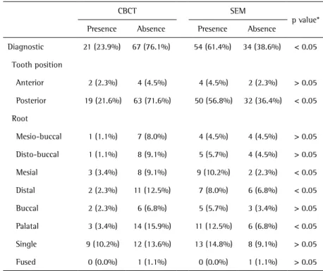
A total of 88 root apexes were analyzed. Table 1 shows the identification of AIRR according to the different methods, tooth position and dental root. AIRR was detected in 23.9% and 61.4% of cases by CBCT and SEM images, respectively (p<0.05). The results of chi-square test revealed significant differences between the study methods in the diagnosis of AIRR regarding tooth position and dental root (p<0.05). The kappa coefficient for inter-observer agreement was 0.83. Figure 1 shows clinical aspects of AIRR evaluated with CBCT and SEM methods.
Discussion
The high discrepancy between the evaluation methods (CBCT and SEM) to detect AIRR associated with root canal infection and apical periodontitis was observed in this study. AIRR was detected in 61.4% of the teeth associated with periapical lesion when SEM was used, and 23.9% by CBCT images.
Based on the accuracy and
Table 1. Prevalence of apical inflammatory root resorption (AIRR) according to imaging method, tooth position and dental root (n=88)
CBCT SEM
p value* Presence Absence Presence Absence
Diagnostic 21 (23.9%) 67 (76.1%) 54 (61.4%) 34 (38.6%) < 0.05 Tooth position
Anterior 2 (2.3%) 4 (4.5%) 4 (4.5%) 2 (2.3%) > 0.05 Posterior 19 (21.6%) 63 (71.6%) 50 (56.8%) 32 (36.4%) < 0.05 Root
Mesio-buccal 1 (1.1%) 7 (8.0%) 4 (4.5%) 4 (4.5%) > 0.05 Disto-buccal 1 (1.1%) 8 (9.1%) 5 (5.7%) 4 (4.5%) > 0.05 Mesial 3 (3.4%) 8 (9.1%) 9 (10.2%) 2 (2.3%) < 0.05 Distal 2 (2.3%) 11 (12.5%) 7 (8.0%) 6 (6.8%) < 0.05 Buccal 2 (2.3%) 6 (6.8%) 5 (5.7%) 3 (3.4%) > 0.05 Palatal 3 (3.4%) 14 (15.9%) 11 (12.5%) 6 (6.8%) < 0.05 Single 9 (10.2%) 12 (13.6%) 13 (14.8%) 8 (9.1%) > 0.05 Fused 0 (0.0%) 1 (1.1%) 0 (0.0%) 1 (1.1%) > 0.05 CBCT: cone beam computed tomography. SEM: scanning electron microscopy.*Chi-square test.
Apical root resorption detection diagnostic performance of CBCT to detect apical
periodontitis, and root resorption associated with history of traumatic dental injury or orthodontic treatment when compared with conventional radiographs (13-23), it was sought to compare the detection potential of AIRR in infected root canals and apical periodontitis using CBCT scans and SEM.
The identification of AIRR developing as a consequence of apical periodontitis in periapical radiographs and microscopic analysis has shown little accuracy for the two-dimensional image methods. Laux et al. (10) reported that the routine single radiographs are not sufficiently accurate or sensitive to consistently diagnose apical root resorptive defects developing as a consequence of apical periodontitis. Considering the imaging method by periapical radiographs, 19% of the teeth were diagnosed as having AIRR, whereas histologically, 81% of the teeth revealed AIRR.
Some studies have shown promising results in detecting resorption lesions on imaging methods using CBCT scans (18-23). Silveira et al. (18) analyzed the diagnostic ability of CT to detect simulated external root resorption defects. Several external root resorption defects of different sizes and different locations were simulated in 59 human mandibular incisors. Axial CT was used to obtain cross-sectional images of the teeth, and 177 root thirds were assessed. Of the 131 cavities, 117 were detected (89%). Thirty-two of the 44 (72.72%) cavities located in the apical third were identified. Difference was found between the sizes of defects examined in the apical third. The evaluation of CT diagnostic ability revealed high sensitivity and excellent specificity. Small cavities located in the apical third were more difficult to detect than all other cavities. Lermen et al. (22) evaluated the accuracy of coronal and sagittal CT sections to detect cavities simulating root resorption in 60 mandibular incisors with cavities with different diameter and depth. No significant difference was observed in the diagnosis of simulated resorption cavities among the apical, middle, and coronal thirds. When the axial plane was assessed separately, diagnoses differed among the three thirds of the root. Diagnostic errors were more often observed in the apical third, as compared with the cervical and middle thirds. When tomographic sections are required for the diagnosis of buccal or lingual external root resorption, sagittal sections afford the best image characterization of the resorption process. Patel et al. (19) compared the accuracy of digital intraoral radiographs with CBCT for the detection and management of resorption lesions in patients with internal resorption, external cervical resorption and no resorption. The detection of resorption lesions using CBCT showed to be effective. The authors found that the intraoral digital radiography resulted in an acceptable level of accuracy,
and that CBCT images enabled increased probability of correct management of resorption lesions. Estrela et al. (20) evaluated a methodology to measure IRR associated with traumatic dental injury or orthodontic treatment by CBCT scans. IRRs were better detected by CBCT images than periapical radiographs when root third, root surface, and extension were analyzed. This method provided an accurate diagnosis with high-resolution images and little observer interference.
Contemporary imaging methods such as CBCT combined with map-reading strategy in various planes has shown high accuracy to identify apical periodontitis, resorptive lesions associated with traumatic dental injury or orthodontic treatments (13-23).
When periapical and panoramic radiographs, and CBCT scans were compared for identification of the periapical radiolucencies, the CBCT images provided greater scores. It was considered as standard reference in this imaging methods evaluation (15). Silva et al. (24) evaluated the accuracy of periapical radiography (PR), CBCT scans in diagnosing AP using histopathological findings as a gold standard, in 83 treated or untreated roots of dogs' teeth. PR detected AP in 71% of roots, a CBCT scan detected AP in 84%, and AP was histologically diagnosed in 93%.
In this study, AIRR associated with root canal infection and apical periodontitis was found in 61.4% of the cases studied by SEM, and at least half of the cases by CBCT. The microscopic analysis remains the reference standard against imaging methods to identify AIRR. It could be speculated that CBCT may improve with time and, in the future, may allow increased diagnostic power for this very challenging clinical situation.
Resumo
O objetivo deste estudo foi detectar reabsorção radicular inflamatória apical (RRIA) associada à lesão periapical utilizando tomografia computadorizada de feixe cônico (TCFC) e microscopia eletrônica de varredura (MEV). Este estudo clínico avaliou RRIA em 88 ápices radiculares de 52 dentes permanentes de 14 pacientes, extraídos por diferentes motivos. Os pacientes foram submetidos a uma entrevista clínica, revisão da história médica/dental, exames clínicos e de imagem para o plano de tratamento. Todos os dentes selecionados apresentaram condição não restaurável devido à extensa perda de estrutura dental associada a lesões cariosas, e infecção do canal radicular associada a lesões periapicais. TCFC foram obtidas dos pacientes com o objetivo de diagnosticar as alterações periapicais que se mostraram complexas ou duvidosas. Dois examinadores avaliaram a presença ou ausência de RRIA. Os ápices foram também analisados por MEV. O teste do qui-quadrado foi usado para comparar os métodos de detecção de RRIA. O nível de significância foi estabelecido em 5%. RRIA associada à infecção do canal radicular e periodontite apical foi encontrada em 61,4% dos casos estudados usando MEV, e pelo menos metade dos casos utilizando TCFC. A análise microscópica continua a ser o padrão frente a métodos de imagens para a identificação de RRIA.
Acknowledgements
408
C. Estrela et al.
Scientific and Technological Development (CNPq grant 306394/2011-1 to C.E.).
References
1. Andreasen JO, Andreasen FM. Essentials of traumatic injuries to the teeth. 2nd ed. Copenhagen: Munksgaard, 2001:188.
2. Tronstad L. Root resorption. Etiology, terminology and clinical manifestations. Endod Dent Traumatol 1988;4:241–245.
3. Nance RS, Tyndall D, Levin LG, Trope M. Diagnosis of external root resorption using TACT (tuned-aperture computed tomography). Endod Dent Traumatol 2000;16:24–28.
4. Pierce A. Pathophysiological and therapeutic aspects of dentoalveolar resorption. Aust Dent J 1989;34:437–448.
5. Ne RF, Witherspoon DE, Gutmann JL. Tooth resorption. Quintessence Int 1999:9–25.
6. Gunraj M. Dental root resorption. Oral Surg Oral Med Oral Pathol Oral RadiolEndod 1999;88:647–653.
7. Andreasen FM, Sewerin I, Mandel U, Andreasen JO. Radiographic assessment of simulated root resorption cavities. Endod Dent Traumatol 1987;3:21–27.
8. Delzangles B. Apical periodontitis and resorption of the root canal wall. Endod Dent Traumatol 1988;4:273-277.
9. Ferlini-Filho J, Garcia RB. Radiographic and microscopic study of root resorption in the presence of chronic apical periodontitis (optical and scanning electron microscopy). Rev Fac Odontol Porto Alegre 1999;40:60-64.
10. Laux M, Abbott PV, Pajarola G, Nair PNR. Apical inflammatory root resorption: a correlative radiographic and histological assessment. Int Endod J 2000;33:483–493.
11. Vier FV, Figueiredo JAP. Prevalence of different periapical lesions associated with human teeth and their correlation with the presence and extension of apical external root resorption. Int Endod J 2002;35:710-719.
12. Felippe WT, Ruschel MF, Felippe GS, Pozzobon MH, Felippe MCS. SEM evaluation of the apical external root surface of teeth with chronic periapical lesion. Aust Endod J 2009;35:153–157.
13. Cotton TP, Geisler TM, Holden DT, Schwartz SA, Schindler WG. Endodontic applications of cone beam volumetric tomography. J Endod 2007;33:1121–1132.
14. Patel S, Dawood A, Pitt Ford T, Whaites E. The potential applications of cone beam computed tomography in the management of endodontic
problems. Int Endod J 2007;40:818-813.
15. Estrela C, Bueno MR, Leles CR, Azevedo B, Azevedo JR. Accuracy of cone beam computed tomography and panoramic and periapical radiography for detection of apical periodontitis. J Endod 2008;34:273-279. 16. Estrela C, Bueno MR, Azevedo B, Azevedo JR, Pécora JD. A new
periapical index based on cone beam computed tomography. J Endod 2008;34:1325-1331.
17. Durack C, Patel S. Cone beam computed tomography in endodontics. Braz Dent J 2012;23:179-191.
18. Silveira HL, Silveira HE, Liedke GS, Lermen CA, Santos RB, Figueiredo JA. Diagnostic ability of computed tomography to evaluate external root resorption in vitro. Dentomaxillofac Radiol 2007;36:393-396. 19. Patel S, Dawood A, Wilson R, Horner K, Mannocci F. The detection and
management of root resorption lesions using intraoral radiography and cone beam computed tomography – an in vivo investigation. Int Endod J 2009;42:831-838.
20. Estrela C, Bueno MR, Alencar AH, Mattar R, Valladares-Neto J, Azevedo BC, et al.. Method to evaluate inflammatory root resorption using cone beam computed tomography. J Endod 2009;35:1491–1497.
21. Liedke GS, Silveira HE, Silveira HL, Dutra V, Figueiredo JA. Influence of voxel size in diagnostic ability of cone beam tomography to evaluate simulated external root resorption. J Endod 2009;35:233-235. 22. Lermen CA, Liedke GS, Silveira HED, Silveira HLD, Mazzola AAM,
Figueiredo JAP. Comparison between two tomographic sections in the diagnosis of external root resorption. J Appl Oral Sci 2010;18:303-307. 23. Gunst V, Mavridou A, Huybrechts B, Van Gorp G, Bergmans L,
Lambrechts P. External cervical resorption: an analysis using cone beam and microfocus computed tomography and scanning electron microscopy. Int Endod J 2013;46:877–887.
24. Silva FWGP, Wu MK, Leonardo MR, Silva LAB, Wesselink PR. Accuracy of periapical radiography and cone-beam computed tomography in diagnosing apical periodontitis using histopathological findings as a gold standard. J Endod 2009;35:1009-1012.
25. American Association of Endodontists and the American Academy of Oral and Maxillofacial Radiology. Use of cone-beam computed tomography in endodontics. Joint Position Statement. Oral Surg Oral Med Oral Pathol Oral RadiolEndod 2011;111:234–237.