Rev Odontol UNESP. 2018 Mar-Apr; 47(2): 85-91 © 2018 - ISSN 1807-2577
ORIGINAL ARTICLE
Doi: http://dx.doi.org/10.1590/1807-2577.10617
This is an Open Access article distributed under the terms of the Creative Commons Attribution License, which permits unrestricted use, distribution, and reproduction in any medium, provided the original work is properly cited.
Prevalence evaluation and classification of bifid mandibular canals
in CBCT exams in different facial types
Avaliação da prevalência e classificação dos canais mandibulares bífidos em exames de TCFC
nos diferentes tipos faciais
George Borja de FREITAS
a*, Alessandra de FREITAS e SILVA
a,
Luiz Roberto Coutinho MANHÃES JÚNIOR
b, José Luiz Cintra JUNQUEIRA
aaSão Leopoldo Mandic, Faculdade de Odontologia e Centro de Pesquisas Odontológicas, Campinas, SP, Brasil bUNESP – Universidade Estadual Paulista, Instituto de Ciência e Tecnologia, Departamento de Diagnóstico
e Cirurgia, São José dos Campos, SP, Brasil
Resumo
Objetivo: O presente trabalho objetiva verificar a prevalência e classificação das alterações do canal da mandíbula por meio de exames de tomografia computadorizada de feixe cônico nos diferentes tipos faciais. O trabalho foi submetido e aprovado ao comitê de ética e pesquisa através do parecer n˚ 2.065.839. Material e método: A amostra foi composta por 90 tomografias, divididas em três grupos de acordo com os tipos faciais, braquicefálico, dólicocefálico e mesocefálico. Todas as imagens foram obtidas no banco de dados da Faculdade São Leopoldo Mandic, Campinas-SP. Resultado: Dos 90 pacientes pesquisados, 23 apresentaram os canais da mandíbula bífidos, ou seja, 25,6% da amostra total. Desses, 60,9% pertenciam ao sexo masculino e 39,1% ao sexo feminino. Em 39,1% o direcionamento era para região retromolar (classe D), enquanto em 21,7%, a trajetória era no sentido alveolar ou superior (classe C). Com menor proporção, 13,1% foi constatada a classificação C-E. Para as demais classificações encontradas (A, E, F e A-E), as frequências foram na faixa de 8,7% a 4,3%. Em nenhum dos 23 casos de bifurcação do canal da mandíbula foi encontrada classificação (B), ou seja, em direção mesial. Conclusão: De acordo com os resultados obtidos nesse estudo, encontrou-se uma prevalência de 25,6% de canais da mandíbula bífidos, o tipo de canal bífido mais prevalente foi classe D para região retromolar e a maior ocorrência dos canais mandibulares bífidos foi unilateral esquerda. Quando avaliada a ocorrência dos canais da mandíbula bífidos em relação aos tipos faciais, os pacientes braquifaciais foram os mais acometidos.
Descritores: Cirurgia; anatomia; mandíbula.
Abstract
Objetive: The aim of this study is to establish the prevalence and classification of mandibular canal alterations using cone beam computed tomography (CBCT) in different facial types. This research was submitted and approved by the research ethics committee, registration number 2.065.839. Material and method: The sample consisted of 90 CBCTs from the São Leopoldo Mandic Dental School database (Campinas-SP), divided into three groups according to brachycephalic, dolichocephalic and mesocephalic facial types. Result: Of the 90 patients, 23 presented bifid mandible canals (25.6%), of which 60.9% were in males and 39.1% in females. In 39.1%, the canal bifurcation occurred towards the retromolar region (class D), 21.7% had a trajectory to an alveolar or upper direction (class C) and 13.1% were classified as C-E. For the remaining classifications (A, E, F and A-E), the frequencies were in the range of 8.7% to 4.3%. None of the 23 cases of mandibular canal bifurcation was classified as B (mesial direction).
Conclusion: According to the results obtained from this study, the prevalence of bifid mandibular canals was found to be 25.6%, with class D being the most prevalent for the retromolar region and the highest occurrence was unilaterally on the left side. When evaluating the occurrence of bifid mandibular canals in relation to facial types, brachycephalic patients were the most affected.
INTRODUCTION
Mandibular canals are intraosseous conduits, usually presenting as a single structure on each side of the mandible, beginning on the medial aspect of the ramus in the mandibular foramen and out through the mental foramen, with or without a continuous intraosseous path towards the anterior region as a single canal. Topographically, it is located close to the internal compact bone until it reaches the mesial surface of the first molar, where it approaches the external compact bone until the mental foramen1.
Such canals house the inferior alveolar neurovascular bundle, the largest branch of the mandibular division of the trigeminal nerve, responsible for innervating the posterior teeth, adjacent bone and the oral mucosa of the mandibular region2,3.
Radiographically, the mandibular canal is characterized by a radiolucent band, delimited by two radiopaque lines4, generally as
a single structure, assuming different positions inside the body of the mandible, both in the upper-lower and mid-lateral directions, occasionally showing duplications or bifurcations along its path and, in some cases, trifurcations. Because of this considerable variation in its course, it is difficult to predict the exact position of the inferior alveolar nerve5,6.
Anatomically, the mandibular canal often presents as a single conduit though in some cases the presence of an accessory canal can be identified, known as bifid canal. According to Langlard et al.7 the
mandibular canal may vary in shape as oval, circular or piriform. Many dental surgeons are unaware of the existence of anatomical variants of this canal and thus may not be able to visualize them in conventional panoramic and tomographic images8. As a consequence,
trans and postoperative surgical complications, anesthetic failures and implant placement failures may occur9.
Panoramic radiographs can be used to assess bone height and horizontal distances1. However, image enlargement and distortion
should be taken into account when these distances are critical to treatment planning. Thus, computed tomography should be performed to overcome the limitations of the two-dimensional image provided by conventional radiographs10,11.
Facial typology should be considered in studies involving anthropometry and cephalometrics, since the values of the measurements may vary depending on facial type and is directly related to craniofacial growth patterns, with the configuration of orofacial structures, musculature, stomatognathic functions and occlusion12,13. There are three facial types established in the
literature: mesocephalic, brachycephalic and dolichocephalic12,14,15.
There are no previous reports on the association between anatomical variations of the mandibular canal and facial types using cone beam computed tomography (CBCT). Hence the importance of the present study to verify the prevalence of the different classifications of anatomical variations of the mandibular canal using CBCT and correlating them with facial types.
MATERIAL AND METHOD
This study was approved by the research ethics committee of the Sao Leopoldo Mandic Dental School, registration number 2.065.839. It was designed as a descriptive observational survey that
included 90 CBCT images from the archives of the Department of Radiology and Imaging of the São Leopoldo Mandic Dental School, Campinas-SP. Image selection was based on examinations taken using the equipment Classic I-Cat (Imaging Sciences
International, USA), with a 0.25 mm standard voxel, Fov (Field of view) of 13 cm and acquisition time of 40 pulsating seconds, according to the manufacturer’s standards, with a useful radiation time of 6.6 seconds. The factors used for the acquisitions were those pre-established by the equipment that works at fixed 120 kV with 5 to 7 mA, depending on the resolution used.
All images were processed on the XoranCat software (Xoran
Technologies, USA) of the equipment itself. For the analysis of the tomographic images, the anatomical planes were first corrected on the tomographer’s own workstation using the multiplanar reconstruction page (MPR). From the axial slice (0.25 mm thick), a plane was drawn along the alveolar ridge of each patient, from which the panoramic image and the cross-sectional slices were formed, standardized at 1.00 mm in thickness with a distance of 1.00 mm in between. In the panoramic reconstruction, the slices were 5.25 mm thick, according to Figure 1.
Only images with were tomographic quality were selected from both genders. Patients with a history of mandibular trauma, bone lesions in the lower arch and orthognathic or restorative surgery in the posterior region of the mandible were excluded.
The sample was divided into 3 groups according to facial types:
• GROUP A: Dolicocephalic (n=30);
• GROUP B: Mesocephalic (n=30);
• GROUP C: Brachycephalic (n=30).
CT scans were all obtained from the I-Cat (Imaging Science,
Hatfield, PA) equipment with Fov 20 cm, voxel 0.25 mm and 20s, Kvp120 and 36mA. The sample size was based on previous recently published studies16-18.
For the determination of facial type, the cephalometric analysis of Ricketts14 was used based on the Vert Index. The software used
was Dolphin Imaging version 11.0, which facilitates marking of
the cephalometric points, including the sequence to be followed for tracing, as well as the possibility of zooming in to the area of interest. After joining the dots, digital tracings and the linear and angular values were calculated automatically.
A single examiner performed the standardization and tomographic measurements for the classification of the facial types. This step was performed on 30 randomly selected CT scans and then repeated 30 days later. The results were analyzed using intra-examiner agreement statistics. The intraclass correlation test (ICC) revealed that intra-examiner agreement was excellent for both linear measures (ICC> 0.9, p <0.0001) and nominal values (kappa = 1.0) obtained at two different times.
The Xoran 3.0.34 program (Xoran Tecnologies, USA) was used to carry out all evaluations. The three slicing planes were axial, coronal and sagittal, with a sharpen 3x3 filter.
percentage frequency was used, with a descriptive analysis of the results. In cases where bifid canals were present, these were classified according to the study by Freitas et al.9. According to this study,
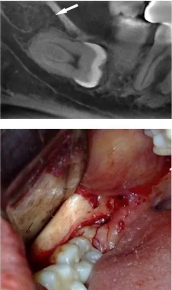
bifid mandibular canals are classified according to the direction they follow into Class A - buccal direction, Class B - mesial direction, towards the mesial or anterior aspects, Class C - alveolar direction, towards the alveolar or upper directions, Class D - retromolar direction (according to Figure 2), Class E - lingual direction and
Class F - towards the base of the mandible.
The prevalence of mandibular canal bifurcation identified in CBCT scans according to gender, age, facial type and location were described as absolute and relative frequencies and analyzed statistically using the G test and chi-square.
To check for age differences between the males and females as well as between the brachycephalic, dolichocephalic and mesocephalic subjects, the Student t-test for independent samples and analysis of variance, followed by Tukey’s test, were used. Statistical calculations were performed using SPSS 23 (SPSS INC., Chicago, IL, USA) and BioEstat 5.0 (Mamirauá Foundation, Belém, PA, Brazil), with a significance level of 5%.
RESULT
Examination of the 90 CBCT scans that composed this study verified the presence of mandibular canal bifurcation in 23, indicating, therefore, a prevalence of 25.6%.
The 90 images came from 51 (56.7%) males and 39 (43.3%) females. Table 1 shows that 14 of the 51 males (27.5%) and 9 of the 39 females (23.1%) presented this type of bifurcation of the mandibular canal, which was not significantly different between genders (chi-square, p = 0.820).
Figure 1. Demonstrative images illustrating the evaluation methods for the tomographic images; A - Axial image with tracing of the mandibular contour to obtain the cross-sectional slices; B - Panoramic Reconstruction; C - Cross-sectional slices.
When investigating whether the prevalence of mandibular canal bifurcation was associated with age, the G test (p = 0.500) showed that there was no statistically significant difference in the frequency of bifurcation when comparing the age groups < 20 years, 21 to 40 years and > 41 years (Table 2).
Table 2 also shows that the presence of bifurcation of the mandibular canal was present in 9 of the 30 brachycephalic subjects, in 8 of 30 dolichocephalics and 6 of 30 mesocephalics. Therefore, the prevalence of bifurcation of the mandibular canal between brachycephalic, dolichocephalic and mesocephalic patients was 30.0%,
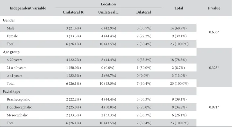
Table 2. Absolute (n) and relative (%) frequencies of mandibular canal bifurcation, according to gender, age group and facial type
Independent variable Location Total P value
Unilateral R Unilateral L Bilateral
Gender
0.635*
Male 3 (21.4%) 6 (42.9%) 5 (35.7%) 14 (60.9%)
Female 3 (33.3%) 4 (44.4%) 2 (22.2%) 9 (39.1%)
Total 6 (26.1%) 10 (43.5%) 7 (30.4%) 23 (100.0%)
Age group
0.325*
≤ 20 years 4 (22.2%) 8 (44.4%) 6 (33.3%) 18 (78.3%)
21 a 40 years 1 (50.0%) 0 (0.0%) 1 (50.0%) 2 (8.7%)
≥ 41 years 1 (33.3%) 2 (66.7%) 0 (0.0%) 3 (13.0%)
Total 6 (26.1%) 10 (43.5%) 7 (30.4%) 23 (100.0%)
Facial type
0.971*
Brachycephalic 2 (22.2%) 4 (44.4%) 3 (33.3%) 9 (39.1%)
Dolichocephalic 2 (25.0%) 4 (50.0%) 2 (25.0%) 8 (34.8%)
Mesocephalic 2 (33.3%) 2 (33.3%) 2 (33.3%) 6 (26.1%)
Total 6 (26.1%) 10 (43.5%) 7 (30.4%) 23 (100.0%)
Legend: * p values obtained from the G test.
Table 1. Absolute (n) and relative (%) frequencies of mandibular canal bifurcation, according to gender, age group and facial type
Independent Variable
Bifurcação de canal mandibular
Total P Value
Present Absent
Gender
0.820*
Male 14 (27.5%) 37 (72.5%) 51 (56.7%)
Female 9 (23.1%) 30 (76.9%) 39 (43.3%)
Total 23 (25.6%) 67 (74.4%) 90 (100.0%)
Age group
0.500**
≤ 20 years 18 (26.9%) 49 (73.1%) 67 (74.4%)
21 to 40 years 2 (14.3%) 12 (85.7%) 14 (15.6%)
≥ 41 years 3 (33.3%) 6 (66.7%) 9 (10.0%)
Total 23 (25.6%) 67 (74.4%) 90 (100.0%)
Facial type
0.664*
Brachycephalic 9 (30.0%) 21 (70.0%) 30 (33.3%)
Dolichocephalic 8 (26.7%) 22 (73.3%) 30 (33.3%)
Mesocephalic 6 (20.0%) 24 (80.0%) 30 (33.3%)
Total 23 (25.6%) 67 (74.4%) 90 (100.0%)
26.7% and 20.0%, respectively. Comparing these proportions using the chi-square test revealed that the bifurcation of the mandibular canal was not significantly associated with facial type (p = 0.664). The relative frequency of mandibular canal bifurcation according to its location indicates that in 69.6%, i.e., 16 of the 23 bifurcation carriers, this was unilateral. Specifically, of the 16 cases with bifurcation of the mandibular canal this variation was unilateral, 6 of the 23 cases (26.1%) occurred on the right side and 10 of the 23 cases (43.5%) on the left side. In 7 of the 23 cases (30.4%), bifurcation of the mandibular canal was bilateral.
The unilateral location (right or left) and bilateral bifurcation of the mandibular canal were not significantly associated with gender (p = 0.635), age group (p = 0.325) and facial type (p = 0.971) (Table 2). The relative frequency of mandibular canal bifurcation according to the classification suggested by Freitas et al.9, Class D,
was the commonest (retromolar direction), since it occurred in 9 of the 23 cases (39.1%). The second most prevalent classification was “C” (alveolar direction), which occurred in 5 of the 23 cases (21.7%). With a lower proportion (13.1%, i.e., 3 in 23 cases), the C-E classifications were observed. For the remaining classifications (“A”, “E”, “F” and “A-E”), frequencies ranged from 8.7% to 4.3%. None of the 23 cases of bifurcation of the mandibular canal was classified as B (mesial direction).
DISCUSSION
The literature points to the importance of knowing the anatomy of the mandibular canal and its possible variations for the success of many dental procedures performed on the mandible, such as anesthesia, endodontics, periodontics, pediatric dentistry and more invasive procedures such as surgical exodontia, orthognathic surgeries, implant placement, among others9,16,19-21.
The dentist should be aware that the mandibular canal may undergo anatomical variations in shape, size, number, as well as in vertical and horizontal positions. For these reasons, several authors have emphasized the importance of studying the precise location of such landmarks and possible anatomical variations therein using CT scans3,5,8,9.
In most cases, the mandibular canal presents as a single structure, though canal duplications, also known as bifid canals, are among the commonest anatomical variations of the mandibular canal and can be grouped in different classifications, as proposed by several authors6,9,19. Chávez-Lomeli et al.20 questioned the definition of
the term bifid attributed to the mandibular canal, as the authors state that there is no division of the mandibular canal into two or more canals as such, but the persistence or non-fusion of the embryologically defined branches of the mandibular canal.
Kang et al.10 defended the use of CBCT, which is a diagnostic
imaging method that uses X-ray to reproduce a section of the human body in one of three planes (axial, sagittal or coronal). Unlike conventional radiographs, which project an image onto a single plane and many the structures crossed by X-rays fatally overlap, the CBCT exposes the in-depth relationships between structures, thus creating images in “slices” of the human body3, disposing all
the structures in layers, mainly the mineralized tissues, allowing
delimitation of three-dimensional irregularities and facilitating the diagnosis of anatomical alterations.
According to de Sanchis et al.22 the vast majority of dental
surgeons are unaware of the anatomical variations of the mandibular canal, and the presence of such variations has a number of clinical implications when not identified prior to surgical interventions. According to several authors, it is up to the dental surgeon to know the possible anatomical variations of the mandibular canal to reduce the risk of failure during surgical or anesthetic procedures3,6,9.
In the present study, bifid mandibular canals were observed in 25.6% of the cases that composed the sample. Previous studies with panoramic radiographs have reported incidences of less than 1%6,23 though CBCT-based studies have shown a much higher
values with prevalence rates ranging from 15.6% to 65% 2,3,9,24,25.
Thus, conventional radiographs are not reliable for detection of anatomical variations of the mandibular canal. In addition, differences in incidence may be related to ethnic, geographical, as well as methodological variations.
When evaluating the relationship between the prevalence of bifid mandibular canals and facial types, a prevalence of 30.0% was found in brachycephalic, 26.7% in dolichocephalic and 20.0% in mesocephalic individuals. No studies were found investigating a possible correlation between anatomical variations of the mandibular canal and the different facial types. Therefore, further studies for ratification of these results are recommended.
Bearing in mind the idea of the clinical relevance of the results obtained in this study, only bifid canals with a diameter greater than 1 mm were included. Moreover, the inclusion of “false bifid or pseudobifid mandibular canals” as described by Kim et al.25
was carefully avoided. According to Sanchis et al.22, an image
mimicking a bifid mandibular canal can be produced by the impression of the mylohyoid nerve onto the inner surface of the mandible. Such images may lead to misdiagnoses, especially in panoramic reconstructions, hence the importance of combining different reconstructions and tomographic slices when evaluating the anatomy of the mandibular canal.
According to Orhan et al.2 there are several classifications of
the anatomical variations of the mandibular canal that consider width, extension, direction and presence of additional foramens. Naitoh et al.23 suggested a classification of bifid mandibular
canals into four types based on trajectory: buccal-lingual (type I), mesial direction (type II), alveolar ridge direction (type III) and retromolar direction (type IV). Freitas et al.9 suggested a more
elaborate classification that took into account directions of the bifid mandibular canal that were not contemplated in previous classifications, Class A (buccal direction), Class B (mesial direction), Class C (alveolar direction), Class D retromolar direction), Class E (lingual direction) and Class F (body of the mandible direction).
When observing the relative frequency of mandibular canal bifurcation according to its classification in the present study, class D (retromolar direction) was the most prevalent with 39.1% of cases of mandibular canal bifurcation. Freitas et al.17 found a prevalence
classified as C-E. For the remaining classifications (A, E, F and A-E), frequencies ranged from 8.7% to 4.3%. None of the 23 cases of bifurcation was found classified as B (mesial direction). These data diverge from the study by Freitas et al.9 that evaluated the prevalence
of bifid mandibular canals without distinguishing between facial types and observed that the most prevalent type of bifurcation was class B (mesial direction). No studies have been found classifying mandibular canal variations in relation to facial types.
With respect to the affected side, it was observed that bifid mandibular canals occurred bilaterally in 30.4% of the cases and unilaterally in 69.6%. When the unilateral occurrence was evaluated alone, the left side was the most affected (43.5%), against 26.1% on the right side. The data found in this study diverge from those reported by Orhan et al. 2 and Freitas et al.9 who reported a higher
prevalence of bifid mandibular canals on the right side.
In the present study, no statistically significant difference were observed regarding the prevalence of bifid mandibular canals between genders, which is consistent with the findings by Freitas et al.9
In addition, some authors2,6,23 have reported a higher prevalence
of bifid mandibular canals in women.
According to Orhan et al.2, regardless of type and classification,
bifid mandibular canals may be associated with an increased risk of accidents and complications during surgical approaches in the
mandibular region and difficulty in obtaining inferior alveolar nerve block, especially in cases where two mandibular foramens or supranumerary retromolar foramens are observed. The importance of understanding the neurovascular anatomy of the mandibular canal and its possible variations is therefore highlighted herein to minimize possible anesthetic failures, as well as sensory and hemorrhagic complications during surgical approaches in the mandible.
CONCLUSION
According to the results obtained in this study, a prevalence of 25.6% of bifid mandibular canals was found, with class D (retromolar direction) being the most prevalent and the highest occurrence of bifid mandibular canals was unilateral on the left side. When evaluating the occurrence of bifid mandibular canals in terms of facial types, brachycephalic individuals were the most affected. When face type was taken into account, brachyfacial individuals were the most affected. The importance of knowing the neurovascular anatomy of the the mandibular canal and its possible variations is herein highlighted in order to minimize possible failures in anesthetic procedures, accidents and both sensory and bleeing complications during surgical approaches within the mandible.
REFERENCES
1. Haas LF, Dutra K, Porporatti AL, Mezzomo LA, De Luca Canto G, Flores-Mir C, et al. Anatomical variations of mandibular canal detected by panoramic radiography and CT: a systematic review and meta-analysis. Dentomaxillofac Radiol. 2016;45(2):20150310.http://dx.doi. org/10.1259/dmfr.20150310. PMid:26576624.
2. Orhan K, Aksoy S, Bilecenoglu B, Sakul BU, Paksoy CS. Evaluation of bifid mandibular canals with cone beam computed tomography in a Turkish adult population: a retrospective study. Surg Radiol Anat. 2011 Aug;33(6):501-7.http://dx.doi.org/10.1007/s00276-010-0761-y. PMid:21161224.
3. Kuribayashi A, Watanabe H, Imaizumi A, Tantanapornkul W, Katakami K, Kurabayashi T. Bifid mandibular canals: cone beam computed tomography evaluation. Dentomaxillofac Radiol. 2010 May;39(4):235-9. http://dx.doi.org/10.1259/dmfr/66254780. PMid:20395465. 4. Correr GM, Iwanko D, Leonardi DP, Ulbrich LM, Araújo MR, Deliberador TM. Classification of bifid mandibular canals using cone beam
computed tomography. Braz Oral Res. 2013 Dec;27(6):510-6. http://dx.doi.org/10.1590/S1806-83242013000600011. PMid:24346049. 5. Gerlach NL, Meijer GJ, Maal TJ, Mulder J, Rangel FA, Borstlap WA, et al. Reproducibility of 3 different tracing methods based on cone beam
computed tomography in determining the anatomical position of the mandibular canal. J Oral Maxillofac Surg. 2010 Apr;68(4):811-7.http:// dx.doi.org/10.1016/j.joms.2009.09.059. PMid:20036043.
6. Nortjé CJ, Farman AG, de V. Joubert JJ. The radiographic appearance of the inferior dental canal: an additional variation. Br J Oral Surg. 1977 Nov;15(2):171-2. http://dx.doi.org/10.1016/0007-117X(77)90050-6. PMid:271020.
7. Langlard OE, Langlais RP, McDadid WD, Delbalso A. Panoramic radiology. J Am Dent Assoc. 1989;2:183-223.
8. Villaça-Carvalho MF, Manhães LR Jr, Moraes ME, Lopes SL. Prevalence of bifid mandibular canals by cone beam computed tomography. Oral Maxillofac Surg. 2016 Sep;20(3):289-94. http://dx.doi.org/10.1007/s10006-016-0569-y. PMid:27417545.
9. Freitas GB, Silva AF, Morais LA, Silva MBF, Silva TCG, Manhães LRC Jr. Incidence and classification of bifid mandibular canals using cone beam computed tomography. Braz J Oral Sci. 2015 Dec;14(4):294-8. http://dx.doi.org/10.1590/1677-3225v14n4a08.
10. Kang JH, Lee KS, Oh MG, Choi HY, Lee SR, Oh SH, et al. The incidence and configuration of the bifid mandibular canal in Koreans by using cone-beam computed tomography. Imaging Sci Dent. 2014 Mar;44(1):53-60. http://dx.doi.org/10.5624/isd.2014.44.1.53. PMid:24701459. 11. Tsunori M, Mashita M, Kasai K. Relationship between facial types and tooth and bone characteristics on the mandible obtained by CT
scannering. Angle Orthod. 1998 Dec;68(6):557-62. http://dx.doi.org/10.1043/0003-3219(1998)068<0557:RBFTAT>2.3.CO;2. PMid:9851354. 12. Schmidt APG, Rossi AC, Freire AR, Groppo FC, Prado FB. Association between facial type and mandibular canal morphology - analysis in
digital panoramic radiographs. Braz Dent J. 2016 Sep-Oct;27(5):609-12. http://dx.doi.org/10.1590/0103-6440201600973. PMid:27982243. 13. Benedicto EN, Kairalla SA, Kajeda AK, Miranda SL, Torres FC, Paranhos LR. Determinação do padrão esquelético vertical da face. Rev Bras
14. von Arx T, Friedli M, Sendi P, Lozanoff S, Bornstein MM. Location and dimensions of the mental foramen: a radiographic analysis by using cone-beam computed tomography. J Endod. 2013 Dec;39(12):1522-8. http://dx.doi.org/10.1016/j.joen.2013.07.033. PMid:24238440. 15. Voljevica A, Talović E, Hasanović A. Morphological and morphometric analysis of the shape, position, number and size of mental foramen
on human mandibles. Acta Med Acad. 2015;44(1):31-8. http://dx.doi.org/10.5644/ama2006-124.124. PMid:26062695.
16. Vujanovic-Eskenazi A, Valero-James JM, Sánchez-Garcés MA, Gay-Escoda C. A retrospective radiographic evaluation of the anterior loop of the mental nerve: comparison between panoramic radiography and cone beam computerized tomography. Med Oral Patol Oral Cir Bucal. 2015 Mar;20(2):e239-45. http://dx.doi.org/10.4317/medoral.20026. PMid:25549693.
17. Freitas GB, Freitas e Silva A, Manhães LRC Jr. The prevalence of mandibular retromolar canals on cone beam computed tomography and its clinical repercussions. Rev Odontol UNESP. 2017 Jun;46(3):231-6. http://dx.doi.org/10.1590/1807-2577.00117.
18. Saito K, Araújo NS, Saito MT, Pinheiro JJV, Carvalho PL. Analysis of the mental foramen using cone beam computerized tomography. Rev Odontol UNESP. 2015 Aug;44(4):226-31. http://dx.doi.org/10.1590/1807-2577.0067.
19. Langlais RP, Broadus R, Glass BJ. Bifid mandibular canals in panoramic radiographs. J Am Dent Assoc. 1985 Jun;110(6):923-6. http://dx.doi. org/10.14219/jada.archive.1985.0033. PMid:3860553.
20. Chávez-Lomeli ME, Mansilla-Lory J, Pompa JA, Kjaer I. The human mandibular canal arises from three separate canals innervating different tooth groups. J Dent Res. 1996 Aug;75(8):1540-4. http://dx.doi.org/10.1177/00220345960750080401. PMid:8906121.
21. Liang X, Jacobs R, Corpas LS, Semal P, Lambrichts I. Chronologic and geographic variability of neurovascular structures in the human mandible. Forensic Sci Int. 2009 Sep;190(1-3):24-32.http://dx.doi.org/10.1016/j.forsciint.2009.05.006. PMid:19525074.
22. Sanchis JM, Peñarrocha M, Soler F. Bifid mandibular canal. J Oral Maxillofac Surg. 2003 Apr;61(4):422-4. http://dx.doi.org/10.1053/ joms.2003.50004. PMid:12684957.
23. Naitoh M, Yoshida K, Nakahara K, Gotoh K, Ariji E. Demonstration of the accessory mental foramen using rotational panoramic radiography compared with cone-beam computed tomography. Clin Oral Implants Res. 2011 Dec;22(12):1415-9. http://dx.doi.org/10.1111/j.1600-0501.2010.02116.x. PMid:21382086.
24. Oliveira-Santos C, Souza PH, Azambuja Berti-Couto S, Stinkens L, Moyaert K, Rubira-Bullen IR, et al. Assessment of variations of the mandibular canal through cone beam computed tomography. Clin Oral Investig. 2012 Apr;16(2):387-93. http://dx.doi.org/10.1007/s00784-011-0544-9. PMid:21448636.
25. Kim MS, Yoon SJ, Park HW, Kang JH, Yang SY, Moon YH, et al. A false presence of bifid mandibular canals in panoramic radiographs. Dentomaxillofac Radiol. 2011 Oct;40(7):434-8. http://dx.doi.org/10.1259/dmfr/87414410. PMid:21960401.
CONFLICTS OF INTERESTS
The authors declare no conflicts of interest.
*CORRESPONDING AUTHOR
George Borja de Freitas, Faculdade de Odontologia e Centro de Pesquisas Odontológicas, São Leopoldo Mandic, Campinas, SP, Brasil, Tel: (87) 99657-0044; (87) 3844-2453, e-mail: [email protected]