Increase in upper airway volume in patients
with obstructive sleep apnea using a
mandibular advancement device
Luciana Baptista Pereira Abi-Ramia*, Felipe Assis Ribeiro Carvalho**, Claudia Torres Coscarelli***, Marco Antonio de Oliveira Almeida****
Introduction: Diagnosis, treatment and monitoring of patients with obstructive sleep apnea syndrome (OSAS) are crucial because this disorder can cause systemic changes. The effective-ness of OSAS treatment with intraoral devices has been demonstrated through cephalometric studies. Objective: The purpose of this study was to evaluate the effect of a mandibular ad-vancement device (Twin Block, TB) on the volume of the upper airways by means of Cone-Beam Computed Tomography (CBCT). Sixteen patients (6 men and 10 women) with mild to moderate OSAS, mean age 47.06 years, wore a mandibular advancement device and were fol-lowed up for seven months on average. Methods: Two CBCT scans were obtained: one with and one without the device in place. Upper airway volumes were segmented and obtained using Student’s paired t-tests for statistical analysis with 5% significance level. Results: TB use increased the volume of the upper airways when compared with the volume attained without TB (p<0.05). Conclusion: It can be concluded that this increased upper airway volume is as-sociated with the use of the TB mandibular advancement device.
Abstract
Keywords: Obstructive sleep apnea syndrome. Mandibular advancement device. Cone-Beam Computed Tomography.
* MSc in Orthodontics, School of Dentistry, Rio de Janeiro State University (FO-UERJ). ** PhD Student in Orthodontics, FO-UERJ.
A B C
introduction
With the increase in respiratory sleep disor-ders, such as snoring, upper airway resistance syndrome (UARS) and obstructive sleep apnea syndrome (OSAS), the need for better diagnos-tics and treatment of these disorders became apparent.4,11 Treatment of OSAS is impor-tant11,15,21,25 as it is considered a high morbidity, progressive disease.11,28
The effectiveness of mandibular protru-sion appliances has been demonstrated in sev-eral studies.13,25 Although cephalometric ra-diography is a simple method, widely used in dentistry and in studies of obstructive sleep apnea,2,3,4,5,10,13,25,26 this method generates two-dimensional images of three-two-dimensional struc-tures, which limits the validity and reproduc-ibility of airway measurements.14,16,24
Three-dimensional studies14,21 to determine the effectiveness and action mechanism of oral appliances have shown that such appliances can modify pharyngeal geometry,21 significantly en-larging the minimum pharyngeal area.14
The aim of this study was to evaluate, using Cone-Beam Computed Tomography (CBCT), the effects of mandibular advancement, performed with a modified Twin Block type appliance, on the volume of OSAS patients’ upper airways.
MAtEriAL And MEtHodS
This research was submitted to the Ethics Committee of Pedro Ernesto University Hospital and approved under number 1366-CEP/HUPE.
Patients were referred to the Orthodontics postgraduate clinic, School of Dentistry, Rio de Ja-neiro State University (FO-UERJ) by specialists in Sleep Medicine after undergoing a nocturnal poly-somnography examination and being diagnosed with mild to moderate OSAS (AHI<30).
Other inclusion criteria were used: Only pa-tients with a body mass index (BMI) of less than 27; having at least ten teeth in each arch to en-sure adequate device retention; having an over-jet of at least 4 mm so as to enable mandibular advancement.
Sixteen patients, 6 men and 10 women, mean age of 47.06 years, received modified Twin Block (TB) type oral appliances for mandibular ad-vancement (Fig 1). They were instructed to wear the appliance at night and were monitored for an average period of seven months. The mandibular advancement achieved with TB was approximate-ly 75% of maximum protrusion.12 To participate in the sample the patients signed a form of free and informed consent after being given informa-tion about the research.
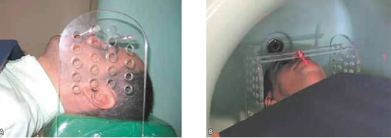
At the end of the follow up period each pa-tient underwent two CBCT scans (NewTom 3G, Verona, Italy) with field of view of 9 inches and slice thickness of 0.2 mm. Both scans were per-formed on the same day, one without and one with the mandibular advancement appliance in place. The patients were awake, lying supine, with the Frankfort plane perpendicular to the floor.24
The scanning method was standardized with the aid of an acrylic positioner (Fig 2) and the
B A
NewTom 3G laser beam itself, to position the fa-cial midline. Moreover, the distances between pa-tient and scanner, and the height of the stretcher were recorded in the first examination to ensure that the two scans were as similar as possible. This position was verified on the computer with the aid of a scanogram before the start of the second examination.
After the primary reconstruction of the pro-jections in the three orthogonal planes (axial, coronal and sagittal) and images of the entire cra-niofacial complex volume were obtained in DI-COM format (Digital Imaging Communications in Medicine), the images were manipulated with
ITK SNAP 1.8.030 software to obtain volumetric reconstructions of the relevant structures. The software allows semiautomatic segmentation6,7 of the area of interest, which was limited in the an-terior and superior regions by the posan-terior nasal spine (PNS)24,27 while in the inferior region, the limits were the anterior-most and inferior-most regions of the third cervical vertebra (C3)13 (Fig 3). The volume in mm3 of the three-dimensional model of the upper airway (Fig 4) was obtained with the software.
The statistical data were tabulated in a statistical program (Biostat 2.0, Belém, Pará State, Brazil). Method error was used only to
FIGURE 2 - Cone-Beam Computer Tomography scans: A) Patient positioned at NewTom 3G with acrylic positioner and Frankfort horizontal plane perpendicular to the floor; B) Using the laser beam to position the facial midline.
FIGURE 3 - Points used to determine upper airway volume. PNS (poste-rior nasal spine), C3 (ante(poste-rior-most and infe(poste-rior-most portions of the third vertebra).
15000
5000 10000
0
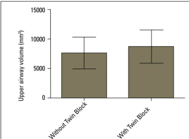
FIGURE 5 - Volume values of upper airway (mm3) of patients without and with TB.
TABLE 1 - Mean, standard deviation and p value for comparing airway volume (in mm3) between patients with and without TB.
circumscribe the structure because it is a semi-automatic method. Two examiners delimited the area of interest twice at intervals of two days, and the intraclass correlation coefficient (ICC) for nominal or quantitative variables was utilized to assess the correlation between re-peated measurements in the same patient. The ICC showed excellent intra and interexaminer repeatability, which allows the authors to assert that the method used to segment and obtain up-per airway volume is reliable (p<0.0001).
After using the Shapiro-Wilk normality test, the paired t-test was applied to compare the volumes with and without TB. To be considered significant, p value was set at 0.05.
rESuLtS
The mean airway volumes with and
with-out TB were 8710±2813 mm3 and 7601±2659
mm3, respectively (Fig 5). There was a statisti-cally significant difference (p=0.0494) in air-way volume between patients with and with-out TB (Table 1), demonstrating that TB was successful in increasing upper airway volume in the TB patients.
diScuSSion
Upper airway three-dimensional assessment was performed using CBCT given its low radia-tion dose.16,27 According to Aboudara et al,1 al-though CBCT is not usually indicated for evaluat-ing soft tissues the contrast between the airway lumen and the soft and hard tissue enhances segmentation accuracy when quantifying airway volume. The NewTom 3G scanner used in this study enabled the assessment of the upper airways while the patient was lying down and, although it failed to reproduce the exact sleeping position, positioning the pharyngeal tissues is important in determining the severity of the syndrome.18 How to position the patient during follow-up exami-nations is a much debated issue, since air flow is influenced by changes in head position,8,29 which
may result from the reduced dimensions of the upper airways in the retropalatal region.1
While assessing the images in the three planes of space with ITK-SNAP software reference points were selected for defining the area of interest ac-cording to previous studies.24,27 The reference points used in this study were the ENP1,4,13,24,27 and the most anterior and inferior point of the third cervical vertebra.13
The statistically significant difference found in this study between patients with and without TB in place (p<0.05) shows that the upper airways expanded as a result of the mandibular advance-ment caused by the TB. This mechanism is still under debate. It is believed, however, that the more anterior position of the mandible and hy-oid bone and the consequent stimulation of the pharyngeal muscles and tongue are responsible
Mean Standand
deviation P value
Volume
without TB 7601 2659
p = 0.0494 Volume
with TB 8710 2813
U
p
p
e
r
a
ir
w
a
y
vo
lu
m
e
(
m
m
3)
With out T
win B lock
With Twin
for increasing airway volume.30 According to the cephalometric study by Fransson et al13, the in-crease in pharyngeal area occurred because of the more anterior position of the hyoid bone as a result of increased activity in the genioglossus and lateral pterygoid muscles.
Only two patients had lower airway volume with TB than without TB, which may be ex-plained by the amount of mandibular advance-ment in these patients or the width of their soft palate. The amount of mandibular advancement varies widely between different studies, ranging from 2.0 mm20 to 9.5 mm.12,13 Despite the dif-ferences, a protrusion of 75% of each patient’s maximum capacity is very often used by research-ers.9,10,13,14,15,17,19,22,23,25 This advancement yields ad-equate success rates and can usually be endured by most patients.
CBCT comparison between healthy patients and patients presenting with OSAS has shown that the anteroposterior dimensions and mini-mum oropharyngeal dimensions of patients with OSAS were significantly lower compared to pa-tients who did not have this syndrome.24
Three-dimensional studies in patients with SAOS14,21,30 have shown increased upper airways, predominantly in the oropharyngeal21 and velo-pharyngeal30 regions. However, most of these stud-ies21,30 assessed the airways using linear measure-ments only, i.e., using two-dimensional data ob-tained through three-dimensional examinations.
According to Zhao, Liu, Gao30 and Kyung, Park, Pae,21 airway augmentation is achieved at the ex-pense of an increase in transverse diameter. Gale et al14 found an increase in the pharyngeal area using a mandibular advancement device but with substantial individual variability.
In the present study, preference was given to conducting two CBCT scans on the same day after the monitoring period due to image acqui-sition standardization, since each patient’s ide-al position is unique. Moreover, there could be changes in patients’ BMI and health status dur-ing follow-up, as well as climate changes. These factors would render impracticable any compari-sons between soft tissues and upper airway vol-umes at different times.
OSAS studies using Cone-Beam Computed Tomography and three-dimensional models re-quire further research and improved standard-ization of assessment methods, in addition to a better understanding of the action mechanisms underlying mandibular advancement devices and their results, if these devices are to become the treatment of choice for OSAS patients.
concLuSionS
1. Aboudara C, Nielsen I, Huang JC, Maki K, Miller AJ, Hatcher D. Comparison of airway space with conventional lateral headilms and 3-dimensional reconstruction from cone-beam computed tomography. Am J Orthod Dentofacial Orthop. 2009 Apr;135(4):468-79.
2. Almeida FR, Lowe AA, Sung JO, Tsuiki S, Otsuka R. Long-term sequellae of oral appliance therapy in obstructive sleep apnea patients: Part 1. Cephalometric analysis. Am J Orthod Dentofacial Orthop. 2006 Feb;129(2):195-204.
3. Battagel JM, Johal A, Kotecha B. A cephalometric comparison of subjects with snoring and obstructive sleep apnoea. Eur J Orthod. 2000 Aug;22(4):353-65.
4. Blanco J, Zamarrón C, Abeleira Pazos MT, Lamela C, Suarez Quintanilla D. Prospective evaluation of an oral appliance in the treatment of obstructive sleep apnea syndrome. Sleep Breath. 2005 Mar;9(1):20-5.
5. Cevidanes LH, Styner MA, Profit WR. Image analysis and superimposition of 3-dimensional cone-beam computed tomography models. Am J Orthod Dentofacial Orthop. 2006 May;129(5):611-8.
6. Cevidanes LH, Bailey LJ, Tucker GR Jr, Styner MA, Mol A, Phillips CL, et al. Superimposition of 3D cone-beam CT models of orthognathic surgery patients. Dentomaxillofac Radiol. 2005 Nov;34(6):369-75.
7. Choi JK, Goldman M, Koyal S, Clark G. Effect of jaw and head position on airway resistance in obstructive sleep apnea. Sleep Breath. 2000;4(4):163-8.
8. Clark GT, Arand D, Chung E, Tong D. Effect of anterior mandibular positioning on obstructive sleep apnea. Am Rev Respir Dis. 1993 Mar;147(3):624-9.
9. Cooke ME, Battagel JM. A thermoplastic mandibular advancement device for the management of non-apnoeic snoring: a randomized controlled trial. Eur J Orthod. 2006 Aug;28(4):327-38.
10. Dal Fabbro C, Chaves C Jr, Tuik S. A Odontologia na medicina do sono. 1ª ed. Maringá: Dental Press; 2010.
11. Ferguson KA, Ono T, Lowe AA, al-Majed S, Love LL, Fleetham JA. A short term controlled trial of an adjustable oral appliance for the treatment of mild to moderate obstructive sleep apnoea. Thorax. 1997 Apr;52(4):362-8.
12. Fransson AM, Tegelberg A, Svenson BA, Lennartsson B, Isacsson G. Inluence of mandibular protruding device on airway passages and dentofacial characteristics in obstructive sleep apnea and snoring. Am J Orthod Dentofacial Orthop. 2002 Oct;122(4):371-9.
13. Fransson AM, Tegelberg A, Johansson A, Wenneberg B. Inluence on the masticatory system in treatment of obstructive sleep apnea and snoring with a mandibular protruding device: a 2-year follow-up. Am J Orthod Dentofacial Orthop. 2004 Dec;126(6):687-93.
14. Gale DJ, Sawyer RH, Woodcock A, Stone P, Thompson R, O’Brien K. Do oral appliances enlarge the airway in patients with obstructive sleep apnoea? A prospective computerized tomographic study. Eur J Orthod. 2000 Apr;22(2):159-68. 15. Garib DG, Raymundo R Jr, Raymundo MV, Raymundo DV,
Ferreira SN. Tomograia computadorizada de feixe cônico (Cone Beam): entendendo este novo método de diagnóstico por imagem com promissora aplicabilidade na Ortodontia. Rev Dental Press Ortod Ortop Facial. 2007 mar-abr; 12(2):139-56.
16. Horiuchi A, Suzuki M, Ookubo M, Ikeda K, Mitani H, Sugawara J. Measurement techniques predicting the effectiveness of an oral appliance for obstructive sleep apnea hypopnea syndrome. Angle Orthod. 2005 Nov;75(6):1003-11. 17. Ingman T, Nieminen T, Hurmerinta K. Cephalometric
comparison of pharyngeal changes in subjects with upper airway resistance syndrome or obstructive sleep apnoea in upright and supine positions. Eur J Orthod. 2004 Jun;26(3):321-6.
rEfErEncES
18. Johnston CD, Gleadhill IC, Cinnamond MJ, Peden WM. Oral appliances for the management of severe snoring: a randomized controlled trial. Eur J Orthod. 2001 Apr;23(2):127-34.
19. Jureyda S, Shucard DW. Obstructive sleep apnea: an overview of the disorder and its consequences. Semin Orthod. 2004 Mar;10(1):63-72.
20. Kato J, Isono S, Tanaka A, Watanabe T, Araki D, Tanzawa H, et al. Dose-dependent effects of mandibular advancement on pharyngeal mechanics and nocturnal oxygenation in patients with sleep-disordered breathing. Chest. 2000 Apr;117(4):1065-72.
21. Kyung SH, Park YC, Pae EK. Obstructive sleep apnea patients with the oral appliance experience pharyngeal size and shape changes in three dimensions. Angle Orthod. 2005 Jan;75(1):15-22.
22. Marklund M, Franklin KA, Persson M. Orthodontic side-effects of mandibular advancement devices during treatment of snoring and sleep apnoea. Eur J Orthod. 2001 Apr;23(2):135-44. 23. Ogawa T, Enciso R, Shintaku WH, Clark GT. Evaluation of
cross-section airway coniguration of obstructive sleep apnea. Oral Surg Oral Med Oral Pathol Oral Radiol Endod. 2007 Jan;103(1):102-8.
24. O’Sullivan RA, Hillman DR, Mateljan R, Pantin C, Finucane KE. Mandibular advancement splint: an appliance to treat snoring and obstructive sleep apnea. Am J Respir Crit Care Med. 1995 Jan;151(1):194-8.
25. Otsuka R, Almeida FR, Lowe AA, Ryan F. A comparison of responders and non-responders to oral appliance therapy for the treatment of obstructive sleep apnea. Am J Orthod Dentofacial Orthop. 2006 Feb;129(2):222-9.
26. Tso HH, Lee JS, Huang JC, Maki K, Hatcher D, Miller AJ. Evaluation of the human airway using cone-beam computerized tomography. Oral Surg Oral Med Oral Pathol Oral Radiol Endod. 2009 Nov;108(5):768-76.
27. Walker-Engström ML, Tegelberg A, Wilhelmsson B, Ringqvist I. Four-year follow-up of treatment with dental appliance or uvupalatopharyngoplasty in patients with obstructive sleep apnea. Chest. 2002 Mar;121(3):739-6.
28. Yildirim N, Fitzpatrick MF, Whyte KF, Jalleh R, Wightman AJ, Douglas NJ. The effect of posture on upper airway dimensions in normal subjects and in patients with the sleep apnea/hypopnea syndrome. Am Rev Respir Dis. 1991 Oct;144(4):845-47.
29. Yushkevich PA, Piven J, Hazlett CHC, Smith CRG, Ho CS, Gee JC, et al. User-guided 3D active contour segmentation of anatomical structures: Signiicantly improved eficiency and reliability. Neuroimage. 2006 Jul;31(3):116-28.
30. Zhao X, Liu Y, Gao Y. Three-dimensional upper-airway changes associated with various amounts of mandibular advancement in awake apnea patients. Am J Orthod Dentofacial Orthop. 2008 May;133(5):661-8.
contact address
Luciana Baptista Pereira Abi-Ramia
Rua Franz Weissman, 530 Bl 02/ 305 – Barra da Tijuca CEP: 22775-051 – Rio de Janeiro/RJ, Brazil
E-mail: [email protected] Submitted: June 2010