The aim of this study was to isolate Enterobacteria and Pseudomonas from the oral cavity
of hospitalized newborns (NB) and determine their prevalence and the sensitivity profile to most commonly used antibiotics for this age group. Samples from the oral cavity of NB from 24 to 48 h age were collected using swabs. The samples were inoculated on MacConkey agar, incubated and the colonies counted and identified. For each strain, the minimum inhibitory concentration (MIC) was determined using agar dilution test. Tests for enterobacteria producing extended spectrumβ-lactamases (ESBL) were performed
using agar diffusion. Descriptive statistics was used for data analysis. Two of the isolated strains were submitted to the susceptibility test in biofilm. Of the collected samples, 8% presented Enterobacteria (mean of 6,141 CFU/mL) and no Pseudomona species was
isolated. Positive samples were from NB in accommodation set or in the NB nursery. Enterobacter was the most prevalent genus and some strains were resistant to ampicillin, gentamicin and cephalothin. No ESBL strain was detected. Microorganisms in biofilms were resistant to all antibiotics, with concentrations four times higher than MIC. The presence of enterobacteria in the oral cavity of newborns, especially some strains resistant
to normally usedantibiotics, warns to the need for care to avoid the early colonization of this niche and the occurrence of a possible hospital infection in this age group.
P r e v a l e n c e a n d S e n s i t i v i t y
o f B a c i l l i a n d P s e u d o m o n a s
i n t h e N e w b o r n ’ s O r a l C a v i t y
Priscila Vitor Alves Ferreira1, Isabela Amêndola1, Luciane Dias de Oliveira2, Célia Regina Gonçalves e Silva1, Mariella Vieira Pereira Leão1, Silvana Soléo Ferreira dos Santos1
1UNITAU - Universidade de
Taubaté, Taubaté, SP, Brazil
2Institute of Science and Technology,
UNESP - Univ Estadual Paulsta, São José dos Campos, SP, Brazil
Correspondence: Isabela Amêndola, Avenida Tiradentes, 500, Centro, 12030-180 Taubaté, SP, Brasil. Tel: +55-12-3635-3466. e-mail: [email protected]
Key Words: newborn,
Enterobacteriaceae, Pseudomonas, multidrug resistance, biofilm.
Introduction
The increase of drug resistant bacteria worldwide has caused concern among health professionals and in the general community, especially the growing resistance of Gram-negative bacteria (1,2).
Nosocomial infection in newborns is one of the greatest problems in NICU, for being closely associated with increased morbidity and mortality in this age group (3,4). Among them are Enterobacteria, responsible for an
increasing number of hospital infections. Their persistence in hospitals is favored by the constant use of antibiotics and presence of patients with diminished organic defenses.
These frames of in-hospital origin are hard treated and have limited therapeutic options. Species producing extended spectrum β-lactamases (ESBL), have been frequently observed, among them Klebsiella pneumoniae
and Escherichia coli (5-7).
Neonatal units, places where sick newborns (NB) and premature infants stay, are risk locals for the development of opportunistic and pathogenic infections, as Enterobacteria
infections. These microorganisms can survive in the environment and also temporarily on the hands of persons handling newborns, making the child-child transmission easier. Another important risk factor for NB infections
is the limited protection of the immune system during this period of life (8,9). These and other pathogenic microorganisms may remain viable in areas associated and non-associated with the newborn, increasing the risk of infection outbreaks (9).
There is also an increasing number of cases of neonatal infections caused by Pseudomonas aeruginosa,
opportunistic Gram negative bacilli with resistance to many antibiotics, especially among hospitalized newborns in the intensive care units (6,11,12). These microorganisms are found in various environments, including damp reservoirs of any hospital and even in disinfectant solutions. This wide environmental distribution is due to its simple growth requirements. The clinical syndromes most described in newborns caused by this species include bloodstream infections, pneumonia, endophthalmitis and associated infections with ventricular-peritoneal catheters (13).
P
.V
.A. Ferreira et al.
to antimicrobials most commonly used for this age group at the hospital.
Material and Methods
This research was approved by the Research Ethics Committee (CEP/UNITAU 554/10) and followed the determinations of the Resolutions 196/96 and 251/97 of the National Health Council, which establishes the guidelines and regulatory standards for research involving human subjects.
Newborns, 24-48-hour old, born at term or prematurely, accommodated in nursery or rooming-in (mother and child) or admitted to neonatal intensive care units (NICUs) of a University hospital were included in this study. Caregivers were informed of the study’s purpose and signed an informed consent form.
Sample size calculation was performed based on the monthly average of births (n=115) using the BioEstat 2.0 program, with 0.8 (80%) power, 0.01% significance level and standard deviation of 7.76. For these data, the Student t test was applied for one sample, which generated a number of 36 newborns/month, totaling 72 as minimum.
Samples of 75 newborns were collected from the oral cavity with sterile swabs (Absorve, Jiangsu, China) wet in sterile saline (0.85% NaCl), by a rotating mode (360º).
The tip of the swab was cut with sterile scissors, placed in a test tube containing 2 mL of sterile phosphate buffered solution (PBS, pH 7.4), stored in coolers and sent to the microbiology laboratory to be spread within at most 3 hours after collection.
After shaking for 30 s (Vortex; Phoenix, Araraquara, SP, Brasil), 100 µL of each sample were plated on MacConkey agar (Oxoid, Basingstoke, UK). All samples were plated in duplicate and incubated at 37 °C for 24 h.
The plates that showed no growth after this period, remained for 24 h more at 37 °C. After obtaining the microbial growth, the macro-morphological characteristics were observed, by visualization of the colonies and micromorphological characteristics, by the smear and Gram staining technique. The colony forming units per plate were counted and the number of colony-forming units was calculated per milliliter (CFU/mL). The isolated colonies were identified by API 20E system (BioMerieux, Marcy-l’Étoile, France).
Standard strain of Escherichia coli (ATCC 25922) was
used as control in the identification tests, as well as the susceptibility tests.
The susceptibility to the antimicrobials was determined by the agar dilution method (11), which indicates the minimum inhibitory concentration (MIC) in µg/mL. Five of the most suitable antimicrobials for this age group (15) and also those often used for newborns at the site of
research (Ampicillin, Cephalothin, Gentamicin, Amikacin and Cefepime), were tested for Enterobacteriaceae. The tests were performed with positive and negative controls growth. From the results obtained from the MIC, microorganisms were considered sensitive or resistant based on CLSI - M100-S22 breakpoints criteria (16).
From a culture of 24 h of each sample or standard strain, dilutions were made in sterilized physiological solution (0.9% NaCl) until the concentration of 1.5 x 10⁸ cells/mL (range 0.5 McFarland). The antibacterials, in pre-prepared concentrations (2-256 µg/mL in multiple by 2 sequential dilutions), sterilized by filtration (Millipore Membrane 0.22 µm), were added to Mueller-Hinton agar (Himedia, Mumbai, India) at 50 °C and then plated. The bacterial suspensions were seeded with Steers replicator and incubated at 37 °C for 24 h. Reading was performed by observing presence or absence of colonies growing on the agar surface. The strains of Enterobacteriaceae were also investigated for extended spectrumβ-lactamases (ESBL), using the agar diffusion test with cefotaxime, ceftazidime antimicrobials and combination discs of amoxicillin and clavulanic acid (CLSI - M100-S20) (14). As control, a strain of Klebsiella pneumoniae positive ESBL (ATCC 700603) was used.
For the biofilm formation, the selected strains were grown at 37 °C for 18 h in brain and heart infusion agar (BHI, Himedia). Next, the microorganisms were inoculated into BHI mixture (Himedia) plus 5% saccharose. After 16 h of incubation, the microorganisms were centrifuged at 5000 ×g for 5 min and washed twice with 5 mL of sterilizing PBS. Then were centrifuged again and re-suspended in BHI mixture. The suspension was standardized at a 107 cells/ mL concentration in spectrophotometer (B582; Micronal, São Paulo, B SP, Brazil) with a wavelength of 530 nm and optical density of 0.381.
Two hundred microliters of organism suspension were placed into a 96-well microtiter plate. The plate was incubated under rotation of 75 rpm at 37 °C for 90 min, for initial stage of adhesion. After this period, the mixture was gently aspirated and each well washed with 200 µL of PBS. This procedure was repeated twice to remove the non-adhered cells. Then, 200 µL of BHI broth were added and the plates incubated at 37 °C for 48 h under stirring. The culture medium was exchanged each 24 h.
After the incubation period, the medium was aspirated and then 200 µL of PBS were added and the plates placed under stirring for 5 min to remove the non-adhered cells from the biofilm. After shaking, the PBS was removed.
Enterobacteria in newborns’ oral cavity
After this period, the antimicrobial was gently aspirated and each well washed with 200 µL of PBS. This procedure was repeated twice to remove residual antimicrobial. Then, 200 µL of PBS was added and the bottom of each well of the plate was gently scraped with a sterile toothpick. Then, 10 µL of the resulting contents of each well were aspirated and seeded into plates containing MacConkey agar and incubated for 24 h at 37 °C.
The tests to determine the viability of the biofilm microorganisms were performed in triplicate. The microorganism was considered viable if it grew on the surface of agar.
Descriptive statistics were used for data analysis.
Results
The samples were collected from the oral cavity of 75
newborns, 42 of them were in rooming, 10 in nursery and 23 in NICU. The newborns had 24-48 h of life, (average 33±8.5 h).
Pseudomonas was absent in the mouth of all evaluated neonates and enterobacteria present in 8% (n=6). Among the neonates with positive samples for enterobacteria, 83.3% (n=5) were in rooming and 16.7% (n=1) in nursery. All the samples of the NICU neonates were negative.
Sixteen (21.3%) of the neonates were using antibiotics at the time of collection: penicillin (n=1), cephalexin (n=1) and association of penicillin and amikacin (n=14). All these samples were negative.
Among the positive samples of rooming (n=5), one was from normal labor with 36 h of life and the other four samples were by Cesarian delivery and at 24, 32, 36 and 48 h of life, respectively. For the positive sample in nursery, the infant was born from vaginal birth and had 24 h of life.
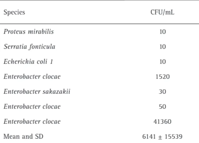
Enterobacter was the most prevalent enterobacteria gender. The species identified in each neonate and the numbers of forming units per milliliter (CFU/mL) are shown in Table 1.
During the susceptibility tests to antimicrobials was observed the occurrence of Enterobacter strains, which were resistant to ampicillin, gentamicin and cephalotin. The minimum inhibitory concentration (MIC) for each antimicrobial to each identified species is presented in Table 2.
None of the clinical strains presented extended
spectrum strains β-lactamases (negative ESBL).
In biofilm, both E. coli 1 and Enterobacter sakazakii
were resistant to four concentrations above the minimum inhibitory concentration of each antimicrobial agent.
Discussion
The oral environment is sterile at birth, but the introduction of microorganisms can occur during labor, through microbial invasion of amniotic cavity and aspiration of contaminated amniotic fluid, or by contact with microorganisms present in the birth canal or genital secretions or maternal feces (9,18,19). Thus, the oral cavity can harbor various microbial species, including microorganisms of the Enterobacteriaceae and Pseudomonadaceae multiresistant families. In and/or from this niche, these microorganisms may cause various infections that could interfere with breastfeeding and compromise the newborn’s health.
Table 1. Enterobacteriaceae species identified in neonates of 24 to 48 hours hospitalized and the number of colony forming units (CFU) per milliliter
Species CFU/mL
Proteus mirabilis 10
Serratia fonticula 10
Echerichia coli 1 10
Enterobacter clocae 1520
Enterobacter sakazakii 30
Enterobacter clocae 50
Enterobacter clocae 41360
Mean and SD 6141 ± 15539
Table 2. Minimum Inhibitory Concentration (MIC) of each antimicrobial (in micrograms per milliliter - µg /mL) for the recovered species and E. coli standard strains (ATCC 25922).
Species
MIC (µg/mL)
amikacin ampicillin cefalotin cefepime gentamicin
E. clocae 1 (S) 8 (S) >256 (R) 1 (S) 2 (S)
E. clocae 1 (S) 4 (S) 64 (R) 1 (S) 8 (R)
E. clocae 1 (S) 128 (R) >256 (R) 1 (S) 4 (S)
E. sakazakii 4 (S) 16 (I) >256 (R) 1 (S) 4 (S)
S. fontícula 2 (S) 4 (S) 2 (S) 1 (S) 4 (S)
E. coli 1 4 (S) 8 (S) 8 (S) 1 (S) 4 (S)
P. mirabilis 1 (S) 4 (S) 4 (S) 1 (S) 4 (S)
P
.V
.A. Ferreira et al.
In this study, newborns’ oral enterobacteriaceae strains were isolated. However, considering what was mentioned above, it was expected a higher positivity in babies born from vaginal birth rather and not from Caesarean delivery as observed. In Cesarean delivery, the direct contact of the newborn’s oral cavity with intestinal and vaginal microbiota is replaced by exposure to exogenous bacteria that usually do not colonize the mother (19). As the collections took place between 24 and 48 h of life, it must be considered that after birth, the child is exposed to a wide range of microorganisms from a variety of sources, including the maternal ones. Although the literature shows that the birth mode interferes with the establishment of the microbiota and later in the child’s health, most part of the research focuses on intestinal microbiota (20), leaving a gap in the knowledge of the initial phase of microbial acquisition by the oral cavity.
Comparing the lifetime, the occurrence of enterobacteria in infants with more than 32 h (n=4) was more common. According to Cezário et al. (21), hospital stay increases the risk of colonization by multiresistant enteric bacilli, an information that corroborates the results obtained in this work.
Nelson-Filho et al. (9) obtained similar results, collected samples from the oral cavity of newborns within 10 min to 53 h after birth, found a greater number of Gram-negative bacilli in NB with 24 to 53 h of life, confirming that these microorganisms increase with increased age.
Regarding the local where the newborns stayed, positive samples were found in those accommodated in nursery or rooming-in (mother and child). The greater care of newborns admitted to NICUs may have contributed to the absence of positive samples in these babies. These results may also be related to fewer samples from newborns in NICU and nursery, due to lower turnover in NICU and shorter stay in nurseries, which are indicated for special cases only, such the maternal abandonment or need for monitoring while waiting for vacancy in NICU.
Bokulich et al. (10) identified samples collected from areas in NICU associated or not to newborns before and after cleaning of premises, noting that the number of positive samples for Enterobacteria was lower compared with other identified microorganisms. This factor may be associated with the small number of positive samples for oral Enterobacteria in the present study, since microorganisms in the environment may be associated with the patient’s microbiota.
Regarding the identified enterobacteriaceae gender, higher prevalence of Enterobacter in the oral cavity of newborns reflects a tendency in the studied region (22).
All newborns that were treated with antibiotics at sample collection showed no positivity for the
studied microorganisms. This fact may demonstrate the effectiveness of these antimicrobials, especially amikacin, according to the results of this work.
Although none of the strains were ESBL positive, some were resistant to ampicillin, gentamicin and cephalothin. The resistance of all the strains Enterobacter to cephalothin found in the present study confirms the low sensitivity of this genre to this antimicrobial. The resistance to ampicillin and to gentamicin draws special attention to the possibility of difficulties of treating infections in newborns and to the need for further investigation regarding the profile of sensibility of these microorganisms.
Regarding the biofilm, the tested clinical strains were resistant to all tested antimicrobials at a concentration four times higher than the MIC obtained for the planktonic form. This result only reinforces the existing literature that demonstrates the persistence and the greater resistance of microorganisms when in biofilm (22).
Thus, it must be considered that occurrence of enterobacteria in the oral cavity of newborns, especially of some strains resistant to normally used antibiotics, warns to the need for greater care to avoid the early colonization of this niche and the occurrence of a possible hospital infection in this age group.
Resumo
O objetivo foi isolar enterobactérias e Pseudomonas da cavidade oral de recém-nascidos hospitalizados (RN) e determinar a prevalência e o perfil de sensibilidade aos antibióticos mais comumente utilizados para este grupo etário. Foram coletadas amostras da cavidade oral de NB com idade de 24-48 horas, usando swab. As amostras foram inoculadas em ágar MacConkey, incubadas e, as colônias contadas e identificadas. Para cada cepa, a concentração inibitória mínima (CIM) foi determinada utilizando teste de ágar diluição. Testes para enterobactérias produtoras de β-lactamases de espectro estendido (ESBL) foram realizados utilizando difusão em ágar. Estatística descritiva foi utilizada para análise dos resultados. Duas das cepas isoladas foram submetidas ao teste de susceptibilidade em biofilme. Das amostras coletadas, 8% apresentaram enterobactérias (média de 6,141 UFC / ml) e nenhuma espécie de Pseudomonas foi isolada. As amostras positivas foram de RN de alojamento conjunto ou RN de berçário. Enterobacter foi o gênero mais prevalente e algumas cepas foram resistentes à ampicilina gentamicina e cefalotina. Não foi detectada cepa ESBL. Micro-organismos em biofilme foram resistentes a todos os antibióticos, em concentrações quatro vezes superiores ao MIC. A presença de enterobactérias em cavidade oral de recém-nascidos, especialmente algumas cepas resistentes aos antibióticos normalmente utilizados, alerta para a necessidade de cuidados, evitando a colonização precoce deste nicho e a ocorrência de possível infecção nosocomial neste grupo etário.
Acknowledgements
To the Foundation of Research of the State of São Paulo - FAPESP - for granting the scientific initiation scholarship, which enabled this work.
References
1. Boucher HW, Talbot GH, Bradley JS, Edwards JE, Gilbert D, Rice LB, et al.. Bad bugs, no drugs: no ESKAPE! An update from the Infectious Diseases Society of America. Clin Infect Dis 2009;48:1–12.
Enterobacteria in newborns’ oral cavity
Enterobacteriaceae. Am J Infect Control 2006;34:S20-S28.
3. Tapia-Rombo CA, Ugarte-Torres RG, Alvarez-Vázquez A, Salazar-Acuña AH. Risk factors for intrahospital infection in newborns. Arch Med Res 2001;32:304-311.
4. Serrano M, Barcenilla F, Limón E. Nosocomial infections in long-term health care facilities. Enferm Infecc Microbiol Clin 2014;32:191-198. 5. Kim YK, Pai H, Lee HJ, Park SE, Choi EH, Kim J, et al.. Bloodstream
infections by extended-spectrum beta-lactamase-producing Escherichia coli and Klebsiella pneumoniae in children: epidemiology and clinical outcome. Antimicrob Agents Chemother 2002;46:1481-1491.
6. Vijayakanthi N, Bahl D, Kaur N, Maria A, Dubey NK. Frequency and characteristics of infections caused by extended-spectrum beta-lactamase-producing organisms in neonates: a prospective cohort study. Biomed Res Int 2013; 2013:756209.
7. Guzmán-Blanco M, Labarca JA, Villegas MV, Gotuzzo E. Extended spectrum -lactamase producers among nosocomial Enterobacteriaceae in Latin America. Braz J Infect Dis 2014;18:421-433. doi: 10.1016/j. bjid.2013.10.005.
8. Kristóf K, Szabó D, Marsh JW, Cser V, Janik L, Rozgonyi F, et al.. Extended-spectrum beta-lactamase-producing Klebsiella spp. in a neonatal intensive care unit: risk factors for the infection and the dynamics of the molecular epidemiology. Eur J Clin Microbiol Infect Dis 2007;26:563-570.
9. Nelson-Filho P, Borba IG, Mesquita KSF, Silva RAB, Queiroz AM, Silva LAB. Dynamics of microbial colonization of the oral cavity in newborns. Braz Dent J 2013;24:415-419.
10. Bokulich NA, Mills DA, Underwood MA. Surface microbes in the neonate intensive care unit: changes with routine cleaning and over time. J Clin Microbial 2013;51:2617-2624.
11. Smith RS, Iglewski BH. Pseudomonas aeruginosa quorum sensing as a potential antimicrobial target. J Clin Invest 2003;112:1460-1465. 12. Kipnis E, Sawa T, Wiener-Kronish J. Targeting mechanisms of
Pseudomonas aeruginosa pathogenesis. Med Mal Infect 2006;36:78-91.
13. Foca MD. Pseudomonas aeruginosa infections in the neonatal intensive care unit. Semin Perinatol 2002;26:332-339.
14. National Commitee for Clinical Laboratory Standards. Methods for Dilution Antimicrobial Susceptibility Tests for Bacteria that Grow Aerobically; Approved Standard - Sixth Edition - NCCLS document M7-A6 [ISBN 1-56238-486-4]. Wayne, PA, USA;2003.
15. Gerdes JS . Diagnosis and management of bacterial infections in the neonate. Pediatr Clin N Am 2004;51-939-959.
16. Clinical and Laboratory Standards Institute. Performance Standards for Antimicrobial Susceptibility Testing; 22th Informational Supplement: Approved Standard M100-S22. Wayne, PA, USA:NCCLS, 2012. 17. Clinical and Laboratory Standards Institute: Performance Standards
for Antimicrobial Susceptibility Testing; Twentieth Informational Supplement: Approved Standard M100-S20. Wayne, PA, USA: NCCLS, 2010.
18. Neu J, Rushing J. Cesarean versus vaginal delivery: long term infant outcomes and the hygiene hypothesis. Clin Perinatol 2011;38:321-331. 19. Guarino A, Wudy A, Basile F, Ruberto E, Buccigrossi V. Composition and
roles of intestinal microbiota in children. J Matern Fetal Neonatal Med 2012;25:S63-S66.
20. Dominguez-Bello MG, Costello EK, Contreras M, Magris M, Hidalgo G, Fierer N, et al.. Delivery mode shapes the acquisition and structure of the initial microbiota across multiple body habitats in newborns. Proc Natl Acad Sci USA 2010;107:11971-11975.
21. Cezário RC, Ribas RM, Abdallah VOS, Carneiro CL, Gontijo Filho PP. Infection and colonization by gram-negative bacilli in neonates hospitalized in high risk nursery at Uberlandia Federal University hospital: etiology, resistant phenotypes and risk factors. Braz J Microbiol 2004;35:193-198.
22. Conti S, Santos SSF, Koga-Ito CY, Jorge AOC. Enterobacteriaceae and Pseudomonadaceae on the dorsum of the human tongue. J Appl Oral Sci 2009;17:375-380.