238 239
238 239
Braz Oral Res 2004;18(3):238-41
Effect of the diameter on Cu-Al post retention
Efeito do diâmetro na retenção de pinos de Cu-Al
Celso Bernardo de Souza Filho* Silvana Maria Paulino**
Edson Alfredo*
Manoel Damião de Sousa Neto** Luiz Pascoal Vansan**
ABSTRACT: This study compared the resistance to removal by traction of abraded cylindrical metal cast posts of Cu-Al (Goldent-LA). The posts had constant length (9 mm) and three different diameters (0.9, 1.3 and 1.7 mm), and were cemented with zinc phosphate cement. The crowns of 36 sound maxillary canines were sectioned, the roots were immersed in resin blocks and the root canals were endodontically treated. The teeth were divided into three groups to be prepared and standardized with the use of a parallelometer with the following burs: Group 1 - Largo n. 2; Group 2 - Largo n. 4; Group 3 - Largo n. 6. The posts were molded with chemically activated resin and after casting they were abraded and their dimensions were confirmed with a digital caliper. After cementation of the posts in the prepared root canals, the samples were kept at 37ºC in distilled water for 7 days and subsequently submitted to the traction test in a universal testing machine (Instron 4444). The results showed no statistical difference between the groups. Diameter variation (0.9 mm, 1.3 mm and 1.7 mm) in abraded cylindrical posts cemented with zinc phosphate did not affect resistance to removal.
DESCRIPTORS: Tensile strength; Post and core technique.
RESUMO: Este estudo in vitro comparou a resistência à remoção por tração de núcleos metálicos fundidos em liga de Cu-Al (Goldent-LA) cilíndricos, jateados, de comprimento constante igual a 9 mm, cimentados com cimento de fosfato de zinco e com três diferentes diâmetros: 0,9 mm, 1,3 mm e 1,7 mm. Trinta e seis caninos superiores hígi-dos tiveram suas coroas seccionadas, sendo as raízes incluídas em blocos de resina acrílica, e os canais, tratahígi-dos endodonticamente. Os dentes foram divididos em três grupos para serem preparados e padronizados com o auxílio de um paralelômetro com as seguintes brocas: Grupo 1 - Largo nº 2; Grupo 2 - Largo nº 4; Grupo 3 - Largo nº 6. Os núcleos foram moldados com resina acrílica ativada quimicamente e, após a fundição, foram jateados e tiveram as suas dimensões conferidas com um paquímetro digital. Após a cimentação, os corpos-de-prova foram arma-zenados em água destilada durante 7 dias, em estufa a 37ºC e, posteriormente, submetidos a teste de tração em uma máquina universal de ensaios Instron 4444. Com a análise estatística dos resultados, pôde-se concluir que não houve diferença estatisticamente significante entre os grupos testados. A variação do diâmetro em núcleos cilíndricos jateados cimentados com fosfato de zinco não afetou a resistência à remoção.
DESCRITORES: Resistência à tração; Técnica para retentor intra-radicular.
INTRODUCTION
Technical and scientific evolution in endodon-tics has led to the preservation of an increasing number of teeth. Studies have shown a high rate of success after endodontic treatments6,7,16;
how-ever, the type of restoration used for recovery of the shape, function and esthetics and protection of the dental remnant play an important role in the recovery of these teeth3,6.
The manipulation of the pulp cavity leads to greater fragility of endodontically treated teeth. The configuration of the roof of the pulp chamber, which is in the form of an arch, provides resistance to pressure and compression. With the removal of
this roof for endodontic access, this resistance is reduced and a restoration that provides internal resistance and external support for the remaining walls is needed.
The root canal filling does not provide resist-ance to this area; however, an intraradicular post should not be used for reinforcement. According to Sorensen, Martinoff19,20 (1984) and Hansen et al.10
(1990), the use of an intraradicular post to reinforce or increase tooth resistance is useless. The use of posts to improve resistance of the dental element does not have proven efficacy; studies have shown different results for this treatment4,15,16. Fracture
* Adjunct Professors; **Head Professors – Department of Dentistry, University of Ribeirão Preto.
238 239
238 239
Souza Filho CB, Paulino SM, Alfredo E, Sousa Neto MD, Vansan LP. Effect of the diameter on Cu-Al post retention. Braz Oral Res 2004;18(3):238-41.
resistance of endodontically treated teeth differs only slightly when compared to fracture of non-treated teeth; however, studies on teeth with posts report less resistance of teeth and show that the quantity of remaining root dentine is more impor-tant than the radicular contention for prevention of root fracture.
Researchers are concerned that the dental wear may be excessive during preparation of tooth diameter for metal cast posts. Thus, we evaluated the resistance to removal of abraded cylindrical Cu-Al metal cast posts cemented with zinc phosphate cement by traction, with different diameters.
MATERIALS AND METHOD
Thirty-six maxillary human canines from the Dental Research Laboratory, University of Ribeirão Preto were used. These teeth were selected accord-ing to the shape and length of the roots (saccord-ingle canal and straight root, approximately 15 mm in length). These teeth also had a root diameter less than 0.9 mm at the cervical level because this was the smallest diameter of the posts tested.
Teeth were sectioned transversely at the cervi-cal level next to the cemento-enamel junction with carborundum discs (Deutorium Ltda., New York, Brazil) and water spray cooling, and the crowns were discarded. The roots were embedded in acryl-ic resin (Artigo Odontológacryl-ico Clássacryl-ico, SP, Brazil) using a rectangular aluminum mold and kept in a hermetic sealed container with distilled water.
The root canals were instrumented to a work-ing length of 14 mm (1 mm from the anatomical apex) with K-files (Dentsply Maillefer, Ballaigues, Switzerland) up to #40 (master apical file). Irriga-tion was performed with 1% sodium hypochlorite (Indústria Farmacêutica Rioquímica, São José do Rio Preto, Brazil) between files. Root canals were sealed with gutta-percha points (Dentsply, Petrópolis, Brazil) and Sealer-26 (Dentsply De
Trey GmBH, Konstanz, Germany), using the lat-eral condensation technique. After obturation, the canals were sealed with Coltosol (Vigodent, Rio
de Janeiro, RJ, Brazil) and samples were kept in distilled water at 37ºC for 7 days.
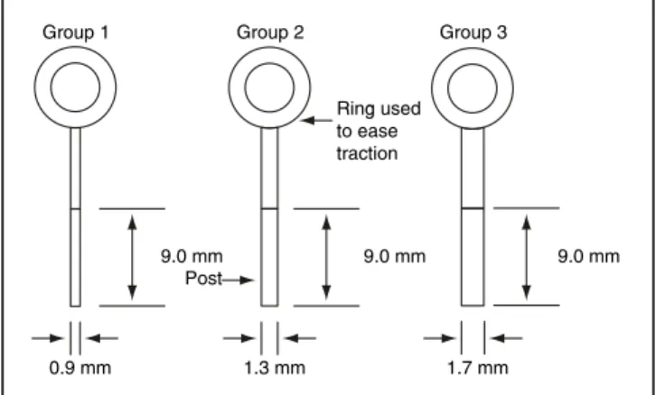
The samples were randomly divided into 3 groups (Figure 1) of 12 teeth and cylindrical post spaces with 9 mm in length were prepared with the following diameters: Group 1: 0.9 mm (prepared with #2 Largo bur - Dentsply Maillefer, Ballaigues, Switzerland); Group 2: 1.3 mm (prepared with a #4 Largo bur); Group 3: 1.7 mm (prepared with a #6
Largo bur). A new bur was used for each prepared tooth, totalizing 36 burs.
The root canals were prepared with a low-speed straight handpiece (Dabi Atlante, Ribeirão Preto, Brazil) attached to a parallelometer (Bio Art Ltda., São Carlos, Brazil), so that the root prepara-tions were parallel to the long axis of the roots not allowing the introduction of horizontal forces when applying traction to the post-core system.
After the post space preparation, the root ca-nals were molded using the chemically activated acrylic resin Duralay (Reliance Dental Mfg. Co.,
Worth, IL, USA). These impressions were then included in silicon rings (Polidental, São Paulo, Brazil) (with one sample from each group in each ring) with Termocast phosphate investment
(Poli-dental Ind. Com. Ltda., São Paulo, SP, Brazil) and cast in copper-aluminum alloy (Goldent-LA, São Paulo, SP, Brazil), according to the manufacturer’s instructions.
The root canals were cleaned with a detergent solution (tergensol, Inodon, Porto Alegre, RS, Bra-zil), dried with paper points (Dentsply, Petropolis, Brazil) and air jet, and cemented with zinc phos-phate (Vigodent, Rio de Janeiro, Brazil) by incre-ments, according to the manufacturer’s instruc-tions.
The cement was placed in the canal with the help of a reamer and spread on the core with a spatula. The core was properly placed in the canal and small spinning movements were made to as-sist the cement flow. The core was kept in place with digital pressure for 5 minutes and the excess of cement was removed with a dental probe.
After cementation, the samples were kept at 37ºC for 7 days.
All samples were subsequently placed in an Instron 4444 Universal Testing Machine (Instron Corporation, Canton, MA, USA), attached to a
0.9 mm 1.3 mm 1.7 mm
9.0 mm Post
Ring used to ease traction
9.0 mm
Group 1 Group 2 Group 3
9.0 mm
Souza Filho CB, Paulino SM, Alfredo E, Sousa Neto MD, Vansan LP. Effect of the diameter on Cu-Al post retention. Braz Oral Res 2004;18(3):238-41.
240 241
240 241
device developed to minimize lateral forces. The sample was maintained in vertical position in or-der to apply traction along the root axis. The posts were submitted to increasing traction (1 mm/min) until displacement from the root occurred. The maximum traction values, in kgf, were submitted to statistical analysis by ANOVA.
RESULTS
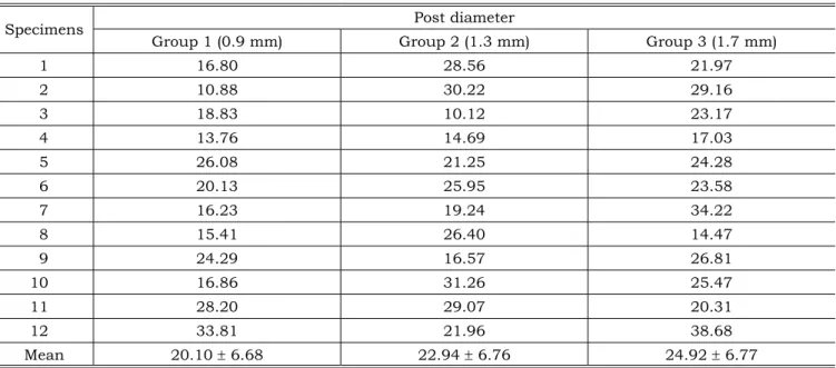
Using the test of resistance to removal by trac-tion of the posts, the means (± standard deviatrac-tions) of the experimental data of the 36 numeric val-ues of strength (kgf) were: Group 1: 20.10 ± 6.68; Group 2: 22.94 ± 6.76; Group 3: 24.92 ± 6.77 (Table 1).
Preliminary tests indicated normality and ho-moscedasticity. The ANOVA analysis was indicated and results can be seen in Table 2.
There were no statistically significant differ-ences among the 3 groups (ANOVA, p > 0.05).
DISCUSSION
Currently, endodontic therapy has shown a high success rate (95%) and when correctly re-stored, the function of endodontically treated teeth does not differ from that of teeth with no treat-ment7,16.
The intraradicular post and core system, used for endodontically treated teeth restoration, is composed of 2 parts: the post, responsible for root retention and resistance4,11,15,19, and the crown
extension, the core, responsible for crown reten-tion. The post had a length of 9 mm, corresponding to 2/3 of the length of most human teeth15 and
considering that 4-5 mm of the root canal sealing should remain to protect the apical seal.
Despite scientific and technologic advances, the intraradicular post does not meet all the needs of a tooth with no pulp2. Resistance of an
endo-dontically treated tooth is directly related to the quantity of remaining dental structure and is not reinforced by the post4,8,19. In fact, preparation for
post insertion may lead to the weakening of the root9,18. These results are clinically important
be-cause the wear of the dental structure, during endo-dontic treatments, occurs during the endoendo-dontic access and the chemical-mechanical preparation, especially with cervical preparation techniques. The loss of tooth structure because of caries and trauma leads to a more fragile endodontically treat-ed tooth6,17,22. Therefore, the clinician must abrade
the least amount of dentinal structure possible TABLE 1 - Tension values to remove posts (kgf).
Specimens Post diameter
Group 1 (0.9 mm) Group 2 (1.3 mm) Group 3 (1.7 mm)
1 16.80 28.56 21.97
2 10.88 30.22 29.16
3 18.83 10.12 23.17
4 13.76 14.69 17.03
5 26.08 21.25 24.28
6 20.13 25.95 23.58
7 16.23 19.24 34.22
8 15.41 26.40 14.47
9 24.29 16.57 26.81
10 16.86 31.26 25.47
11 28.20 29.07 20.31
12 33.81 21.96 38.68
Mean 20.10 ± 6.68 22.94 ± 6.76 24.92 ± 6.77
TABLE 2 - ANOVA results for tensile strength.
Sum of
squares DF squareMean (F) Sig Between
groups 140.9727 2 70.4863 1.55 22.57% Within
goups 1499.336 33 45.4344 Total 1640.309 35
240 241
240 241
Souza Filho CB, Paulino SM, Alfredo E, Sousa Neto MD, Vansan LP. Effect of the diameter on Cu-Al post retention. Braz Oral Res 2004;18(3):238-41.
or only remove gutta-percha from the root canal, respecting the 4-5 mm apical limit and take ad-vantage of the chemical-mechanical preparation already performed by the endodontist for place-ment of the intraradicular post; thus reducing the risk of fracture1,14.
The diameter of the core was evaluated be-cause the preparation of larger posts than nec-essary can result in a weaker dental structure. According to some authors5,13,16,21,22, the diameter
variation of posts to enhance resistance and re-tention leads to a higher risk of root perforation and fracture.
The chosen diameters (0.9 mm, 1.3 mm and 1.7 mm) were selected based on the mean diameter of human roots15. At 4 mm from the root apex, the
maximum diameter of maxillary central incisors is 1.7 mm and 0.7 mm for mandibular central inci-sors. For the other groups of teeth, the diameter ranges between these values. The post diameter
must not allow deviation and must never exceed 1/3 of the root diameter12,15.
The results of the present study show that the tensile strength of Cu-Al cylindrical posts did not depend on the diameter and there were no significant statistical differences between the three compared groups. Table 2 indicates the probability of 22.57% to the equality hypothesis.
Clinically, regardless of the type of intracanal post used, root canal abrasion must be minimal, though sufficient for post placement, taking into account that post diameter does not affect reten-tion and that the resistance of the dental element is directly related to the quantity of remaining dentin.
CONCLUSIONS
The variation in diameter (0.9 mm, 1.3 mm and 1.7 mm) of cylindrical posts cemented with zinc phosphate did not affect their tensile strength.
REFERENCES
1. Akkayan B, Gulmez T. Resistance to fracture of endodonti-cally treated teeth restored with different post systems. J Prosthet Dent 2002;87:431-7.
2. Assif D, Oren E, Marshak BL, Aviv I. Photoelastic analysis of stress transfer by endodontically treated teeth to the supporting structure using different restorative techniques. J Prosthet Dent 1989;61:535-43.
3. Berekally T. Contemporary perspectives on post-core sys-tems. Aust Endod J 2003;29:120-7.
4. Bonilla ED, Anderkvist T, Miller GD. A customized acrylic resin shell for fabricating an amalgam core on the coronally debilitated, endodontically treated posterior tooth. Quintes-sence Int 1995;26:317-24.
5. Cooney JP, Caputo AA, Trabert KC. Retention and stress distribution of tapered-end endodontic posts. J Prosthet Dent 1986;55:540-6.
6. Fernandes AS, Shetty S, Coutinho I. Factors determin-ing post selection: a literature review. J Prosthet Dent 2003;556-62.
7. Freeman MA, Nicholls JI, Kydd WL, Harrington GW. Leak-age associated with load fatigue-induced preliminary fail-ure of full crowns placed over three different post and core systems. J Endod 1998;24:26-32.
8. Gomes APM, Pagani C, Araújo MAM, Serikaku AL, Gui-marães MP. Avaliação da resistência à tração de pinos cerâmicos Cosmopost cimentados com dois diferentes ci-mentos resinosos. Rev Odontol UNESP 2002;31:127-39. 9. Gutmann JL. The dentin-root complex: anatomic and
bio-logic considerations in restoring endodontically treated teeth. J Prosthet Dent 1992;67:458-67.
10. Hansen EK, Asmussen E, Christiansen NC. In vivo
fractures of endodontically treated posterior teeth restored with amalgam. Endod Dent Traumatol 1990;6:49-55. 11. Nergiz I, Schmage P, Platzer U, Mcmullan-Vogel CG.
Effect of different surface textures on retentive strength of tapered posts. J Prosthet Dent 1997;78:451-7.
12. Perel ML, Muroff FI. Clinical criteria for posts and cores. J Prosthet Dent 1972;28:405-11.
13. Pettiette MT, Phillips C, Trope M. Effect of endodontic instrument taper on post retention. J Endod 2003;29:65-8.
14. Robbins JW. Guidelines for the restoration of endo-dontically treated teeth. J Am Dent Assoc 1990;120:558-66.
15. Shillingburg HT. Fundamentals of fixed prosthodon-tics. 3rd ed. Chicago: Quintessence Publishing Co. Inc.;
1998.
16. Sidoli GE, King PA, Setchell DJ. An in vitro evaluation of a carbon fiber-based post and core system. J Prosthet Dent 1997;78:5-9.
17. Sokol DJ. Effective use of current core and post con-cepts. J Prosthet Dent 1984;52:231-4.
18. Sorensen JA, Engelman MJ. Effect of post adaptation on fracture resistance of endodontically treated teeth. J Prosthet Dent 1990;64:419-24.
19. Sorensen JA, Martinoff JT. Clinically significant fac-tors in dowel design. J Prosthet Dent 1984;52:28-35. 20. Sorensen JA, Martinoff JT. Intracoronal reinforcement
and coronal coverage: a study of endodontically treated teeth. J Prosthet Dent 1984;51:780-4.
21. Standlee JP, Caputo AA, Hanson EC. Retention of endodontic dowels: effects of cement, dowel length, diam-eter, and design. J Prosthet Dent 1978;39:400-5.
22. Trabert KC, Cooney JP. The endodontically treated tooth. Restorative concepts and techniques. Dent Clin North Am 1984;28:923-51.