I
ABSTRACT
ASSOCIATION BETWEEN INTRA-RADICULAR POSTS
AND PERIAPICAL LESIONS IN ENDODONTICALLY
TREATED TEETH*
Cíntia Gonçalves Carvalho ROSALEM1, Claudia Machado de Almeida MATTOS2, Selva Maria Gonçalves GUERRA2
1- DDS, Professor, Course on Dental Prosthodontics, Osseointegrated Implant-Retained Prostheses and Esthetic, Brazilian Dental Association - ES, Vitória, ES, Brazil.
2- DDS, Professor, Department of Dental Prosthodontics, Health Sciences Center, Federal University of Espírito Santo, Vitória, ES, Brazil.
Corresponding address: Cíntia Gonçalves Carvalho Rosalem - Rua José Alexandre Buaiz, 190, sala 1209, Enseada do Suá, Vitória, ES, Brasil
Phone / Fax: 55 27 3314-4015 - e-mail: [email protected] / [email protected]
Received: December 22, 2006 -Modification: March 09, 2007 - Accepted: May 08, 2007
*Paper submitted to the Clinical Prosthesis Course as part of the requirements to obtain the certificate of Continued Education in Dental Prosthesis, Universidade Federal do Espírito Santo, Brazil.
ntroduction: A significant number of endodontically treated teeth restored with posts have associated periapical lesions, and several authors have discussed the probable causes of the development of these. Attention has been focused on restorative procedures performed after endodontic treatment and their association with the prognosis of endodontic therapy because a number of root-filled teeth will require post- and core-retained restorations. Purpose: The purpose of this study was to evaluate, by examination of periapical radiographs, whether the placement of intra-radicular posts in endodontically treated teeth may act as a risk factor for development of periapical lesions. Material and Methods: This case-control study analyzed periapical radiographs of 72 endodontically treated teeth with coronal restorations. All radiographs were obtained from a single private practice. Specimens were assigned to 2 groups: Group 1 (control) was composed of teeth without periapical lesions and Group 2 (case) was composed of teeth with periapical lesions. The number of teeth with and without posts in each group was recorded. Three calibrated examiners analyzed the radiographs visually under X4 magnification. Results: In Group 1, 28 (65.1%) out of 43 teeth were restored with posts. In Group 2, 24 (82.8%) out of 29 teeth had intra-radicular posts. The interpretation of chi-square test showed that these percentages were not significantly different (x2=2.687; p=0.101). Odds ratio was 2.571
(0.815-8.118), which indicates that there was no statistically significant association between periapical lesions and posts. Conclusion: Intra-radicular posts placed in endodontically treated teeth were not a significant risk factor for development of periapical lesions in the practice where the cohort of patients was treated.
Uniterms: Nonvital teeth; Dental posts; Periapical diseases.
INTRODUCTION
The goal of endodontic therapy is to clean, disinfect and obturate the root canals4. Root canal obturation is aimed
at preventing that bacteria from the oral cavity reach the periapical region and induce the formation of periapical lesions. However, the currently available root canal filling materials do not always yield a complete canal sealing and thus a well adapted coronal restoration is recommended to protect the canal and the apical region against oral bacterial recontamination30. Several types of restorations for
endodontically treated teeth have been described1,21,24,26.
These restorations should assure that the conditions achieved during root canal cleaning and shaping are
maintained.
The biological basis of endodontic therapy has been extensively studied. Attention has been focused on the restorative procedures conducted after endodontic treatment and their effects on case prognosis. Such procedures may fail and microorganisms and their byproducts may reach the periradicular region and the adjacent alveolar bone17.
Several studies have investigated the possible causes of the formation of periapical lesions associated with the roots of endodontically treated teeth that were restored with post-retained cores14,20,26. According to Zuolo, et al.31 (1996),
suggested that root canal preparation for the placement of a post may cause displacement of remaining filling material, when this material is insufficient or not correctly condensed. Other authors2,23 have reported that endodontically treated
teeth in which part of the root filling material has been removed have a poorer sealing ability than those where the root filling is intact. In a study that evaluated radiographically the periapical region of endodontically treated teeth, Boucher, et al.6 (2002) found that teeth with
radicular posts were significantly more associated with periapical lesions than teeth without posts. However, in a radiographic study that evaluated the association between the quality of coronal restorations, root filling and periapical conditions of endodontically treated teeth, Tronstad, et al.30
(2000) observed that the quality of endodontic treatment was significantly more important than the quality of coronal restorations when the periapical region was examined. In the same study, these authors observed that posts did not have any negative effects on the success of endodontic therapy in any situation.
Considering the large number of patients who present with endodontically treated teeth that require the placement of post- and core-retained restoration, the purpose of this study was to evaluate, by examination of periapical radiographs, whether the placement of intra-radicular posts in endodontically treated teeth may act as a risk factor for development of periapical lesions.
MATERIAL AND METHODS
Study Design
This case-control study analyzed a control group (teeth without periapical lesions) and a case group (teeth with periapical lesions), to determine whether the placement of intra-radicular posts could be considered a risk factor for development of periapical lesions.
Sample
In an attempt to ensure that the treatment procedures were standardized, the patient dental records were selected from the files of a single private practice. Data were gathered from the analysis of periapical radiographs obtained using the paralleling radiographic technique and processed manually in fresh chemicals between the years 1990 and 2001.
A total of 65 radiographs from 47 patients (72 teeth) were selected according to the information contained in the patient dental records with respect to the following inclusion criteria: all teeth should be endodontically treated and have coronal restorations; root canal fillings should be homogenous along the canal, extending up to at least 1 mm within the anatomic apex3; the intra-radicular posts should be metal cast posts.
From the 72 selected teeth, 43 were assigned to the control group (Group 1) and 29 were assigned to the case group (Group 2). Figures 1 and 2 summarize the characteristics of each group.
Radiographs were included in the study after patients granted their authorization and signed an informed consent form.
Examination of Radiographs
All analyses of the radiographs were conducted by visual examination with the aid of an X4 magnifying lens and a transparent flexible millimeter ruler.
The radiographs were examined by 3 examiners according to the following sequence and preestablished criteria: Examiner 1 - The first examiner was calibrated by a radiologist. Kappa statistics was used to check agreement between the calibrator and the examiner. After calibration, this examiner selected the radiographs for the study sample according to the inclusion criteria; Examiner 2 - The second examiner was calibrated by the same radiologist, and kappa statistics was again used to check agreement between the calibrator and the examiner. After calibration, this examiner assigned the
FIGURE 1- Group 1 (control) and number of teeth with and without posts
selected radiographs to either the case group (teeth with periapical lesions) or the control group (teeth without periapical lesions). Examiner 3 - The third examiner was calibrated by the same radiologist, and kappa statistics was again used to check agreement between the calibrator and the examiner. After calibration, this examiner rated the radiographs according to the presence or absence of posts. This examiner was blinded to the classification conducted by Examiner 2, and therefore did not know which radiographs belonged to the control or case group. Examiner 1 collected all data obtained after the analyses of Examiners 2 and 3.
The radiographs were examined on the basis of 5 criteria: 1. Length of Apical Endodontic Obturation: the length of apical endodontic obturation was assessed by measuring the amount of remaining filling material in the apical region of the root canal. This measurement was obtained by Examiner 1; 2. Quality of Endodontic Treatment: quality was assessed by Examiner 1. Endodontic treatment was considered adequate when the canal filling was dense and homogeneous and extended to at least within 1 mm of the anatomic apex3,30, 3. Marginal Adaptation of Coronal
Restorations: the marginal adaptation of the coronal
restorations was considered adequate when there was no evidence of any radiolucent images between the restoration margin and the remaining crown7,25. This visual evaluation
was conducted by Examiner 1 using an X4 magnifying lens; 4. Presence or Absence of Periapical Lesion: periapical lesion was defined as any radiolucent image larger than 1
mm3,30. Examiner 2 measured the radiolucent area in millimeters
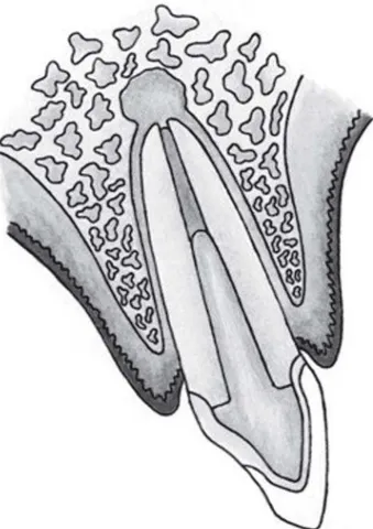
and obtained the greatest diameter of the lesion from the root apex to its opposite border (Figure 3); 5. Presence or
Absence of Intra-radicular Posts: the presence of a post
was detected by comparing the patient’s clinical records to radiographic findings. Posts were identified by an area of greater radiopacity in the middle and cervical thirds of the root canal compatible with the image of the post described in the file. This analysis was conducted by Examiner 3.
Statistical Analysis
Frequency tables were used for the descriptive analysis of data, and results are reported dichotomously. The chi-square test was used to check statistical differences in quantitative parameters between groups. Odds ratio was used to evaluate the association between the risk factor (placement of post) and the occurrence of lesion. Calculations were performed using SPSS statistical software (Statistical Package for Social Sciences for Windows 8.0; SPSS Inc, Chicago, IL, USA). Significance level was established at 5%.
RESULTS
From the 72 teeth evaluated, 52 (72.2%) had intra-radicular posts.
As shown in Table 1, from the 43 teeth in the Group 1 (teeth without periapical lesion), 28 (65.1%) had post-retained restorations. From the 29 teeth in Group 2 (teeth with periapical lesion), 24 (82.8%) had post-retained restorations against 5 (17.2%) without posts.
Chi-square test results showed that these percentages were not significantly different (x2=2.687; p=0.101). Odds
ratio was 2.571 (0.815-8.118), which indicates that there was no statistically significant associations between periapical lesions and intra-radicular posts.
DISCUSSION
In order to grant more reliability to the collected data, this retrospective case-control study analyzed periapical radiographs retrieved from the files of a single private practice, and evaluated teeth whose posts were all placed by the same specialist.
Because this study was based exclusively on radiographic data, only two-dimensional success could be assessed. Nevertheless, the sample was selected according
Group 1 Group 2
(n=43) (n=29)
With post Without post With post Without post 28 (65.1%) 15 (34.9%) 24 (82.8%) 5 (17.2%)
TABLE 1- Results of the control and case groups
to literature-based criteria3,5,7,8,10,16,22,25,30. The type of material
was standardized to ensure that it would not have any influence on the results of the study. No radiolucent images were found between the restoration margin and the tooth7
because any failure in the coronal restorations might result in leakage of oral fluids and affect the health of periapical tissues17,30.
In vitro studies with similar goals have demonstrated
that the literature is not conclusive regarding the role of intra-radicular posts in the etiology of periapical lesions. Dalat and Spangberg11 (1993),evaluating the presence of
dye microleakage between endodontically treated teeth with post preparations and teeth with intact obturation, found no statistically significant differences between the studied groups. However, Metzger, et al.23 (2000) evaluated the seal
provided by root canal fillings after post space preparation and observed that post prepared endodontically treated teeth had a significantly lower apical sealing ability than teeth with an intact root filling.
Even thought several studies have shown that 3-5 mm of remaining filling material is necessary to ensure an effective apical seal in endodontically treated teeth5,8,10,22,
Abramovitz, et al.2 (2001), in a study evaluating the
effectiviness of apical seals after post preparation in endodontically treated teeth, found that teeth with 3 to 6 mm of endodontic filling material remants had a lower sealing ability than teeth with an intact root filling. However, their study was limited by the fact that the teeth were not previously evaluated to check the quality of endodontic treatments. Therefore, establishing the real cause of apical seal failure is doubtful. Sunay, et al.29 (2007), in a study
evaluating the periapical status in a selected population of urban Turkish adults, observed that 91% of root-filled teeth with periapical pathosis presented inadequate root fillings. Tronstad, et al.30 (2000) conducted an in vivo study to
investigate the association between quality of coronal restoration, root canal filling and periapical health of endodontically treated teeth. The criteria used to evaluate the quality of endodontic treatment and adjacent structures were similar to those used in the present study. In agreement with our results, these authours reported that the presence of posts in root-filled teeth were not a risk factor for development of apical periodontitis. Hommez, et al.18 (2002),
in their report about the impact of the quality of coronal restorations and root fillings on the periapical health, also shared the same conclusions.
It may be pointed out that posts did not represent a statistically significant risk in the present study because the sample was relatively small (n=72). However, Tronstad, et al.30 (2000) evaluated a considerably more comprehensive
tooth sample (n=1001) selected according to strict criteria and reached similar conclusions.
In spite of all discussion in the current literature, coronal leakage has been pointed out as an important cause of endodontic treatment failure9,13,27. Additionally, the exposure
of root canal fillings to saliva within a relatively short period of time (30 days or longer) may be considered as indicative for endodontic retreatment28.
Therefore, it is important to highlight that the restoration of endodontically treated teeth, with or without intra-radicular posts, should be carefully performed to avoid coronal leakage in temporary or permanent restorations at any stage of the restorative treatment12. We agree with Jamani, et al.19
(2005), who stated that dentists should be better prepared to perform both endodontic treatments and restorations of endodontically treated teeth.
CONCLUSION
Intra-radicular posts placed in endodontically treated teeth were not a significant risk factor for development of periapical lesions in the practice where the cohort of patients was treated.
REFERENCES
1- Abou-Rass M, Donovan TE. The restoration of endodontically treated teeth. J Calif Dent Assoc. 1993;21(12):61-7.
2- Abramovitz L, Lev R, Fuss Z, Metzger Z. The unpredictability of seal after post space preparation: a fluid transport study. J Endod. 2001;27(4):292-5.
3- American Association of Endodontists. Quality assurance guidelines. Chicago: The Association; 1987.
4- Antoniazzi JH, Polo I, Marques JLL, Cardoso RJA. Selamento marginal simples e duplo em endodontia. Rev Assoc Paul Cir Dent. 1996;50(5):435-9.
5- Assif D, Aviv I, Himmel R. A rapid dowel core construction technique. J Prosthet Dent. 1989;61(1):16-7.
6- Boucher Y, Matossian L, Rilliard F, Machtou P. Radiographic evaluation of the prevalence and technical quality of root canal treatment in a French subpopulation. Int Endod J. 2002;35(3):229-38.
7- California Dental Association. Quality evaluation for dental care guidelines for the assessment of clinical quality and professional performance. Sacramento: The Association; 1995.
8- Caputo AA, Standlee J P. Pins and posts - why, where and how. Dent Clin North Am. 1976;20(2):299-311.
9- Cheung GS. Endodontic failures - changing the approach. Int Endod J. 1996;46(3):131-8.
10- Cohen S, Burns RC. Caminhos da polpa. 7. ed. Rio de Janeiro: Guanabara Koogan; 2000.
11- Dalat DM, Spangberg LS. Effect of post preparation on the apical seal of teeth obturated with plastic thermafil obturators. Oral Surg Oral Med Oral Pathol. 1993;76(6):760-5.
12- Fox K, Gutteridge DL. An in vitro study of coronal microleakage in root-canal-treated teeth restored by the post and core technique. Int Endod J. 1997;30(6):361-8.
14- Gish SP, Drake DR, Walton RE, Wilcox L. Coronal leakage: bacterial penetration through obturated canals following post preparation. J Am Dent Assoc. 1994;125(10):1369-72.
15- Grieve AR, Radford JR .Radiographic observations of posts crowns: some problems and solutions. Dent Update. 1995;22(9):370-2.
16- Gutmann JL. Clinical, radiographic, and histologic perspectives on success and failure in endodontics. Dent Clin North Am. 1992;36(2):372-92.
17- Heling I, Gorfil C. Endodontic failure caused by inadequate restorative procedures: review and treatment recommendations. J Prosthet Dent. 2002;87(6):674-8.
18- Hommez GM, Coppens CR, de Moor RJ. Periapical health related to the quality of coronal restorations and root fillings. Int Endod J. 2002;35(8):680-9.
19- Jamani KD, Agrabawi J, Fayyad MA. A radiographic study of the relationship between technical quality of coronoradicular posts and periapical status in a Jordanian population. J Oral Sci. 2005;47(3):123-8.
20- Karapanou V, Cabrera P, Vera J, White R, Goldman M. Effect of immediate and delayed post preparation on apical dye leakage using two different sealers. J Endod. 1996;22(11):583-5.
21- Manning KE, Yu DC, Yu HC, Kwan EW. Factors to consider for predictable post and core build-ups of endodontically treated teeth, Part I: basic theoretical concepts. Restorative Dent. 1995;61(8):685-8,690,693-5.
22- Mattison GD, Delivanis PD, Thacker RW, Hassell KJ. Effect of post preparation on the apical seal. J Prosthet Dent. 1984;51(6):785-9.
23- Metzger Z, Abramovitz R, Abramovitz L, Tagger M. Correlation between remaining length of root canal fillings after immediate post space preparation and coronal leakage. J Endod. 2000;26(12):724-8.
24- Morgano SM, Brackett SE. Foundation restorations in fixed prosthodontics: current knowledge and future needs. J Prosthet Dent. 1999;82 (6):643-57.
25- Ray HA, Trope M. Periapical status of endodontically treated teeth in relation to the technical quality of the root canal filling and the coronal restoration. Int Endod J. 1995;28(1):12-8.
26- Robbins JW. Restoration of the endodontically treated tooth. Dent Clin North Am. 2002;46(2):367-84.
27- Saunders WP, Saunders EM. Coronal leakage as a cause of failure in root canal therapy: a review. Endod Dent Traumatol. 1994;10(3):105-8.
28- Siqueira JF. Aetiology of root canal treatment failure: why well-treated teeth can fail. Int Endod J. 2001;34(1):1-10.
29- Sunay H, Tanalp J, Dikbas I, Bayirli G. Cross-sectional evaluation of the periapical status and quality of root canal treatment in a selected population of urban Turkish adults. Int Endod J. 2007;40:139-45.
30- Tronstad L, Asbjornsen K, Doving L, PedersenI, Heriksen H. Influence of coronal restorations on the periapical health of endodontically treated teeth. Endod Dent Traumatol. 2000;16:218-21.