CASE REPORT
OSTEOID OSTEOMA IN THE ILIAC BONE: REPORT ON TWO CASES
Elmano de Araújo Loures1, Bruno Fajardo do Nascimento2, Marcelo de Carvalho Amorim2, Clarice Naya Loures3
1 – Specialist in Orthopedics and Traumatology; Supervisor of the Medical Residence Program in Orthopedics and Traumatology, Department of Orthopedics and Traumatology, University Hospital, Federal University of Juiz de Fora, Juiz de Fora, MG, Brazil.
2 – Resident in the Department of Orthopedics and Traumatology, University Hospital, Federal University of Juiz de Fora, Juiz de Fora, MG, Brazil.
3 – Undergraduate Medical Student, Department of Morphology, Institute of Biological Sciences and School of Medicine, Federal University of Juiz de Fora, Juiz de Fora, MG, Brazil.
Work performed at the University Hospital, Federal University of Juiz de Fora, and the Orthopedics Center, Rio Branco Medical Center, Juiz de Fora, MG.
Correspondence: Av. Olegário Maciel 297/1101, 36015-350 Juiz de Fora, MG. E-mail: [email protected]
Work received for publication: May 24, 2011; accepted for publication: June 3, 2011. INTRODUCTION
Osteoid osteoma was first described by Jaffe in 1935 and is a benign lesion(1) that preferentially affects
adolescents and young adults, in the approximate
ma-le-to-female proportions of 2:1(2,3). The lesions are
generally small, not exceeding one centimeter in dia-meter, and present a well delimited rim and usually a peripheral zone of reactive bone neoformation. They predominantly affect the appendicular skeleton,
espe-cially the femur and tibia(4). Their presence has been
reported in practically all the bones of the skeleton,
including the cranium, face and spine(5). They only
rarely occur in the pelvis: just 1 to 3% of all cases are located in this region, and the majority of pelvic
lesions are found in the acetabulum(6).
Macroscopically, they are characterized by a fria-ble vascular niche (nidus) with sandy consistency, surrounded by sclerotic bone in association with thick vascular periosteum. Microscopically, the nidus con-sists of osteoid tissue, with vascular stroma
surroun-ded by dense bone(7).
The diagnosis can be made by means of simple
radiography in 75% of the cases(8). The typical image
comprises a nidus that appears in the form of a small oval or rounded focus that is generally radiotranspa-rent. This is surrounded by a zone of variable density corresponding to reactive bone sclerosis and, on some occasions, this zone may be very intense and cause difficulty in viewing the nidus. In such cases, bone scintigraphy, computed tomography or magnetic re-sonance can be used to determine the exact location of the nidus(9,10).
The typical clinical evolution is characterized by pain, predominantly during the night, probably because of increased synthesis of prostaglandins triggered by the tumor. In most cases, aspirin and other non-steroidal anti-inflammatory drugs (NSAIDs) that act by blocking prostaglandin synthesis provide transitory but significant pain relief. It is rare for the
lesions to be painless(11). Regarding their location
within the bone, subperiosteal, intracortical, endosteal and intramedullary lesions can be identified, among which intramedullary lesion are the least frequent(12).
Rev Bras Ortop. 2012;47(2):260-62
The authors declare that there was no conflict of interest in conducting this work
This article is available online in Portuguese and English at the websites: www.rbo.org.br and www.scielo.br/rbort
ABSTRACT
Osteoid osteoma is a benign bone tumor that generally pre-sents with nighttime pain among young adults and is relieved by rest and salicylates. It can affect any bone, but occurrences in the iliac are unusual. The authors describe two cases of in-tramedullary osteoid osteoma next to the sacroiliac joint, with symptoms that simulated sciatic pain. The cases were diag-nosed late, although the initial radiographs showed sclerotic lesions in both cases. The diagnosis was confirmed by means
of CT scan and the nidus was excised surgically through en bloc resection. The definitive diagnosis was given by means of histopathological examination. Over long-term follow-up, both cases remained asymptomatic and complete bone remo-deling at the surgical site was observed. The authors highlight the typical characteristics of the tumor, the unusual location, the differential diagnosis and the treatment.
261
The present study had the aim of presenting a re-port on two cases of intramedullary osteoid osteoma with unusual locations and clinical presentations. Re-ference is also made to the importance of the diffe-rential diagnosis (in these cases, with lumbalgia and pain of sciatic origin), in comparison with the data in the literature.
CASE REPORTS
Case 1
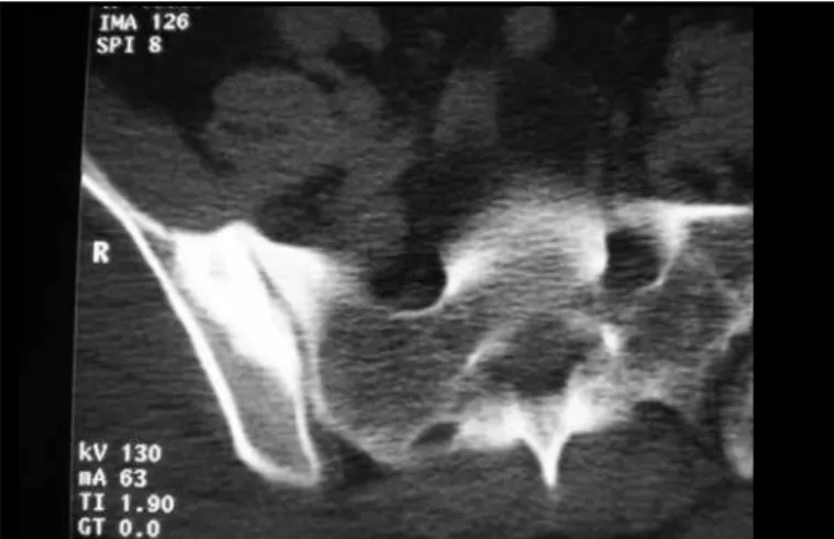
The patient was a 38-year-old male carpenter. His pain started in the lumbar and sacroiliac region, with intermittent irradiation to the right buttock, i.e. a pic-ture similar to pain of sciatic origin. He underwent clinical treatment with physiotherapy and analgesics for one year, with any satisfactory result. Imaging examinations on the lumbar spine showed normal re-sults. There was a partial improvement through using NSAIDs, but the pain worsened through exertion re-lating to his profession. He described nocturnal pain and insomnia. Simple radiography on the pelvis in AP view showed a lesion with central radiopacity in the iliac wing, with an intense sclerotic halo that exten-ded as far as the sacroiliac joint. The diagnosis was confirmed by means of computed tomography. HLA B-27 investigation and other laboratory tests produced normal results (Figures 1 and 2). The treatment imple-mented was en-bloc resection of the lesion, in February 1998. The nidus was located intraoperatively by means of radiographs. There was complete remission of the symptoms, and follow-up examinations after 10 years of evolution showed that complete bone remodeling had taken place at the site of the operation, and the individual remained free from symptoms.
Figure 1 – Case 1: computed tomography.
Figure 2 – Case 1: radiograph of the surgical specimen from the right iliac bone. Note the resected nidus and the bone sclerosis.
Case 2
The patient was a 46-year-old female housewife. She had had constant pain in the lumbar and right sacroiliac regions for eight months, with poorly defined irradiation to the buttock and posterior face of the right thigh, thus simulating pain of sciatic origin. Clinical treatment during this period, with physiotherapy sessions and analgesics, was fruitless. Imaging examinations in the lumbar region showed a mild degree of spondylodiscoarthrosis. Simple radiography on the pelvis showed a sclerotic lesion in the iliac that impinged in the margin of the sacroiliac joint, without a clear nidus. The diagnosis was confirmed by means of computed tomography (Figure 3). Laboratory tests showed normal results, including in relation to HLA B-27. The treatment proposed was en-bloc resection of the lesion, which was performed in May 2000. The nidus was located intraoperatively by means of radiographs. These was complete remission of the symptoms, and follow-up examinations after 10 years of evolution showed that complete bone remodeling had taken place at the site of the operation, and the individual remained free from symptoms.
OSTEOID OSTEOMA IN THE ILIAC BONE: REPORT ON TWO CASES
262
DISCUSSION
In the cases reported here, the patients consulted several specialists until a confirmed diagnosis was achieved, proven through histopathological analysis. The diagnostic hypothesis of osteoid osteoma as the cause of insidious nocturnal bone pain that is of long duration and is alleviated by salicylates and NSAIDs should be considered in the differential diagnosis of
any painful state involving the skeleton(13). The lesion
can often only be identified by means of conventional radiographic examination, but computed tomography and magnetic resonance imaging are necessary to elu-cidate and/or confirm the diagnosis, and they are useful
for surgical planning(10). In a bibliographic review in
Medline and Latin American Lilacs, only 14 reports on cases of osteoid osteoma located in the iliac bone between 1945 and 2010 were found.
Spontaneous cure has been well documented in some studies. The pain and radiological findings were observed to gradually disappear, although some
de-gree of bone sclerosis remained(14). The natural history
of this disease is unpredictable and prolonged, and surgical intervention is indicated(15).
Radiofrequency ablation guided by computed tomo-graphy is an alternative that has been used with very promising results, especially in regions that are difficult to access through conventional surgery(16,17).
The literature indicates that the preferred treatment
is en-bloc surgical resection, including the nidus(18).
Consequently, determining the exact location of the nidus during the operation is crucial for the success of the intervention, which was done in the present cases using conventional radiological markers.
CONCLUSION
The possibility that osteoid osteoma can occur in any bone at any location highlights the overwhelming need for careful assessment of imaging examinations and recognition of the importance of the clinical condi-tion. Involvement of the iliac bone is very uncommon, and osteoid osteoma in unusual locations generally leads to late diagnosis, with the possibility of confoun-ding this with other pathological conditions. In the cases reported here, en-bloc surgical resection of the lesion was shown to be an effective and safe method, as reported in the literature.
Rev Bras Ortop. 2012;47(2):260-62 REFERENCES
1. Broadfoot J, Chapman G. The use of computed tomography in diagnosis of osteoid osteoma. Australas Radiol. 1988;32(3):463-7.
2. Schaefer MP, Smith J. The diagnostic and therapeutic challenge of femoral head osteoid osteoma presenting as thigh pain: a case report. Arch Phys Med Rehabil. 2003;84(6):904-5.
3. Sinha S, Housden P. Discrete synchronous multifocal osteoid osteoma of the femur: a case report and review of literature. Pediatr Radiol. 2004;34(3):280.
4. Radcliffe SN, Walsh HJ, Carty H. Osteoid osteoma: the difficult diagnosis. Eur J Radiol. 1998;28(1):67-79.
5. Iffenecker C, Rocher P, Rabia MH, Dhina Z, Bobin S, Quillard J, et al. Osteoid osteoma of the petrous bone. Neuroradiology. 1997;39(11):821-3.
6. Ishikawa Y, Okada K, Miyakoshi N, Takahashi S, Shimada Y, Itoi E, et al. Osteoid osteoma of the scapula associated with synovitis of the shoulder. J Shoulder Elbow Surg. 2005;14(3):329-32.
7. Akhlaghpoor S, Aziz Ahari A, Arjmand Shabestari A, Alinaghizadeh MR. Radio-frequency ablation of osteoid osteoma in atypical locations: a case series. Clin Orthop Relat Res. 2010;468(7):1963-70.
8. Swee RG, McLeod RA, Beabout JW. Osteoid osteoma. Detection, diagnosis, and localization. Radiology. 1979;130(1):117-23.
9. Pratali R, Zuiani G, Inada M, Hanasilo C, Reganin L, Etchebehere E, et al. Open resection of osteoid osteoma guided by a gamma-probe. Int Orthop. 2009;33(1):219-23.
10. Barros Filho TEP, Oliveira RP, Cristante AF, Barbarini AF. Tratamento de osteoma osteoide de corpo vertebral da coluna lombar por ablação por radiofrequência. Acta Ortop Bras. 2006;14(2):103-5.
11. Lee EH, Shafi M, Hui JH. Osteoid osteoma: a current review. J Pediatr Orthop. 2006;26(5):695-700.
12. Kayser F, Resnick D, Haghighi P, Pereira Edo R, Greenway G, Schweitzer M, et al. Evidence of the subperiosteal origin of osteoid osteomas in tubular bones: analysis by CT and MR imaging. AJR Am J Roentgenol. 1998;170(3):609-14.
13. Hartmann T, Preis C, Gabriel A, Rath T, Ilias W. An osteoid osteoma as an un-diagnosed cause of three years of severe pain. Anesth Analg. 1997;85(6):1344-5. 14. Kneisl JS, Simon MA. Medical management compared with operative treatment
for osteoid-osteoma. J Bone Joint Surg Am. 1992;74(2):179-85.
15. Baptista PPR, Próspero JD, Volpe Neto F, Polesello GC, Barros MGA, Sanmar-tin Fernandez M, et al. Osteoma osteoide: planejamento diagnóstico e técnica cirúrgica. Rev Bras Ortop. 1996;31(11):883-7.
16. Cioni R, Armillotta N, Bargellini I, Zampa V, Cappelli C, Vagli P, et al. CT-gui-ded radiofrequency ablation of osteoid osteoma: long-term results. Eur Radiol. 2004;14(7):1203-8.
17. Sung KS, Seo JG, Shim JS, Lee YS. Computed-tomography-guided percutaneous radiofrequency thermoablation for the treatment of osteoid osteoma-2 to 5 years follow-up.Int Orthop. 2009;33(1):215-8.