Noninvasive positive pressure ventilation after
extubation: features and outcomes in clinical
practice
Liria Yuri Yamauchi1, Maise Figueiroa2, Leda Tomiko Yamada da Silveira2, Teresa Cristina Francischetto Travaglia2, Sidnei Bernardes2, Carolina Fu2
1. Department of Human Movement Sciences, Universidade Federal de São Paulo - Santos (SP), Brazil.
2. Department of Physiotherapy, Communication Sciences & Disorders, and Occupational Therapy, Faculdade de Medicina, Universidade de São Paulo - São Paulo (SP), Brazil.
Objective: To describe post-extubation noninvasive positive pressure ventilation use in intensive care unit clinical practice and to identify factors associated with noninvasive positive pressure ventilation failure.
Methods: his prospective cohort study included patients aged ≥ 18 years consecutively admitted to the intensive care unit who required noninvasive positive pressure ventilation within 48 hours of extubation. he primary outcome was noninvasive positive pressure ventilation failure.
Results: We included 174 patients in the study. he overall noninvasive positive pressure ventilation use rate was 15%. Among the patients who used noninvasive positive pressure ventilation, 44% used it after extubation. he failure rate of noninvasive positive pressure ventilation was 34%. he overall mean ± SD age was 56 ± 18 years, and 55% of participants were male. Demographics; baseline pH, PaCO2 and HCO3; and type of equipment used were similar between groups. All of the noninvasive positive pressure ventilation inal parameters were higher in the noninvasive positive pressure ventilation failure group [inspiratory positive airway pressure: 15.0 versus 13.7cmH2O (p = 0.015), expiratory positive airway pressure: 10.0
Conflicts of interest: None.
Submitted on April 15, 2015 Accepted on August 30, 2015
Corresponding author: Carolina Fu
Rua Cipotânea, 51 - Cidade Universitária Zip code: 05360-000 - São Paulo (SP), Brazil E-mail: [email protected]
Responsible editor: Carmen Valente Barbas
Ventilação não invasiva com pressão positiva pós-extubação:
características e desfechos na prática clínica
ABSTRACT
Keywords: Respiration, artiicial; Positive-pressure respiration/methods; Airway extubation; Ventilator weaning; Treatment outcomes; Intensive care units versus 8.9cmH2O (p = 0.027), and FiO2: 41 versus 33% (p = 0.014)]. he mean intensive care unit length of stay was longer (24 versus 13 days), p < 0.001, and the intensive care unit mortality rate was higher (55 versus 10%), p < 0.001 in the noninvasive positive pressure ventilation failure group. After itting, the logistic regression model allowed us to state that patients with inspiratory positive airway pressure ≥ 13.5cmH2O on the last day of noninvasive positive pressure ventilation support are three times more likely to experience noninvasive positive pressure ventilation failure compared with individuals with inspiratory positive airway pressure < 13.5 (OR = 3.02, 95%CI = 1.01 - 10.52, p value = 0.040).
Conclusions: he noninvasive positive pressure ventilation failure group had a longer intensive care unit length of stay and a higher mortality rate. Logistic regression analysis identiied that patients with inspiratory positive airway pressure ≥ 13.5cmH2O on the last day of noninvasive positive pressure ventilation support are three times more likely to experience noninvasive positive pressure ventilation failure.
INTRODUCTION
Noninvasive positive pressure ventilation (NIPPV) has been widely used in intensive care units (ICU). Despite conlicting scientiic evidence regarding many indications for its use, NIPPV has become a part of routine care in the majority of ICU worldwide.(1-4) According to the
literature, some indications are considered acceptable, but others are still under investigation, such as the use of NIPPV after extubation.
his approach has some diferent nuances, mainly based on timing. Some studies have incorporated NIPPV into the weaning from invasive mechanical ventilation, meaning that NIPPV is applied immediately after extubation as part of a continuous process.(5-8) In these cases, NIPPV can
be applied immediately as a preventive,(9,10) after failure
of a spontaneous breathing trial(11) or after extubation of
high-risk patients.(12)
In other hand, the use of NIPPV after the development of acute respiratory failure (ARF) after extubation has presented conlicting results. While some studies have found that NIPPV may prevent reintubation,(13) others have
shown that it does not seem to diminish the reintubation rate and may even increase the mortality rate.(14)
From a clinical point of view, NIPPV is indispensable in the ICU, and information about its use in practice may raise some important issues not identiied in randomized clinical trials. he present study was undertaken to describe post-extubation NIPPV use in ICU clinical practice and to identify factors associated with NIPPV failure after extubation.
METHODS
Between May and December 2007, a prospective cohort study was conducted at Hospital das Clínicas of
the Faculdade de Medicina of the Universidade de São
Paulo, located in the city of São Paulo, Brazil. he study
was carried out in eleven ICU (140 beds). his study was approved by the hospital Ethical Committee (number 0327/07), and the requirement for informed consent was waived because data were collected from patients’ records, and no intervention was performed.
All adult patients (age ≥ 18 years) consecutively admitted to the ICU who used NIPPV within 48 hours of extubation were included. Patients were excluded if there was any relevant information missing from the charts.
Data were collected from medical charts and directly from the ICU staf. All decisions about NIPPV use were exclusively made by the ICU team; researchers did not intervene in any way. Patients were analyzed as success NIPPV group and failure NIPPV group. he following data were collected: demographics [age, gender and Simpliied Acute Physiology Score (SAPS II) at ICU admission]; day and time of intubation; reason for invasive mechanical ventilation [chronic obstructive pulmonary disease (COPD), asthma, decreased level of consciousness, neuromuscular disease, ARF, cardiac arrest, hemodynamic instability or surgery]; day and time of extubation; and day and time of the start of NIPPV.
Data related to NIPPV collected in the study included the indication for NIPPV [acute respiratory failure after extubation (signs of respiratory distress up to 48 hours after extubation), early weaning (NIPPV immediately after extubation in patients considered at high risk for reintubation, such as COPD patients), and preventive NIPPV (in cases without ARF but with relevant comorbidities)]; the period of NIPPV use; type of equipment used (BIPAP Vision - Respironics®
, BIPAP ST/d Respironics®
, Downs low generator - Vital Signs®
, or double function mechanical ventilator); time from extubation until NIPPV initiation (0 or ≥ 1 day); NIPPV parameters; type of NIPPV interface; arterial blood gas test prior to NIPPV use; mask leakage; intolerance to NIPPV; need for airway suctioning; NIPPV complications; reintubation rate; reasons for reintubation; NIPPV failure rate (deined as reintubation after NIPPV use); ICU mortality rate; and ICU length of stay.
Statistical analysis
A descriptive analysis was carried out. Quantitative variables were presented as the mean and standard deviation (SD) or the median and interquartile range (IQR). Categorical variables were presented as proportions. he predictive capacity of quantitative variables for NIPPV failure was assessed with receiver-operating characteristic (ROC) curves; the area under the curve (AUC) and optimal cutof values (based on best values of sensitivity and speciicity) were calculated.
clinical relevance and were dichotomized based on cutof values calculated by ROC curves. After that, all independent variables were submitted to univariate analysis. Odds ratio and Fisher’s exact test were applied to identify possible associations among independent variables and NIPPV failure. he odds ratio of each independent variable was calculated based on 2 x 2 tables to deine which variables would comprise the initial model of logistic regression. he variables with p-values above 0.30 were not included in the initial model. Multi-collinearity was evaluated by variance inlation factors. he Hosmer-Lemeshow test was applied to verify the goodness of it model.
RESULTS
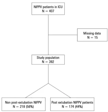
During the study period, 2,773 patients were admitted to the ICU. NIPPV was used on 407 (15%) of them. After excluding 15 patients due to missing data, the study population was 392 patients. hose who used NIPPV only after extubation accounted for 44%, or 174 patients (Figure 1). Baseline characteristics of the study population are presented in table 1.
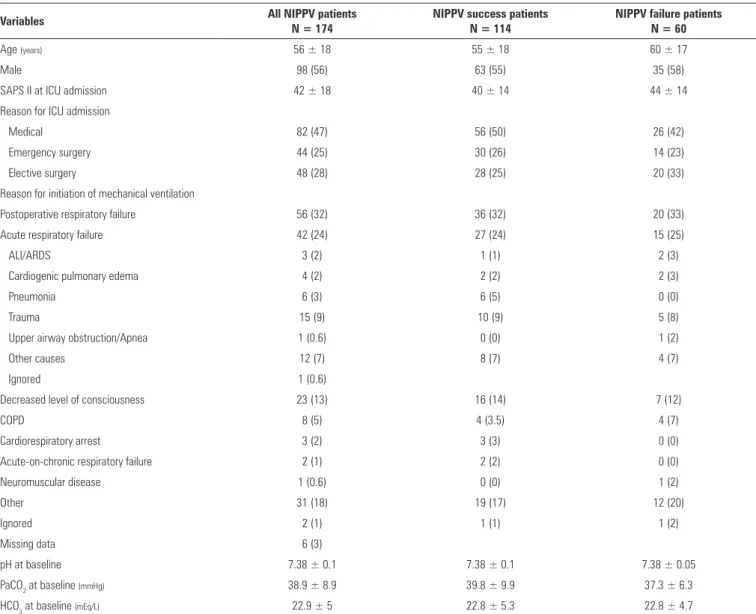
he main reasons for the use of mechanical ventilation prior to the use of NIPPV were hemodynamic instability (33%), acute respiratory failure (24%) and surgery (18%).
he median (IQR) time of use of invasive mechanical ventilation was 4 (1 - 8) days. Noninvasive pressure ventilation features are presented in table 2. BIPAP Vision®
and continuous positive airway pressure low generators were the most commonly used equipment. he main interface was the orofacial mask.
NIPPV after extubation was applied in three situations: a new acute respiratory event [46 cases (26%)], early weaning [17 cases (10%)], and preventive NIPPV application [111 cases (64%)]. he time from extubation to initiation of NIPPV was recorded in days. A total of 121 patients (69%) received NIPPV support on the same day as extubation, and 53 (31%) received NIPPV between one and two days later.
During NIPPV support, the equipment was changed in some cases. At the beginning of NIPPV, the most commonly used device was a continuous positive airway pressure low generator (45%) followed by BIPAP Vision®
(32%). However, on the last day of NIPPV, BIPAP Vision®
was more frequently used (42%).
All of the noninvasive positive pressure ventilation inal parameters were higher in the noninvasive positive pressure ventilation failure group [inspiratory positive airway pressure: 15.0 versus 13.7cmH2O (p = 0.015), expiratory positive airway pressure: 10.0 versus 8.9cmH2O (p = 0.027), and FiO2: 41 versus 33% (p = 0.014)]. he mean intensive care unit length of stay was longer (24 versus 13 days), p < 0.001, and the intensive care unit mortality rate was higher (55 versus 10%), p < 0.001 in the noninvasive positive pressure ventilation failure group.
During the period of NIPPV use, 18% of patients presented with intolerance or excessive low leakage, and treatment impairment occurred in 4%. NIPPV-related complications occurred in seven patients (ive with vomiting, one with abdominal distention and one with skin lesions). he nosocomial pneumonia rate was 6%.
he NIPPV failure rate was 34%. he median time between extubation and reintubation was 2 (1 - 4) days. he main reasons for reintubation (NIPPV failure) were acute respiratory failure (48%) and decreased level of consciousness (22%). NIPPV failure did not difer according to indication: 32% were in the ARF after extubation group, 29% in the early NIPPV group and 35% in the preventive NIPPV group.
Table 1 - Baseline characteristics in patients treated with noninvasive positive pressure ventilation in the intensive care unit according to noninvasive positive pressure ventilation outcome
Variables All NIPPV patients
N = 174
NIPPV success patients N = 114
NIPPV failure patients N = 60
Age (years) 56 ± 18 55 ± 18 60 ± 17
Male 98 (56) 63 (55) 35 (58)
SAPS II at ICU admission 42 ± 18 40 ± 14 44 ± 14
Reason for ICU admission
Medical 82 (47) 56 (50) 26 (42)
Emergency surgery 44 (25) 30 (26) 14 (23)
Elective surgery 48 (28) 28 (25) 20 (33)
Reason for initiation of mechanical ventilation
Postoperative respiratory failure 56 (32) 36 (32) 20 (33)
Acute respiratory failure 42 (24) 27 (24) 15 (25)
ALI/ARDS 3 (2) 1 (1) 2 (3)
Cardiogenic pulmonary edema 4 (2) 2 (2) 2 (3)
Pneumonia 6 (3) 6 (5) 0 (0)
Trauma 15 (9) 10 (9) 5 (8)
Upper airway obstruction/Apnea 1 (0.6) 0 (0) 1 (2)
Other causes 12 (7) 8 (7) 4 (7)
Ignored 1 (0.6)
Decreased level of consciousness 23 (13) 16 (14) 7 (12)
COPD 8 (5) 4 (3.5) 4 (7)
Cardiorespiratory arrest 3 (2) 3 (3) 0 (0)
Acute-on-chronic respiratory failure 2 (1) 2 (2) 0 (0)
Neuromuscular disease 1 (0.6) 0 (0) 1 (2)
Other 31 (18) 19 (17) 12 (20)
Ignored 2 (1) 1 (1) 1 (2)
Missing data 6 (3)
pH at baseline 7.38 ± 0.1 7.38 ± 0.1 7.38 ± 0.05
PaCO2 at baseline (mmHg) 38.9 ± 8.9 39.8 ± 9.9 37.3 ± 6.3
HCO3 at baseline (mEq/L) 22.9 ± 5 22.8 ± 5.3 22.8 ± 4.7
NIPPV - noninvasive positive pressure ventilation; SAPS - Simplified Acute Physiology Score; ICU - intensive care unit; ALI - acute lung injury; ARDS - acute respiratory distress syndrome; COPD - chronic obstructive pulmonary disease; PaCO2 - partial pressure of carbon dioxide; HCO3 - bicarbonate. T-test and chi-square test used as appropriate. The results are expressed in number (percentages) and mean ± standard deviation.
Patients with NIPPV failure presented a higher rate of tracheostomy [14 (23%) versus 0 (0%) patients, p < 0.001], a higher ICU length of stay [24 ± 15 versus 13 ± 7 days, p < 0.001], and a higher ICU mortality rate [33 (55%) versus11 (10%), p < 0.001].
Independent variables were selected based on their clinical relevance, and continuous variables were dichotomized based on cutof values calculated by ROC curves. he predictive power of all variables was not high. Area under the ROC curves, sensitivity and speciicity calculated values are presented in table 3.
Possible associations between the explanatory variables and dependent variables were also investigated. For this reason, the odds ratio of each variable was calculated, as presented in table 4. he multi-collinearity was investigated, and all the variance inlation factors were smaller than 2.
Table 2 - Noninvasive positive pressure ventilation features according to noninvasive positive pressure ventilation outcome
Variables All NIPPV patients
N = 174
NIPPV success N = 114
NIPPV Failure N = 60
Type of equipment
BIPAP Vision 75 (43) 43 (38) 32 (53)
BIPAP ST-D 30 11 (6) 8 (7) 3 (5)
CPAP flow generator 74 (42) 53 (46) 21 (35)
ICU ventilator 12 (7) 7 (6) 5 (8)
Other 2 (1) 2 (2) 0
Type of interface
Oronasal mask 162 (93) 104 (91) 58 (97)
Facial 11 (6) 9 (8) 2 (3)
Nasal 1 (0.6) 1 (0.9) 0 (0)
Duration of NIPPV (hours) 34 (17 - 68) 30 (16 - 55) 50 (22 - 76)
NIPPV parameters in the last day
CPAP (mmHg) 9.6 ± 1.2 9.6 ± 1.1 9.7 ± 1.6
IPAP (mmHg) 14.2 ± 2.3 13.7 ± 2.1 15 ± 2.3
EPAP (mmHg) 9.3 ± 2.1 8.9 ± 1.8 10 ± 2.4
FiO2(%) 36 ± 12 33 ± 9.8 41 ± 15
NIPPV - noninvasive positive pressure ventilation; CPAP - continuous positive airway pressure; ICU- intensive care unit; IPAP - inspiratory positive airway pressure; EPAP - expiratory positive airway pressure; FiO2 - fraction of inspired oxygen. The results are expressed in number (percentages) and mean ± standard deviation.
Table 3 - Receiver operating characteristics curves results
Variables Cutoff values Sensitivity (%)
(95%CI)
Specificity (%) (95%CI)
AUC (95%CI)
Age 59.5 63 (52; 75) 54 (44; 63) 0.56 (0.47; 0.65)
SAPS II score 36.5 70 (58; 80) 45 (36; 53) 0.59 (0.50; 0.68)
EPAP 9.5 71 (55; 84) 50 (36; 64) 0.64 (0.53; 0.76)
IPAP 13.5 81 (64; 93) 42 (28; 54) 0.64 (0.52; 0.76)
FiO2 (%) 37.5 57 (40; 73) 68 (54; 82) 0.65 (0.53; 0.78)
AUC - area under the receiver operating characteristic curves; 95%CI - 95% confidence interval; SAPS - Simplified Acute Physiology Score; EPAP - expiratory positive airway pressure; IPAP - inspiratory positive airway pressure; FiO2 - fraction of inspired oxygen.
Table 4 - Univariate analysis performed prior to the logistic regression
Variable OR (95% CI) p value*
Sex 0.79 (0.39 - 1.57) 0.522
Nasotracheal aspiration (yes or no) 1.69 (0.85 - 3.36) 0.108
Age ≥ 60 2.02 (1.01 - 4.06) 0.037
SAPS II > 36.5 1.88 (0.93 - 3.91) 0.073
Time to NIPPV start (days, 0 versus ≥1) 0.67 (0.30 - 1.41) 0.300
IPAP ≥ 13.5cmH2O 2.98 (0.96 - 10.48) 0.051
EPAP ≥ 9.5cmH2O 2.42 (0.86 - 7.21) 0.069
FiO2≥ 0.37 2.76 (0.96 - 8.18) 0.054
SAPS - Simplified Acute Physiology Score; NIPPV - noninvasive positive pressure ventilation; IPAP - inspiratory positive airway pressure; EPAP - expiratory positive airway pressure; FiO2 - fraction of inspired oxygen. * Fisher’s exact test.
inspiratory positive airway pressure (IPAP) level on the last day of NIPPV (IPAP < 13.5 or ≥ 13.5cmH2O), fraction of inspired oxygen (FiO2) level on the last day of NIPPV
(FiO2 < 0.37 or ≥ 0.37), and time from extubation to NIPPV start (on the same day or ≥ 1 day). he Hosmer-Lemeshow test found a good model it (p = 0.999). After itting, the logistic regression model allowed us to state that patients with IPAP ≥ 13.5cmH2O on the last day of NIPPV support are three times more likely to experience NIPPV failure compared with individuals with IPAP < 13.5 (OR = 3.02, 95%CI = 1.01 - 10.52, p value = 0.040).
DISCUSSION
he use of noninvasive ventilation after planned extubation is part of clinical practice worldwide.(1-4,15) In a
study by Carlucci et al.,(16) the rate of NIPPV in 52 ICUs
concluded that the rate of NIPPV use has increased over time. As we observed, the use of NIPPV after extubation is also high. In our study population, NIPPV after extubation accounted for almost half of all NIPPV use. he literature on this issue has presented conlicting conclusions. In summary, randomized clinical trials with a preventive approach had better results, with lower NIPPV failure or reintubation rates, as shown in some studies(6,10,12,17-20)
that had reintubation rates from 8 to 11%. On the other hand, Esteban et al.(14) found that NIPPV was not efective
for averting ARF after extubation, as they observed a reintubation rate of 48%. Few meta-analyses have focused on NIPPV after extubation. Burns et al.(7) and Zhu et al.(20)
concluded that NIPPV had positive efects on mortality and ventilator-associated pneumonia. hey also found that there is insuicient evidence to deinitively recommend the use of NIPPV to avoid extubation failure(20) and
suggested that the beneits of NIPPV on the weaning process need to be elucidated.(7) Glossop et al.(21) concluded
that NIPPV reduces the ICU length of stay and instances of pneumonia when used in post-surgical patients and as a weaning method. In addition, they found that it reduces the reintubation rate and length of hospital stay in post-surgical patients, suggesting that NIPPV could be useful for patients who may deteriorate after major surgery. Lin et al.(22) corroborates that NIPPV is not beneicial in
those cases, while early NIPPV application after planned extubation decreased the reintubation, ICU mortality and hospital mortality rates.
We estimated that the NIPPV failure rate after extubation was high (34%) and the main cause of NIPPV failure was a new event of ARF. NIPPV failure after extubation did not difer according to NIPPV indication (i.e., ARF initiation).
In randomized clinical trials, we observe that the reintubation rate is lower than in observational studies. Esteban et al.(14) showed a high reintubation rate, but
we noticed that the inclusion criteria difered from other studies; speciically, patients were included after ARF initiation. All other randomized clinical trials had a preventive approach and obtained lower reintubation rates. In cohort studies, we observed a reintubation rate of 40% in two studies.(16,23) We noticed that, except for
the study by Esteban et al.,(14) randomized clinical trials
have presented lower reintubation rates than observational studies. During the period of data collection, the intensive
care units included in our study did not have a standardized protocol of weaning or NIPPV use after extubation, and we did not observe any diference between reintubation rates in a group of patients who used NIPPV at an early stage or immediately after extubation.
Because there was not a standardized protocol of weaning, clinical decisions regarding NIPPV parameters, target physiological parameters and reintubation were made by the ICU team. In the hospital where the study was carried out, the ICU team usually follows the recommendations in the literature,(23) such as reintubation
in the case of a respiratory rate over 25 breaths per minute, peripheral oxygenation under 90% with high FiO2 and pH < 7.25. However, these parameters were not controlled across the units.
Antonelli et al.(24) observed that there are many risk
factors for NIPPV failure in ARF and found that a SAPS II score ≥ 35, the presence of acute respiratory distress syndrome and pneumonia were independent factors of failure.
We estimated that levels of IPAP > 13.5cmH2O are associated with NIPPV failure. Rana et al.(25) did not
ind any association between IPAP and EPAP levels and NIPPV outcome. hey studied a group of acute lung injury patients in a tertiary care center. However, the IPAP and EPAP levels were not high, with a median IPAP of 12 to 13, and EPAP of 5 to 5.5cmH2O. NIPPV parameters were collected from charts during the study course, but we could not identify how that information was managed. Other studies concerning NIPPV after extubation did not evaluate these parameters.(22) Our results showed that
failure group patients presented higher levels of NIPPV parameters at the last day of NIPPV use, suggesting that patients with higher NIPPV pressure levels were more likely to fail.
appropriate to answer this question. On the other hand, these results raise some important questions, such as whether it is possible to identify cutof values of NIPPV parameters to prevent poor NIPPV outcomes, such as late reintubation.
We observed that patients who experienced a NIPPV failure after extubation presented poorer ICU outcomes, such as a higher tracheostomy rate, longer ICU length of stay and greater mortality rate. Data from the literature are conlicting on this issue, but the studies that we researched have some interesting features that can explain these indings. he results of Esteban et al.(14) and Su
et al.(10) are similar. he authors did not ind any diference
in outcomes, but there was a high NIPPV failure rate (48%) in the study by Esteban et al.(14) and a low extubation
failure rate in the study by Su et al.,(10) which was 13% in
the control group and 14.9% in NIPPV group. In both studies, we do not observe any advantages of NIPPV, and, obviously, there was no impact on clinical outcomes. On the other hand, the studies that estimated NIPPV eicacy showed improved ICU outcomes. Girault et al.(6)
showed that NIPPV reduced the duration of weaning; Ferrer et al.(9) estimated that NIPPV improved the 90-day
survival and reduced reintubation rates, and Trevisan et al.(11) found that the use of NIPPV when weaning
patients with spontaneous breathing trial failures reduced the pneumonia rate and the need for a tracheostomy.
CONCLUSIONS
his study was performed at a single university hospital in Brazil, and we believe that our results may not be generalizable. Our results indicate that patients with
inspiratory positive airway pressure ≥ 13.5cmH2O on the last day of noninvasive positive pressure ventilation support are three times more likely to experience noninvasive positive pressure ventilation failure, and that some points should be considered for future research, such as the identiication of a reliable cutof to better indicate noninvasive positive pressure ventilation discontinuation, based on noninvasive positive pressure ventilation parameters and the patient’s severity, to avoid delayed reintubation and the poor outcomes associated with this procedure.
ACKNOWLEDGEMENTS
he authors thank the staf of the Clinics Hospital Intensive Care Units, especially the physiotherapists of these units and the Department of Physiotherapy, Communication Sciences & Disorders, and Occupational herapy, Faculdade de Medicina, Universidade de São Paulo. he authors also thank Mr. Felipe Granado and Mr. Fabio Montesano, who are statisticians at Universidade Federal
deSão Paulo, for statistical support.
Authors’ contributions
L Yamauchi, M Figueroa, TCF Travaglia, S Nobre, and C Fu conceived the study and participated in its design. M Figueroa, TCF Travaglia and S Nobre performed the data collection. L Yamauchi, M Figueroa and C Fu wrote the manuscript. L Yamauchi conducted part of the statistical analysis and reviewed the manuscript. C Fu, L Yamauchi and LTY Silveira also reviewed the manuscript. All authors approved the inal manuscript.
Objetivo: Descrever o uso de ventilação não invasiva com pressão positiva pós-extubação na prática clínica da unidade de terapia intensiva, e identiicar os fatores associados à falência da ventilação não invasiva com pressão positiva.
Métodos: Este estudo prospectivo de coorte incluiu pacientes com idade ≥ 18 anos admitidos consecutivamente à unidade de terapia intensiva e submetidos à ventilação não invasiva com pressão positiva dentro de 48 horas após sua extubação. O desfecho primário foi falência da ventilação não invasiva com pressão positiva.
Resultados: Incluímos um total de 174 pacientes. A taxa global de uso de ventilação não invasiva com pressão positiva foi de 15%. Dentre todos os pacientes que utilizaram ventilação não invasiva com pressão positiva, em 44% o uso ocorreu pós-extubação. A taxa de falência da ventilação não invasiva com pressão positiva foi de 34%. A média de idade (± DP) foi de 56 ± 18 anos, sendo que 55% dos pacientes eram do sexo masculino. Os dados demográicos, níveis basais de pH, PaCO2 e HCO3 além do tipo de equipamento utilizado foram similares entre os grupos. Todos os parâmetros inais de ventilação não invasiva com pressão positiva foram mais elevados no grupo que apresentou falência da ventilação não
Descritores: Respiração artiicial; Respiração com pressão positiva/métodos; Extubação; Desmame do respirador; Resulta-do Resulta-do tratamento; Unidades de terapia intensiva
REFERENCES
1. Bierer GB, Soo Hoo GW. Noninvasive ventilation for acute respiratory failure: a national survey of Veterans Affairs hospitals. Respir Care. 2009;54(10):1313-20.
2. Demoule A, Girou E, Richard JC, Taillé S, Brochard L. Increased use of noninvasive ventilation in French intensive care units. Intensive Care Med. 2006;32(11):1747-55.
3. Sharma S, Agarwal R, Aggarwal AN, Gupta D, Jindal SK. A survey of noninvasive ventilation practices in a respiratory ICU of North India. Respir Care. 2012;57(7):1145-53.
4. Wang S, Singh B, Tian L, Biehl M, Krastev IL, Kojicic M, et al. Epidemiology of noninvasive mechanical ventilation in acute respiratory failure-a retrospective population-based study. BMC Emerg Med. 2013;13:6. 5. Ferrer M, Sellarés J, Valencia M, Carrillo A, Gonzalez G, Badia JR,
et al. Non-invasive ventilation after extubation in hypercapnic patients with chronic respiratory disorders: randomised controlled trial. Lancet. 2009;374(9695):1082-8.
6. Girault C, Bubenheim M, Abroug F, Diehl JL, Elatrous S, Beuret P, Richecoeur J, L’Her E, Hilbert G, Capellier G, Rabbat A, Besbes M, Guérin C, Guiot P, Bénichou J, Bonmarchand G; VENISE Trial Group. Noninvasive ventilation and weaning in patients with chronic hypercapnic respiratory failure: a randomized multicenter trial. Am J Respir Crit Care Med. 2011;184(6):672-9.
7. Burns KE, Meade MO, Premji A, Adhikari NK. Noninvasive ventilation as a weaning strategy for mechanical ventilation in adults with respiratory failure: a Cochrane systematic review. CMAJ. 2014;186(3):E112-22. Review.
8. Agarwal R, Aggarwal AN, Gupta D, Jindal SK. Role of noninvasive positive-pressure ventilation in postextubation respiratory failure: a meta-analysis. Respir Care. 2007;52(11):1472-9.
9. Ferrer M, Valencia M, Nicolas JM, Bernadich O, Badia JR, Torres A. Early noninvasive ventilation averts extubation failure in patients at risk: a randomized trial. Am J Respir Crit Care Med. 2006;173(2):164-70. 10. Su CL, Chiang LL, Yang SH, Lin HI, Cheng KC, Huang YC, et al. Preventive
use of noninvasive ventilation after extubation: a prospective, multicenter randomized controlled trial. Respir Care. 2012;57(2):204-10.
11. Trevisan CE, Vieira SR; Research Group in Mechanical Ventilation Weaning. Noninvasive mechanical ventilation may be useful in treating patients who fail weaning from invasive mechanical ventilation: a randomized clinical trial. Crit Care. 2008;12(2):R51.
12. Nava S, Gregoretti C, Fanfulla F, Squadrone E, Grassi M, Carlucci A, et al. Noninvasive ventilation to prevent respiratory failure after extubation in high-risk patients. Crit Care Med. 2005;33(11):2465-70.
13. José A, Oliveira LR, Dias EC, Fuin DB, Leite LG, Guerra Gde S, et al. [Noninvasive positive pressure ventilation in patients with acute respiratory failure after tracheal extubation]. Rev Bras Ter Intensiva. 2006;18(4):338-43. Portuguese.
14. Esteban A, Frutos-Vivar F, Ferguson ND, Arabi Y, Apezteguía C, González M, et al. Noninvasive positive-pressure ventilation for respiratory failure after extubation. N Engl J Med. 2004;350(24):2452-60.
15. Yamauchi LY, Travaglia TC, Bernardes SR, Figueiroa MC, Tanaka C, Fu C. Noninvasive positive-pressure ventilation in clinical practice at a large university-affiliated Brazilian hospital. Clinics (Sao Paulo). 2012;67(7):767-72. 16. Carlucci A, Richard JC, Wysocki M, Lepage E, Brochard L; SRLF
Collaborative Group on Mechanical Ventilation. Noninvasive versus conventional mechanical ventilation. An epidemiologic survey. Am J Respir Crit Care Med. 2001;163(4):874-80.
17. Harris C, Saskin R, Burns KE. Noninvasive ventilation initiation in clinical practice: A six-year prospective, observational study. Can Respir J. 2010;17(3):123-31.
18. Nery P, Pastore L, Carvalho CR, Schettino G. Shortening ventilatory support with a protocol based on daily extubation screening and noninvasive ventilation in selected patients. Clinics (Sao Paulo). 2011;66(5):759-66. 19. Ornico SR, Lobo SM, Sanches HS, Deberaldini M, Tófoli LT, Vidal AM, et al.
Noninvasive ventilation immediately after extubation improves weaning outcome after acute respiratory failure: a randomized controlled trial. Crit Care. 2013;17(2):R39.
20. Zhu F, Liu ZL, Long X, Wu XD, Zhou J, Bai CX, et al. Effect of noninvasive positive pressure ventilation on weaning success in patients receiving invasive mechanical ventilation: a meta-analysis. Chin Med J (Engl). 2013;126(7):1337-43.
21. Glossop AJ, Shephard N, Bryden DC, Mills GH. Non-invasive ventilation for weaning, avoiding reintubation after extubation and in the postoperative period: a meta-analysis. Br J Anaest. 2012;109(3):305-14. Erratum in Br J Anaesth. 2013;110(1):164. Shepherd, N [corrected to Shephard, N]. 22. Lin C, Yu H, Fan H, Li Z. The efficacy of noninvasive ventilation in
managing postextubation respiratory failure: a meta-analysis. Heart Lung. 2014;43(2):99-104.
23. Schettino G, Altobelli N, Kacmarek RM. Noninvasive positive-pressure ventilation in acute respiratory failure outside clinical trials: experience at the Massachusetts General Hospital. Crit Care Med. 2008;36(2):441-7. 24. Antonelli M, Conti G, Moro ML, Esquinas A, Gonzalez-Diaz G, Confalonieri
M, et al. Predictors of failure of noninvasive positive pressure ventilation in patients with acute hypoxemic respiratory failure: a multi-center study. Intensive Care Med. 2001;27(11):1718-28.
25. Rana S, Jenad H, Gay PC, Buck CF, Hubmayr RD, Gajic O. Failure of non-invasive ventilation in patients with acute lung injury: observational cohort study. Crit Care. 2006;10(3):R79.
invasiva com pressão positiva (pressão inspiratória positiva nas vias aéreas - 15,0 versus 13,7cmH2O; p = 0,015; pressão expiratória positiva nas vias aéreas - 10,0 versus 8,9cmH2O; p = 0,027; e FiO2 - 41 versus 33%; p = 0,014). O grupo que teve falência da ventilação não invasiva com pressão positiva teve tempo médio de permanência na unidade de terapia intensiva maior (24 versus 13 dias; p < 0,001), e taxa de mortalidade na unidade de terapia intensiva mais elevada (55 versus 10%; p < 0,001). Após adequação, o modelo de regressão logística permitiu airmar que pacientes com pressão inspiratória positiva nas vias aéreas ≥ 13,5cmH2O no último dia de suporte com ventilação não invasiva com pressão positiva tiveram risco três vezes maior de se tornarem casos de falência da ventilação não invasiva com pressão positiva, do que os pacientes que tiveram
pressão inspiratória positiva das vias aéreas < 13,5 (OR = 3,02; IC95% = 1,01 - 10,52; p = 0,040).