Right internal jugular vein distensibility appears
to be a surrogate marker for inferior vena cava vein
distensibility for evaluating luid responsiveness
INTRODUCTION
Circulatory failure is often the result of hypovolemia, which therefore must be corrected. Volume expansion improves the prognosis,(1,2) whereas inappropriate
use of vasoconstrictors leads to harmful tissue hypoperfusion.(3,4) However,
volume expansion may prove inefective or even deleterious, by worsening pre-existing heart failure or by degrading gas exchange in a mechanically ventilated
patient.(5) Reliable tools for predicting the eicacy of volume expansion are
therefore essential in critically ill patients. Several tools have proven suiciently reliable, including minimally invasive measurements, such as variation in pulse
pressure.(6,7) he inferior vena cava (IVC) can be visualized by a subcostal
Fabiano Broilo1, Andre Meregalli1, Gilberto Friedman2
1. Central Intensive Care Unit, Complexo Hospitalar Santa Casa - Porto Alegre (RS), Brazil. 2. Postgraduate Program in Pneumological Sciences, Faculdade de Medicina, Universidade Federal do Rio Grande do Sul - Porto Alegre (RS), Brazil.
Objective: To investigate whether the respiratory variation of the inferior vena cava diameter (ΔDIVC) and right internal jugular vein diameter (ΔDRIJ) are correlated in mechanically ventilated patients.
Methods: his study was a prospective clinical analysis in an intensive care unit at a university hospital. hirty-nine mechanically ventilated patients with hemodynamic instability were included. ΔDIVC and ΔDRIJ were assessed by echography. Vein distensibility was calculated as the ratio of (A) Dmax - Dmin/Dmin and (B) Dmax - Dmin/ mean of Dmax - Dmin and expressed as a percentage.
Results: ΔDIVC and ΔDRIJ were correlated by both methods: (A) r = 0.34, p = 0.04 and (B) r = 0.51, p = 0.001. Using 18% for ΔDIVC, indicating luid responsiveness by method (A), 16 patients
Conflicts of interest: None.
Submitted on March 21, 2015 Accepted on June 30, 2015
Corresponding author: Gilberto Friedman
Hospital de Clínicas de Porto Alegre Rua Ramiro Barcelos, 2.350 - Santa Cecília Zip code: 90035-903 - Porto Alegre (RS), Brazil E-mail: [email protected]
Responsible editor: Luciano César Pontes de Azevedo
A distensibilidade da veia jugular interna parece ser uma
alternativa à distensibilidade da veia cava inferior para avaliar
a responsividade a luidos
ABSTRACT
Keywords: Vena cava, inferior/ ultrasonography; Jugular veins/ ultrasonography; Fluid therapy; Respiration, artiicial; Hemodynamics were responders and 35 measurements showed agreement (weighted Kappa = 0.80). he area under the ROC curve was 0.951 (95%CI 0.830 - 0.993; cut-of = 18.92). Using 12% for ΔDIVC, indicating luid responsiveness by method (B), 14 patients were responders and 32 measurements showed agreement (weighted Kappa = 0.65). he area under the ROC curve was 0.903 (95%CI 0.765 - 0.973; cut-of value = 11.86).
Conclusion: he respiratory variation of the inferior vena cava and the right internal jugular veins are correlated and showed signiicant agreement. Evaluation of right internal jugular vein distensibility appears to be a surrogate marker for inferior vena cava vein distensibility for evaluating luid responsiveness.
approach. he IVC is a compliant blood vessel that is easily distended, especially in cases of hypovolemia.(8,9)
Mechanical ventilation induces cyclic variations in vena cava low and diameter that are relected in changes in blood low within the time frame of a few heart beats.(10,11)
hose changes in low have previously been shown to be accurate predictors of luid responsiveness.(5,12)
However, IVC measurements are not possible in 10% to 15% of patients because of large body size, excessive bowel gas, or large amounts of intrathoracic air.(13) It is
well known that pressure and volume changes within the intrathoracic systemic venous compartment are relected by the extrathoracic veins, such as in the extrathoracic internal jugular vein (IJV).(14-16) Ultrasonography of IJV
diameter has been studied in several studies to evaluate
hypovolemia after blood donation.(14,15) Recently,
Guarracino et al. showed that IVJ distensibility accurately
predicts volume responsiveness.(17) hey found that IJV
distensibility more than 18% prior to volume challenge had an 80% sensitivity and 85% speciicity in predicting response. he aim of our study was to test the hypothesis that respiratory changes in right internal jugular vein (RIJV) diameter in mechanically ventilated patients are similar to respiratory changes in IVC and therefore help to predict luid responsiveness when visualization of the IVC is diicult.
METHODS
Patients
his prospective study was conducted over an 11-month period (February -December 2012) in the Central
medical-surgical intensive care unit of the Complexo
Hospitalar Santa Casa. Ventilated patients (> 18 years of age) were included when they presented with circulatory instability and required a rapid volume challenge according to the attending physician. he physician’s decision was based on the presence of clinical signs of acute circulatory failure (low blood pressure or urine output, tachycardia, mottling), and/or clinical signs of organ dysfunction (renal dysfunction, hyperlactacidemia).
Mechanical ventilation was performed in volume-controlled mode using a Servo Ventilator 300 (Siemens, Sweden). he study required perfect adaptation of the
patient to the ventilator before starting the respiration cycle. All patients were in supine position with the head elevated to 30° and with ventilatory parameters adjusted to maintain a tidal volume of 6 - 10mL/kg and a positive
end-expiratory pressure (PEEP) of 5 - 0cmH2O. he Complexo
Hospitalar Santa Casa Research Ethics Committee
approved this study (nº 38077214.1.0000.5335 -
Plataforma Brasil) without the need for a consent form.
Measurements
A single critical care physician with a certiicate
of ultrasound evaluation (basic competence),(18)
performed all of the ultrasound examinations (Siemens ACUSONX150, Korea). An associate critical care professor supervised both examinations. A two-dimensional echographic sector was used to visualize the inferior vena cava (sub-xyphoidal long-axis view), and its M-mode cursor was used to generate a time-motion record of the inferior vena cava diameter (DIVC) approximately 3 cm from the right atrium. Maximum and minimum DIVC values over a single respiratory cycle were collected. To visualize the RIJV a linear transducer was placed over the neck, using the sternocleidomastoid muscle as the external landmark; the IJV was evaluated just below the bifurcation of the sternal and clavicular heads of the muscle. To recognize the IJV, a gentle compression was used to diferentiate it from the carotid artery. hereafter, the probe pressure was relieved to avoid interfering with the IVJ diameters. he internal jugular vein on the transverse axis was recorded over a single respiratory cycle. Patients with evidence of jugular vein thrombosis or atrial ibrillation were excluded.
he distensibility index of inferior vena cava (ΔDIVC) and of the right internal jugular vein (ΔDRIJ), which relect the increase in their diameters on inspiration, was calculated by two methods:
a) Diference (Δ) between the maximum and the minimum diameter value/minimum diameter on expiration. Fluid responsiveness is deined when distensibility value for IVC is > 18%.(9)
Statistical analysis
For each parameter, the diference between values
was compared using the independent sample t test. he
correlation of parameters (crude data and after logarithmic transformation) was evaluated using the Pearson correlation test. P < 0.05 was regarded as statistically signiicant. he agreement between ΔDIVC and ΔDRIJ was assessed using weighted kappa measurement. To compare the predictive ability of ΔDRIJ to discriminate between luid responders and non-responders, a computation of the area under the receiver operating characteristic (AUROC) curve was performed for both methods.
RESULTS
A total of 46 patients were initially enrolled. Five patients were excluded because visualization of the IVC via ultrasound was technically diicult. hree of the patients had undergone laparotomy and the fourth was morbidly obese. Another 2 patients were excluded because RIJV was thrombosed on ultrasound. A total of 39 patients, 23 men (59%) and 16 women (41%), were included in the inal analysis. Demographic characteristics, hemodynamic and ventilatory data are shown in table 1. hirty patients were given norepinephrine and one was given dobutamine. No diferences were observed in vena cava distensibility for central venous pressure (CVP), heart rate (HR), mean arterial pressure (MAP), Acute Physiology and Chronic Health Evaluation II (APACHE II) or Sequential Organ Failure Assessment (SOFA) scores between responders and non-responders by any method of calculation (Table 2).
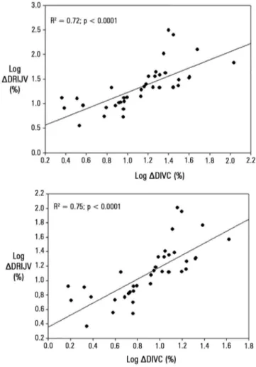
he IVC anteroposterior diameter during inspiration was 21 ± 6mm, and during expiration was 18 ± 6mm (p < 0.0001). he inspiratory RIJV diameter was 11 ± 4mm and expiratory was 9 ± 4mm (p < 0.0001). ΔDIVC and ΔDRIJV were signiicantly correlated by both calculation methods (Figure 1). Correlations did not have a normal distribution, but log transformation revealed a highly signiicant correlation (Figure 2).
Using ΔDIVC of 18% as a cut-of value indicating luid responsiveness for method A, 16 patients were
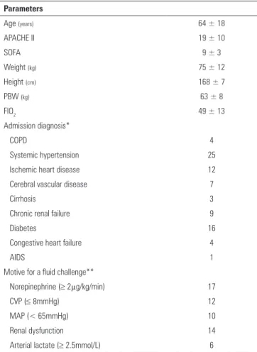
Table 1 - Demographic characteristics
Parameters
Age (years) 64 ± 18
APACHE II 19 ± 10
SOFA 9 ± 3
Weight (kg) 75 ± 12
Height (cm) 168 ± 7
PBW (kg) 63 ± 8
FIO2 49 ± 13
Admission diagnosis*
COPD 4
Systemic hypertension 25
Ischemic heart disease 12
Cerebral vascular disease 7
Cirrhosis 3
Chronic renal failure 9
Diabetes 16
Congestive heart failure 4
AIDS 1
Motive for a fluid challenge**
Norepinephrine (≥ 2µg/kg/min) 17
CVP (≤ 8mmHg) 12
MAP (< 65mmHg) 10
Renal dysfunction 14
Arterial lactate (≥ 2.5mmol/L) 6
APACHE II - Acute Physiology and Chronic Health Evaluation; SOFA - sequential organ failure assessment; PBW - predicted body weight; FIO2 - inspiratory fraction of oxygen; COPD - chronic obstructive pulmonary disease; AIDS - acquired immunodeficiency syndrome; CVP - central venous pressure; MAP - mean arterial pressure. * The total number of diagnoses is greater than 39 because one patient can have two or more diagnosis. ** The total number of motives for a fluid challenge is greater than 39 because according to the assistant doctor, there was more than one reason for a fluid bolus.
responders and 35 measurements showed agreement (15 responders) with a very good weighted Kappa (k = 0.80). Using ΔDIVC of 12% as a cut-of value indicating luid responsiveness for method B, 14 patients were responders and 32 measurements showed agreement (13 responders) with a good weighted Kappa (k = 0.65). Both methods agreed for 31 measurements.
Figure 1 - Distensibility of the inferior vena cava and of the right internal jugular vein are strongly correlated by method 1 (fluid responsiveness cut-off value: 18%) and method 2 (fluid responsiveness cut-off value: 12%). The empty points represent the points disagreeing. Pearson correlation test. ∆DIVC - distensibility of inferior vena cava; ∆DRIJV - distensibility right internal jugular vein.
Table 2 - Comparison of baseline values in responders and non-responders
Method A ∆DIVC cut-off 18%
Method B
∆DIVC cut-off 12% p value*
Responders (N = 16)
Non-responders (N = 23)
Responders (N = 14)
Non-responders
(N = 25) NS
VT (ml/kg/PBW) 8.8 ± 1.8 8.1 ± 1.3 8.6 ± 1.7 8.3 ± 1.5 NS
MAP (mmHg) 73 ± 17 78 ± 15 72 ± 17 78 ± 15 NS
HR (beats/min) 105 ± 23 93 ± 15 107 ± 22 96 ± 116 NS
Norepinephrine #(µg/kg/min) 0.29 ± 0.25 0.37 ± 0.62 0.34 ± 0.25 0.34 ± 0.59 NS
(N = 14) (N = 16) (N = 12) (N = 18)
CVP (mmHg) 14 ± 5 17 ± 8 15 ± 4 16 ± 8 NS
PEEP (cmH2O) 6.8 ± 2.3 7.4 ± 2.1 6.9 ± 2.4 7.2 ± 2.1 NS
∆DRIJV 71 ± 83 13 ± 8 36 ± 29 9 ± 6 p < 0.002
∆DIVC - distensibility of inferior vena cava; NS - not significant; VT - tidal volume; MAP - mean arterial pressure; HR - heart rate; CVP - central venous pressure; PEEP - positive end expiratory pressure; ∆DRIJV - distensibility of the right internal jugular vein. *Independent sample t-test. # 30 patients received an infusion of norepinephrine. The results are expressed as the mean ± standard deviation.
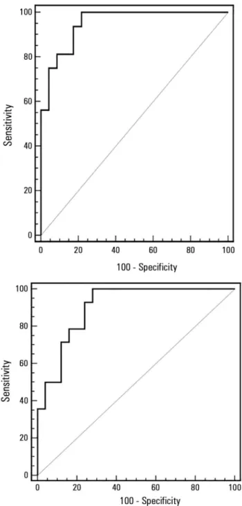
Figure 3 - Receiver operating characteristic curve analysis of the right internal jugular vein distensibility index in predicting fluid responsiveness based on inferior vena cava distensibility values of 18% by method A and 12% by method B. The area under the ROC curve was 0.951 (95%CI 0.830 - 0.993) and 0.903 (95%CI 0.765 - 0.973), respectively. ∆DRIJV - distensibility right internal jugular vein; ∆DIVC - distensibility of inferior vena cava.
DISCUSSION
Our indings demonstrate that ultrasound evaluation of RIJV respiratory diameter changes can serve as a simple
indexes in the evaluation of the appropriateness of volume expansion in mechanically ventilated patients.
Correcting hypovolemia is of paramount importance,(1,2)
but in mechanically ventilated patients, its correction should be guided to avoid inefective or even deleterious volume
expansion and worsening of the respiratory function.(5)
Mechanical ventilation induces cyclic variations in vena cava diameter that have been shown to be accurate predictors of luid responsiveness.(8,9,19) However, IVC measurements are
often not possible.(13)
here are few studies investigating respiratory variations in RIJV diameter in the evaluation of hypovolemia or hemodynamic response to a luid challenge, and these were conducted mainly in spontaneously breathing patients.(14,15,17)
During inspiration, the pressure inside the thorax increases more than the pressure outside the thorax. herefore, the pressure gradient for venous return is reduced, the systemic venous return decreases, the volume of extrathoracic venous blood decreases, and hence the endoluminal diameter of distensible veins, such as the jugular vein increases.(8,10,11) A
greater decrease in venous return during insulation may occur in a hypovolemic patient.
Our study demonstrated that the changes in IJV diameter during inspiration and expiration were signiicant. Similar indings were observed in several studies designed to evaluate IJV changes before and after blood donation(14,15) or
luid challenge.(17) However, in patients who are breathing
spontaneously, the IJV collapse may be inexact.
In critically ill, mechanically ventilated patients, the subject is even less well studied. Recently, Guarracino et al. showed that IJV distensibility accurately predicts volume responsiveness.(17) hey measured cardiac output to
calculate a cut-of of 18% with an 80% sensitivity and 85% speciicity for predicting response. hus, we compared the RIJV with IVC distensibility to predict luid responsiveness, to explore the hypothesis that cyclic respiratory changes in both veins could be similar. In our population of mechanically ventilated patients with hemodynamic instability, we have shown that the IVC distensibility indexes and RIJV distensibility indexes agree and are well correlated. Taken together, despite the diferences in study design, our indings agree with those of Guarracino et al.(17)
In our study, approximately two thirds of the patients were non-responders. his inding is consistent with other studies designed to examine luid responsiveness(7-9,12,20-22)
and strongly emphasize the need for parameters to help with selecting patients who might beneit from a volume load, avoiding inefective or even deleterious volume expansion in non-responder patients.
Our study has several limitations. First, we have not evaluated luid responsiveness after a luid challenge to
identify changes in cardiac output.(17) Second, we did
not evaluate changes in vein diameters before and after a luid challenge. hird, we did not study conditions with high venous pressure or severe right heart failure that could reduce IVJ distensibility even in the presence of preload responsiveness. Fourth, one must be aware that ultrasound of the jugular vein should be performed by a skilled intensivist because even a little pressure could cause a great change in the cross-sectional image and diameter of
the jugular vein during scanning. In patients with shock,
venous scanning becomes even more diicult.(18) Although
all scans were performed by an intensivist certiied in ultrasound, technical errors are possible. In addition, one could criticize that the scans were not repeated by another intensivist. Fifth, several patients were ventilated with low tidal volumes, which is a potential limitation for predicting
luid responsiveness.(7) Although these limitations may
introduce some bias, the consistency of the results implies improved external validity.
CONCLUSION
In conclusion, internal jugular vein cyclic respiratory changes in diameter appear to be a possible surrogate for changes in inferior vena cava diameter in determining luid responsiveness. Further studies should validate these indings by evaluating cardiac output after a luid challenge in several clinical conditions.
Objetivo: Investigar se a variação respiratória no diâmetro da veia cava inferior (ΔDVCI) e no diâmetro da veia jugular interna direita (ΔDVJID) se correlacionam em pacientes submetidos à ventilação mecânica.
Métodos: Estudo clínico prospectivo realizado em uma unidade de terapia intensiva de um hospital universitário. Foram incluídos 39 pacientes mecanicamente ventilados e com instabilidade hemodinâmica. Os valores da variação do diâmetro da veia cava inferior e da variação do diâmetro da veia jugular interna direita foram avaliados por meio de ecograia. A distensibilidade da veia foi calculada como a razão de (A) Dmin - Dmax/Dmin e (B) Dmax - Dmin/média de Dmax - Dmin, e expressa como porcentagem.
Resultados: Com ambos os métodos, observou-se correlação entre a variação do diâmetro da veia cava inferior e a variação do diâmetro da veia jugular interna direita: (A) r = 0,34, p = 0,04 e (B) r = 0,51, p = 0,001. Utilizando o ponto de corte de 18% para indicar responsividade a luidos na variação do
diâmetro da veia cava inferior, pelo o método (A), 16 pacientes foram considerados responsivos e 35 medições mostraram concordância (Kappa ponderado = 0,80). A área sob a curva ROC foi de 0,951 (IC95% 0,830 - 0,993; valor de corte = 18,92). Usando 12% como ponto de corte para a variação do diâmetro da veia cava inferior para indicar capacidade de resposta a luidos, pelo método (B), 14 pacientes foram responsivos e 32 medições mostraram concordância (Kappa ponderado = 0,65). A área sob a curva ROC foi de 0,903 (IC95% 0,765 - 0,973; valor de corte = 11,86).
Conclusão: As variações respiratórias nas dimensões da veia cava inferior e da veia jugular interna direita se correlacionaram e mostraram concordância signiicativa. Avaliação da distensibi-lidade da veia jugular interna direita parece ser uma alternativa à distensibilidade da veia cava inferior para avaliar a responsivi-dade a luidos.
RESUMO
Descritores: Veia cava inferior/ultrassonograia; Veias jugu-lares/ultrassonograia; Hidratação; Respiração artiicial; Hemodi-nâmica
REFERENCES
1. Weil MH, Nishjima H. Cardiac output in bacterial shock. Am J Med. 1978;64(6):920-2.
2. Rivers E, Nguyen B, Havstad S, Ressler J, Muzzin A, Knoblich B, Peterson E, Tomlanovich M; Early Goal-Directed Therapy Collaborative Group. Early goal-directed therapy in the treatment of severe sepsis and septic shock. N Engl J Med. 2001;345(19):1368-77.
3. De Backer D, Biston P, Devriendt J, Madl C, Chochrad D, Aldecoa C, Brasseur A, Defrance P, Gottignies P, Vincent JL; SOAP II Investigators. Comparison of dopamine and norepinephrine in the treatment of shock. N Engl J Med. 2010;362(9):779-89.
5. Pinsky MR, Teboul JL. Assessment of indices of preload and volume responsiveness. Curr Opin Crit Care. 2005;11(3):235-9.
6. Michard F, Boussat S, Chemla D, Anguel N, Mercat A, Lecarpentier Y, et al. Relation between respiratory changes in arterial pulse pressure and fluid responsiveness in septic patients with acute circulatory failure. Am J Respir Crit Care Med. 2000;162(1):134-8.
7. Oliveira-Costa CD, Friedman G, Vieira SR, Fialkow L. Pulse pressure variation and prediction of fluid responsiveness in patients ventilated with low tidal volumes. Clinics (Sao Paulo). 2012;67(7):773-8.
8. Feissel M, Michard F, Faller JP, Teboul JL. The respiratory variation in inferior vena cava diameter as a guide to fluid therapy. Intensive Care Med. 2004;30(9):1834-7.
9. Barbier C, Loubières Y, Schmit C, Hayon J, Ricôme JL, Jardin F, et al. Respiratory changes in inferior vena cava diameter are helpful in predicting fluid responsiveness in ventilated septic patients. Intensive Care Med. 2004;30(9):1740-6.
10. Morgan BC, Martin WE, Hornbein TF, Crawford EW, Guntheroth WG. Hemodynamic effects of intermittent positive pressure respiration. Anesthesiology. 1966;27(5):584-90.
11. Natori H, Tamaki S, Kira S. Ultrasonographic evaluation of ventilatory effect on inferior vena caval configuration. Am Rev Respir Dis. 1979;120(2):421-7. 12. Michard F, Teboul JL. Predicting fluid responsiveness in ICU patients: a
critical analysis of the evidence. Chest. 2002;121(6):2000-8.
13. Nagdev AD, Merchant RC, Tirado-Gonzalez A, Sisson CA, Murphy MC. Emergency department bedside ultrasonographic measurement of the caval index for noninvasive determination of low central venous pressure. Ann Emerg Med. 2010;55(3):290-5.
14. Akilli NB, Cander B, Dundar ZD, Koylu R. A new parameter for the diagnosis of hemorrhagic shock: jugular index. J Crit Care. 2012;27(5):530.e13-8.
15. Unluer EE, Kara PH. Ultrasonography of jugular vein as a marker of hypovolemia in healthy volunteers. Am J Emerg Med. 2013;31(1):173-7. 16. Sankoff J, Zidulka A. Non-invasive method for the rapid assessment of
central venous pressure: description and validation by a single examiner. West J Emerg Med. 2008;9(4):201-5.
17. Guarracino F, Ferro B, Forfori F, Bertini P, Magliacane L, Pinsky MR. Jugular vein distensibility predicts fluid responsiveness in septic patients. Crit Care. 2014;18(6):647.
18. Mayo PH, Beaulieu Y, Doelken P, Feller-Kopman D, Harrod C, Kaplan A, et al. American College of Chest Physicians/La Société de Réanimation de Langue Française statement on competence in critical care ultrasonography. Chest. 2009;135(4):1050-60.
19. Moretti R, Pizzi B. Inferior vena cava distensibility as a predictor of fluid responsiveness in patients with subarachnoid hemorrhage. Neurocrit Care. 2010;13(1):3-9.
20. Huang CC, Fu JY, Hu HC, Kao KC, Chen NH, Hsieh MJ, et al. Prediction of fluid responsiveness in acute respiratory distress syndrome patients ventilated with low tidal volume and high positive end-expiratory pressure. Crit Care Med. 2008;36(10):2810-6.
21. Muller L, Bobbia X, Toumi M, Louart G, Molinari N, Ragonnet B, Quintard H, Leone M, Zoric L, Lefrant JY; AzuRea group. Respiratory variations of inferior vena cava diameter to predict fluid responsiveness in spontaneously breathing patients with acute circulatory failure: need for a cautious use. Crit Care. 2012;16(5):188.