www.eerp.usp.br/rlae
Corresponding Author: Alvisa Palese
Università degli Studi di Udine Viale Ungheria 20
33100 Udine, Italia
E-mail: alvisa.palese@uniud.it
Effectiveness of 10% povidone-iodine drying time before
Peripheral Intravascular Catheter insertion: preliminary results
from an explorative quasi-experimental study
Alvisa Palese
1Federica Cescon
2Aim: to investigate the effectiveness of 10% povidone-iodine after a 30-second or 2-minute
drying time on microbial count reduction at the point of a Peripheral Intravascular Catheter (PIC)
insertion. A quasi-experimental design was adopted. In total, 53 patients were enrolled, 25
were exposed to a 2-m drying time and 28 to a 30-s drying time. From the preliminary results
of this study, no differences in the occurrence of contamination have emerged between patients
receiving 30-s and 2-m drying time for 10% povidone-iodine solutions.
Descriptors: Povidone Iodine/Therapeutic Use; Air Dry; Disinfection; Catheterization Peripheral;
Microbial Count.
Eicácia de iodopovidona a 10% de acordo com tempo de secagem antes da
inserção do cateter intravenoso periférico: resultados preliminares de um estudo exploratório quasi-experimental
Objetivo: investigar a eicácia da solução iodopovidona a 10% sobre a redução da contagem microbiana no ponto de inserção do Cateter Venoso Periférico após tempo de secagem de 30s ou 2 min. Método: desenho quase-experimental. Foram incluídos 53 pacientes no estudo: 25 foram expostos a 2min de secagem e 28 foram expostos a 30s de secagem. Resultados: Os resultados preliminares não apresentaram diferenças na ocorrência de contaminação entre os pacientes que foram submetidos a 30s ou 2min de secagem após desinfecção com solução de iodopovidona a 10%.
Descritores: Iodopovidona/Uso Terapêutico; Desinfecção; Cateterismo Periférico; Contagem Microbiana.
Background
cell wall of the microorganism and to replace the content
with iodine(7).The previous guidelines on intravascular
catheters(5-6) recommend allowing for a drying time of 2
minutes, but no experimental data support this advice.
Previously, commentary that likewise lacks support from
experimental data reported that a 10% povidone-iodine
solution would be effective after drying for 90 seconds(8).
In daily practice, nurses have many doubts about
the time required and different strategies to facilitate
drying are adopted: fanning, using gauze to dry, and
blowing, which seems to be inappropriate as it increases
the risk of infection(7). These strategies are often adopted
in the use of 10% povidone-iodine because its drying
time appears to be longer than for other solutions.
Contributing to knowledge regarding how long the 10%
povidone-iodine should be left in place before applying
the PIC is the main goal of this paper. The Peripheral Intravascular Catheter (PIC) is
widely used in nursing clinical practice. PICs can cause
both local and systemic complications, the most common
being phlebitis, varying from 1–70% in different
observational studies(1-3), and originate most often from skin commensal lora. Recommended strategies to prevent PIC-related infections are hand hygiene, the use
antiseptic techniques and adequate skin preparation(4-6).
Before the placement of a PIC, an effective reduction
of the microbial count is recommended. While different
antiseptic solutions should be used (e.g., >0.5%
chlorhexidine, iodophor), there is a general lack of
recommendations regarding the disinfection drying time,
even in the most recent guidelines(4). In the event of
contraindication of the use of chlorhexidine(4), a solution
with 10% povidone-iodine is recommended and drying
time is necessary for it to release free iodine against the
Eicacia del tiempo de secado de la yodopovidona al 10% antes de la
inserción de catéter venoso periférico: resultados preliminares de un estudio exploratorio casi-experimental
Objetivo: para investigar la eicacia de una solución yodopovidona al 10% tras tiempo de secado de 30 segundos o 2 minutos en la reducción del contaje microbiano en el local de inserción del Catéter Venoso Periférico, fue adoptado un diseño casi-experimental. Al total, fueron incluidos 53 pacientes, 25 expuestos a 2 min. de secado y 28 a 30 segundos. Con base en los resultados preliminares, no se encontraron diferencias en la ocurrencia de contaminaciones entre pacientes sometidos a un tiempo de secado de 30 s. o de 2 min tras desinfección con solución de yodopovidona al 10%.
Objectives and study design
Aiming to investigate the effectiveness of 10%
povidone-iodine after a 30-second or 2-minute drying
time on microbial count reduction at the point of PIC
insertion, a quasi-experimental design was adopted.
Materials and methods
In February 2012, an approachable Emergency
Department (ED) located in northern Italy was involved
after having obtained appropriate authorization from the
Internal Review Board of the Hospital/University. All adults (≥18 years) admitted subsequently to the ED and who were candidates for PIC were eligible. Those
affected by condition(s) contraindicating the adoption
of 10% povidone-iodine (e.g., allergy, pregnancy) or
affected by cardiac arrest or unconsciousness were
excluded. After having obtained written informed
consent from the patients included in the study, they
were divided into two groups according to the priority
given at triage: a) those receiving an ED white code
(=not urgent condition) were exposed to a 2 minute
(2-m) 10% povidone-iodine drying time, while b) those
receiving green or yellow triage codes (=patients in
sub-urgent conditions) were exposed to a 30 second
(30-s) drying time. Clinical nurses disinfected the site with
10% povidone-iodine using sterile gauze, in accordance
with the procedure adopted in the ward.
The microbial count was the primary end-point
of the study, examined at two different points in time: the irst, before skin disinfection, seeking to determine baseline skin contamination (T0), the second after the
drying time (30-s vs. 2-m), in order to measure the
effectiveness of 10% povidone-iodine on the microbial
count at a different drying time (T1). The swabs were
immediately seeded on chocolate agar and conserved
in a thermostat-governed environment at 37° Degree
Celsius (C) for 24 hours and then evaluated by two
researchers in a blind fashion to count the colony-forming units (CFUs). The deinition adopted for skin contamination was the presence of CFUs≥15(9).
A questionnaire investigating patient demographic
characteristics such as age and gender, recent
surgery (yes/no), health problems (cancer, diabetes,
coagulopathies, fever -yes/no-), and antibiotic therapy (yes/no) according to their inluence in the occurrence of PIC-related infection(10-14) was administered by interview.
Patient collaboration (or not) during the procedure,
where the PIC was inserted (e.g., right or left upper
limb, and vein approached), and its size, as well as
factors increasing the risk of contamination(3,10-14), were
also observed and documented by the researcher.
Ultimately, skin preparation procedures adopted by the
clinical nurse performing the PIC insertion (preliminary
hand hygiene, the use of gloves, and the adoption of
aseptic techniques during the procedure, in accordance
with the available guidelines(4)) was then observed, with a grid illed in by the researcher.
Data was processed using the SPSS Statistical
Package (Version 18). Indices of central position (mean,
standard deviation), percentages and frequencies have
been evaluated. Comparison between the two groups
was performed adopting the T-test or non-parametric
tests (according to the normal distribution [or not] of
the variables), and the χ2 test (or Fisher’s Test, when appropriate). Relative Risk (Conidence Interval 95% [95% CI]) was also evaluated. The statistical signiicance level was set at p=0.05.
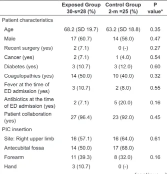
Results
In total, some 53 patients were enrolled, 25 were
exposed to a 2-m drying time and 28 to a 30-s drying
time. Thirty–one patients were male, and the majority
(50/53, 94.3%) collaborated with clinical nurses
during the PIC insertion. The exposed and control
groups were homogeneous in their principal participant
characteristics, as reported in Table 1. The procedure for
PIC insertion and the characteristics of the PIC gauge
and of the site chosen by the clinical nurse were also
homogeneous between the exposed and control groups,
as indicated in Table 1.
Exposed Group 30-s=28 (%)
Control Group 2-m =25 (%)
P value*
Patient characteristics
Age 68.2 (SD 19.7) 63.2 (SD 18.8) 0.35
Male 17 (60.7) 14 (56.0) 0.47
Recent surgery (yes) 2 (7.1) 0 (-) 0.27
Cancer (yes) 2 (7.1) 1 (4.0) 0.54
Diabetes (yes) 3 (10.7) 3 (12.0) 0.60
Coagulopathies (yes) 14 (50.0) 10 (40.0) 0.32
Fever at the time of
ED admission (yes) 3 (10.7) 2 (8.0) 0.55 Antibiotics at the time
of ED admission (yes) 2 (7.1) 5 (20.0) 0.16 Patient collaboration
(yes) 27 (96.4) 23 (92.0) 0.45
PIC insertion
Site: Right upper limb 16 (57.1) 16 (64.0) 0.61
Antecubital fossa 14 (50.0) 17 (68.0)
0.16 Forearm 11 (39.3) 8 (32.0)
Hand 3 (10.7) 0 (-)
Table 1 - Patient demographics and PIC insertion data
Rev. Latino-Am. Enfermagem 2013 Jan.-Feb.;21(Spec):47-51.
At an overall level, 27 out of 53 (50.9%) sites
selected for PIC placements were contaminated at
the baseline (T0; 17 among the exposed group and
10 among the control group); after disinfection (T1),
20 sites (37.7%) were contaminated (13 among
the exposed group and 7 among control groups). A
total of 7 contaminated sites (13.2%) at T0 were not
contaminated at T1 (4 among the exposed group and 3
among the control group). The differences that emerged were not statistically signiicant (Table 2).
Discussion
According to its exploratory nature, this manuscript
has several limitations: a monocentric center was involved
and a limited number of participants were enrolled.
Adopting the perspective of pragmatic trials(15), the
researchers did not standardize the disinfection technique,
given that the quantity of antiseptic solution used in daily
practice for this procedure is adopted heterogeneously
among clinical nurses. Further studies should address
these limits, extending the study design also to those
antiseptic solutions recommended by the up-to-date
guidelines (e.g., >0.5% chlorhexidine, iodophor) and also
at different sites, such as for surgical wound disinfection.
The study involved a homogeneous group of patients
admitted into the ED under different urgency codes where
the insertion of a PIC is considered a routine procedure.
According to the need to have the PIC rapidly in place
(in the case of urgent cases), a group was exposed to a
30-s drying time, while patients admitted in non-urgent
conditions received a 2-m drying time. Considering the lack of evidence available in the ield, the drying time was selected on the basis of the existing literature(7), which
recommended 2 minutes, and on the basis of in vitro
studies(16-17), which applied a 30-s drying time.
The analysis showed that drying time (30-s vs. 2-m) was not signiicantly associated with contamination (CFUs≥15) at T1: in its preliminary phase and among its several limitations, this exploratory study shows that
the drying time should be less than 2 minutes and these
results might help nurses in their practice. For them, waiting for the drying time is particularly dificult in the case of confused, unstable and at risk patients and/or in
turbulent environments such as EDs(18), where multiple
interruptions might threaten the safety of the procedure.
In order to reduce this time, different strategies are
adopted by clinical nurses, such as fanning, using gauze
to dry, or blowing, which seems to be inappropriate as it
increases the risk of infection(7).
Conclusions
Intravenous therapy is largely used for ED patients
via PICs inserted by nurses. PICs can cause both local and
systemic complications, the most common being phlebitis:
aseptic techniques and adequate skin preparation are the
main strategies to reduce contamination at the time of
insertion. To our knowledge, no previous studies developed
evidence regarding the drying time utilized after skin
disinfection or its effectiveness in reducing contamination.
From the preliminary results of this study, no differences Veins approached
Cephalic vein 16 (57.1) 14 (56.0)
0.19 Basilic vein 8 (28.6) 7 (28.0)
Perforating vein 1 (3.6) 1 (4.0)
Dorsal vein 3 (10.7) 0 (-)
Median antebrachial
vein 0 (-) 3 (12.0)
PIC size
18 gauge 20 (71.4) 13 (52.0)
0.24 20 gauge 8 (28.6) 11 (44.0)
22 gauge 0 (-) 1 (4.0)
Skin preparation procedure adopted by the nurse inserting the PIC
Hand hygiene (yes) 2 (7.1) 4 (16.0) 0.31
Gloves (yes) 10 (35.7) 13 (52.0) 0.23
Aseptic technique
respected (yes) 1 (3.6) 5 (20.0) 0.06 Table 1 - (continuation)
*χ2 for categorical variables and U-Mann Whitney for continuous variables
Characteristics Exposed Group 30-s=28 (%) Control Group 2-m=25 (%) P value Number of PIC sites contaminated (CFUs≥ 15) at T0
17 (60.7) 10 (40.0)
RR 1.49
(CI95% 0.87 to 2.54) p = 0.13
Number of PIC sites contaminated (CFUs≥15) at T1
13 (47.4) 7 (28.0) RR 1.43
(CI95% 0.87 to 2.34) p = 0.16
Differences of PIC sites contaminated (CFUs≥ 15) (T1-T0)
4 (14.3) 3 (12.4)
RR 1.10
(CI95% 0.54 to 2.20) p = 0.80 Table 2 - Disinfection with 10% povidone-iodine:
comparison between exposed group and controlled group
Moreover, among the group with a 30-s drying
time, the average of CFUs at T0 were 209 and 80 at
T1 (-24.6%); among the control group (2-m drying
time), the average CFUs at T0 were 4,527 and 502
in the occurrence of contamination have emerged
between patients receiving 30-s and 2-m drying time of
10% povidone-iodine solutions. These preliminary results should be conirmed with further large and multicenter studies addressing the lack of evidence in the ield and the consequent uncertainly of clinical nurses: drying
time increases the length of the procedure and the risk
of accidental contamination of the site. Waiting for a site
to dry for a longer time with some patients (e.g. critical,
agitated patients), and in some turbulent environments
(e.g. emergency departments) is not always advisable.
Acknowledgments
To Dott. Maurizio Ruscio, Beppina Pontarolo
and Vera Persello from San Daniele del Friuli Hospital
(Italy), for having shared the study aim and supported
the research process. We also thank Fabricio Aline,
researcher in Venezia (Italy), for her kindly help in the
manuscript translation in Portuguese language. Special
thank is dedicated to the ED nursing team and to the
patients of the San Daniele Hospital, Udine (Italy).
References
1. Trinh TT, Chan PA, Edwards O, Hollenbeck, Huang B,
Burdick N, et al. Peripheral Venous Catheter-Related
Staphylococcus aureus Bacteremia. Infection Control
Hosp Epidemiol. 2011;32(6):579-83.
2. Martinez JA, Piazuelo M, Almela A, Blecua P, Gallardo
R, Rodrìguez S, et al. Evalutation of add-on device for
the prevention of phlebitis and other complications
associated with the use peripheral catheters in
hospitalized adults: a randomized controlled study. J
Hosp Infection. 2009;73:135-42.
3. Maki DG, Kluger DM, Crnich CJ. The risk of bloodstream
infection in adults with different intravascular devices: a
systematic review of 200 published prospective studies.
Mayo Clin Proc. 2006;81(9):1159-71.
4. Centers for Disease Control and Prevention (CDC).
Guidelines for the Prevention of Intravascular
Catheter-Related Infections. Am J Infection Control. 2011;39:S1-34.
5. Centers for Disease Control and Prevention (CDC).
Guidelines for the Prevention of Intravascular
Catheter-Related Infections. MMWR. 2002;51(RR10):1-26.
6. Registered Nurses’ Association of Ontario. Care and
Maintenance to reduce Vascular Access Complications. Nursing
best practice guidelines program; 2005.[acesso 13 nov 2012]; Disponível em: http://www.rnao.org/Storage/39/3381_ Care_and_Maintenance_to_Reduce_Vascular_Access_ Complications._with_2008_Supplement.pdf.
7. Hadaway L. What you can do to decrease catheter
– related infections: Meticulous cleaning of skin and
insertion site can keep bad bugs out of your patient’s
bloodstream. Nursing. 2002;32:46-8.
8. Aschenbrenner Diane S. A Question of Practice: Skin
Preps and Protocol. Am J Nurs. 2000;100(4):78.
9. Maki DG, Weise CE, Sarafin HW. A semiquantitative
culture method for identifying intravenous – catheter –
related infection. N Engl J Med. 1977;296:1305-9.
10. Grice EA, Segre JA. The skin micro biome. Nature
Rev Microbiol. 2011;9:244-53.
11. Forni C, Loro L, Tremosini M, Trofa C, D’Alessandro
F, Sabbatini T, et al. Studio di coorte sulla popolazione
ortopedica delle complicanze correlate all’utilizzo del
catetere venoso periferico e identificazione dei fattori
predittivi. Assistenza Inferm Ricerca. 2010;29(4):166-73.
12. Lee WL, Chen HL, Tsai TY, Lai IC, Chang WC, Huang CH,
Fang CT. Risk factors for peripheral intravenous catheter
infection in hospitalized patients: a prospective study of
3165 patients. Am J Infect Control. 2009;37:683-6.
13. Nassaji- Zavareh M, Ghorbani R. Peripheral
intravenous catheter –related phlebitis and related risk
factors. Singapore Med J. 2007;48(8):733-8.
14. Tagalakis V, Kahn SR, Libman M, Blostein M. The
epidemiology of peripheral vein infusion thrombophlebitis:
a critical review. Am J Med. 2002;113:146-51.
15. Zwarenstein M, Treweek S, Gagnier J, Douglas G,
Tunis S, Haynes B et al. CONSORT and Pragmatic Trials
in Healthcare (Practihc) groups. Improving the reporting
of pragmatic trials: an extension of the CONSORT
statement. BMJ. 2008;337:a2390.
16. Adams D, Quayum M, Worthington T, Lambert P,
Elliott T. Evaluation of a 2% chlorhexidine gluconate in
70% isopropyl alcohol skin disinfectant. J Hosp Infection.
2005;61:287-90.
17. Federal Register. Department of Health and Human
Services. Food and Drug Administration. Topical
antimicrobials drug products for over-the-counter
human use: tentative final monograph for healthcare
antiseptic drug products – Proposed rule; 1994. Disponível em: http://www.fda.gov/ohrms/dockets/ ac/05/briefing/2005-4098B1_02_03-FDA-TAB1.pdf. 18. Palese A, Cassone A, Kulla A, Dorigo S, Artico M,
Camero F, et al. Factors influencing nurses’ decision
making process on leaving the Peripheral Intravascular
Catheter after 96 hours: a longitudinal study. J Inf Nurs.
2011;34(5):319-26.
Received: Aug. 1st 2012