Inhibition of metastatic potential of B16-F10 melanoma cell line in vivo and in vitro
by bi
fl
orin
Adriana Andrade Carvalho
a,⁎
, Patrícia Marçal da Costa
b, Luciana Gregório Da Silva Souza
c, Telma Leda G. Lemos
c,
Ana Paula Negreiros Nunes Alves
d, Cláudia Pessoa
b, Manoel Odorico de Moraes
baNúcleo de Farmácia, Campus Lagarto, Universidade Federal de Sergipe, 49400-00 Lagarto, Sergipe, Brazil
bDepartamento de Fisiologia e Farmacologia, Universidade Federal do Ceará, Campus do Porangabussu, Caixa Postal 3157, 60.430-270 Fortaleza, Ceará, Brazil cDepartamento de Química Orgânica e Inorgânica, Universidade Federal do Ceará, Caixa Postal-12200, 60021-940 Fortaleza, Ceará, Brazil
dDepartamento de Clínica Odontológica do Curso de Odontologia, Universidade Federal do Ceará, Fortaleza, Ceará, Brazil
a b s t r a c t
a r t i c l e
i n f o
Article history:
Received 17 February 2013 Accepted 21 May 2013
Keywords:
Biflorin
Capraria biflora
Metastasis Melanoma
Aim:The aim of this study was to determine the antimetastatic potential of biflorin using in vivo and in vitro approaches.
Main methods:Biflorin was isolated fromCapraria bifloracollected in Fortaleza, Ceará, Brazil. Adhesion, migra-tion and invasion assays were performed to avail of the antimetastatic potential of this quinone. Experimen-tal metastasis was performed to avail of the antimetastatic potential of bilflorin using in vivo assay. Keyfindings:Treatment with biflorin (25 and 50 mg/kg/day) was shown to be effective in reducing B16-F10 mel-anoma metastasis in C57BL/6 mice. The administration of biflorin at 25 mg/kg/day intraperitoneally inhibited the formation of metastases by about 57% compared to untreated control animals. When the animals were treat-ed with 50 mg/kg/day intraperitoneally, there was a 71% decrease in the number of lung metastases. Morpholog-ical assays showed the presence of hemosiderin and erythrocytes in the lung parenchyma, indicating the occurrence of hemorrhage, probably a side effect of biflorin. Biflorin at non-toxic concentrations (0.5, 1.0 and 1.5 g/mL) was tested directly on B16-F10 cells in vitro, and it inhibited cell adhesion to type I collagen and cell motility using the wound-healing assay.
Significance:These data suggest that biflorin has a promising antimetastatic potential, as shown by its anti-adhesion, anti-migration and anti-invasion properties against a metastatic melanoma cell line. However, further studies are essential to elucidate its mechanism of action.
© 2013 Elsevier Inc. All rights reserved.
Introduction
The spread of cancer cells from the primary tumor to distant loca-tions is known as metastasis. The occurrence of metastasis is the major cause of mortality in cancer patients (Lee et al., 2003; Weiss, 1990), and the treatment of metastasis is still far from satisfactory (Han et al., 2009). Metastasis of cancer cells involves multiple pro-cesses including inhibition of cell-to-cell adhesion, enhancement of cell-to-extracellular matrix (ECM) adhesion, and invasion, which in-volves the degradation of the ECM (Lee et al., 2003, 2006; Cavallaro and Christofori, 2001). Tumor invasion of tissues by penetrating the basement membrane is also an important step, which involves the adhesion of tumor cells to ECM components followed by its degrada-tion (Cavallaro and Christofori, 2001).
Natural products are a rich source of pharmacologically active compounds, in which plant materials hold an important position. One such plantCapraria bifloraL., a perennial shrub of the family Schrophulariaceae, is used to treat various symptoms such as pain, fever, flu, vomiting, childbirth recovery, diarrhea, hemorrhoids, rheumatism, and swelling. The roots of this plant have antibacterial properties (Vasconcellos et al., 2007) and its aqueous extract has demonstrated both peripheral and central analgesic effects (Acosta et al., 2003).
Biflorin is a natural product that can be isolated from the roots ofC. bifloraL., a substance with an o-quinone structure. This quinone has antibiotic activity against Gram-positive bacteria, yeasts and fungi (Aquino et al., 2007). Moreover, biflorin has antifungal and antitumor effects, such as in the melanoma model (Vasconcellos et al., 2011).
Several studies have shown that some compounds isolated from plants can prevent tumor metastasis through inhibition of tumor adhe-sion and migration (Lee et al., 2006; Yang et al., 2007; Huey-Chun et al., 2003). Thus, the aim of this study was to determine the antimetastatic potential of biflorin using in vivo and in vitro approaches.
⁎ Corresponding author at: Núcleo de Farmácia, Campus Lagarto, UFS, Rua Padre Alvares Pitangueira, no 248, Centro, C.E.P.:49.400-000, Lagarto - SE/Brazil. Tel.: + 55 85 2105 6550.
E-mail address:[email protected](A. Andrade Carvalho).
0024-3205/$–see front matter © 2013 Elsevier Inc. All rights reserved. http://dx.doi.org/10.1016/j.lfs.2013.05.018
Contents lists available atSciVerse ScienceDirect
Life Sciences
Material and methods
Plant material
C. bifloraL. (Scrophulariaceae) was collected at a plantation located in Fortaleza, Ceará, Brazil in 2008 and identified by Dr. Edson Nunes. A voucher specimen (No. 30848) was deposited in the Herbarium Prisco Bezerra of the Biology Department of the Federal University of Ceará.
Isolation of biflorin
The isolation of biflorin was performed as described byFonseca et al. (2003). Air-dried powdered roots (4.5 kg) was extracted with light petro-leum (4 L) for 2 days and solvent was evaporated under reduce pressure to yield 2.3 g. The extract was then dissolved in petroleum light 10 mL and kept in a refrigerator for 2 h until precipitating a solid material with a purple color. It was laterfiltered under vacuum and yielded a purple solid material (2 g). The purple solid material was chromatographed on silica gel by isocratic elution using a binary mix-ture of light petroleum/ethyl acetate, 9:1 (v/v). Fractions were pooled according to thin-layer chromatographic (TLC) analysis. Com-bined fractions containing purified biflorin yielded 335.5 mg (purity: 99%). Biflorin was analyzed in high performance liquid chromatography (HPLC) on a Shimadzu SPD-M20A instrument with UV–Vis detector using a 254 nm, C18 column. The eluent was methanol/water (90:10) using isocratic elution and showed a retention time of 6.65 min. Struc-ture determination of biflorin was determined by spectroscopic analy-sis, including one- and two-dimensional nuclear magnetic resonance spectral data, physical properties and comparison with data from the literature (Fonseca et al., 2003). Biflorin is a red intense solid m.p. 154–157 °C, IR (KBr)cm−1 2921, 1684, 1592, 1438, 1236, 1023; 13C NMR (500 MHz, CDCl
3) 7.8(C-17); 18.0(C-14); 22.6 (C-16); 25.2 (C-15); 26.7(C-11); 26.7(C-10); 113.5(C-9); 116.1(C-3); 122.0(C-12); 124.2(C-9b); 126.6(C-6a); 127.8(C-4); 129.0(C-3a); 133.6(C-13); 135.9(C-5); 140.2(C-2); 146.7(C-6); 161.9(C-9a); 178.0(C-8); 182.0(C-7) and1H NMR agree with literature. Melting points were de-termined by a digital Mettler Toledo FP90 apparatus. IR spectra were obtained on a Perkin-Elmer Ft-IR Spectrum 1000. Nuclear magnetic res-onance (NMR) spectra were obtained using CDCl3on a Bruker Avance DRX-500 (500 MHz) spectrometer.
Cell conditions
Mouse melanoma B16-F10 was maintained in RPMI 1640 medium supplemented with 10% fetal bovine serum (FBS), 1.5% MEM vitamin solution, 1% sodium pyruvate, 1% non-essential amino acids and 1% 100× penicillin and streptomycin (Pen-Strep) at 37 °C in a
humidi-fied atmosphere of 5% CO2.
Animals
A total of 48 C57BL/6 mice (female, 25–30 g), obtained from the central animal house of the Federal University of Ceará, Brazil, were used. The animals were housed in cages with free access to food and water. All animals were kept under a 12 h:12 h light–dark cycle (lights on at 6:00 a.m.). The animals were treated according to the eth-ical principles of animal experimentation of SBCAL (Sociedade Brasileira de Ciências em Animais de Laboratório), Brazil. The Animal Studies Committee of the Federal University of Ceará approved the experimen-tal protocols (No. 52/08).
Drugs and reagents
RPMI 1640 media, fetal bovine serum (FBS) were purchased from Cultilab, Pen-Strep,
3-(4,5-dimethyl-2-thiazolyl)-2,5-diphenyl-2H-tetrazolium (MTT), DMSO, Doxorubicin hydrochloride, bovine serum albumin (BSA), mitomycin C and collagen type I were purchased from Sigma.
In vivo experimental lung metastasis and survival assay
B16-F10 cells were harvested, washed with serum-free RPMI 1640 and resuspended to give the appropriate concentrations in phosphate-buffered saline (PBS). An amount of 0.5 mL of the resultant B16-F10 cell suspension (1 × 10⁶cells) was injected into the mice via the tail vein. On the following day (Day 1), mice were randomly divided into three groups (n = 16 for each group). In the control group, the animals received 10% DMSO i.p., and the treatment groups received biflorin (25 mg/kg and 50 mg/kg i.p.) suspended in sterile 10% DMSO in distil-late water. The treatment was continued daily for 21 days, and after-wards, 8 animals from each group were sacrificed. The lungs were excised andfixed in 10% formaldehyde. The number of B16-F10 colonies present on the surface of each set of lungs was determined by visual inspection using a magnifying glass. The other 8 animals in each group were observed for no more than 2 months for a determination of survival rate.
Histopathology and morphological observations
After beingfixed in formaldehyde, the lungs were examined for size or color changes and hemorrhage. Portions of the lungs were then cut into small pieces, and histological sections were stained with hematoxylin and eosin. Histological analyses were performed by light microscopy.
Adhesion assay
Adhesion studies were performed as described previously byHatai et al. (1993)with modifications. Briefly, type I collagen (0.1 mg/mL) was immobilized in 96-well plates containing 0.1% acetic acid, over-night at 4 °C. After immobilization, any uncoated surface was blocked with 1% BSA at 37 °C for 2 h. The cells were trypsinized and suspended in culture medium, pelleted, and resuspended at 2 × 10⁵cells/mL in PBS, and 100μL of the cell suspension was added to each well in the presence or absence of biflorin (0.5, 1.0 and 1.5μg/mL) at 37 °C for 1 h. After incubation, the wells were washed twice with PBS. Next, 200μL of MTT solution (5 mg/mL) was added to each well, and the plates were incubated for 4 h at 37 °C and 5% CO2. After incubation, su-pernatants were removed and DMSO was added. The absorbance was determined at 595 nm (Mosmann, 1983).
Migration assay
Invasion assay
Tumor cell invasion was assayed using transwell chambers coated with Matrigel accordingly as previously described in (Lee et al., 2003, 2006) with some modifications. Briefly, transwell chambers with 24-Multiwell of 8-μm pore size coated with Matrigel (BD BioCoat™, Bio-science) were used. B16-F10 cells (2 × 10⁵cells/well) were treated pre-viously with biflorin (0.5, 1.0 and 1.5μg/mL) during 16 h. After the treatment, the cells were carefully transferred into the upper chambers top well with biflorin (0.5, 1.0 and 1.5μg/mL). Lower chambers were
filled with 10% FBS medium to attract cells. Matrigel chambers were incubated for 8 h at 37 °C with 5% CO2. After 8 h incubation, the non-invaded cells were completely wiped out with a cotton swab, and the lower surface of thefilter wasfixed with 30% methanol, stained with 0.5% crystal violet in 20% methanol. Experiments were performed independently three times. For each replicate, the cells were counted under a microscope at a magnification of 400× in 5 randomly selected
fields and the counts were averaged.
Cell proliferation assay
The cell proliferation assay was performed to test the effect of biflorin against B16-F10 cells using the concentrations used in the adhesion and migration assays. Doxorubicin hydrochloride was used as the positive control. Briefly, cells were grown in RPMI-1640 medium supplemented with 10% FBS, 2 mM glutamine and Pen-Strep, and incubated at 37 °C in 5% CO2atmosphere. For experi-ments, the cells were plated in 96-well plates (0.6 × 10⁵cells/well in 100μL of medium), biflorin was added to each well (final concen-trations of 0.5, 1.0 and 1.5μg/mL and volume of 200μL), and cells were incubated for 24 h. Control groups received the same amount of sterile DMSO. Tumor cell growth was quantified by the ability of
living cells to reduce MTT to a purple formazan product. At the end of the incubation period, plates were centrifuged, the medium was replaced by 150μL of fresh medium containing 0.5 mg/mL MTT, and the cells were re-incubated for 3 h. The formazan produced was dissolved in 150μL of DMSO, and the absorbance was measured using a multiplate reader (Multimode Detector DTX 880, Beckman Coulter) [14, 16]. The growth-inhibitory effect of biflorin was deter-mined as the percentage of control absorbance of reduced dye at 595 nm.
Statistical analysis
Data are presented as a mean ± SEM. The differences between the experimental groups were compared by analysis of variance (ANOVA) followed by Dunnett's multiple comparison test (Pb0.05) using the GRAPHPAD program (Intuitive Software for Science, San Diego, CA). Kaplan–Meier survival curves were plotted to compare survival rates.
Results
Effect of biflorin on experimental lung metastasis and survival
Long-term treatment with biflorin with 25 or 50 mg/kg/day reduced by 57 and 71%, respectively, the number of lung metastases in compar-ison to control group (Figs. 1 and 2). At these dose levels, there was no effect on body weight, nor were there any other clinical signs of toxicity (Table 1).
In addition, the life span of these animals was significantly increased by biflorin treatment (Fig. 3). The control animals survived for 22.3 ± 1.9 days after inoculation of mice with B16-F10 melanoma. Animals treated with biflorin survived for 24.4 ± 0.5 days (25 mg/kg/day) and 25.3 ± 0.42 days (50 mg/kg/day) (Pb0.05).
Histopathological analysis
Histopathological analysis showed the presence of melanoma cells in the lung parenchyma, which proved the presence of metastasis (Fig. 4). In addition, fewer numbers of melanoma cells were seen after
Fig. 1.Photography of the lung of animals inoculated with B16-F10 melanoma (1 × 10⁶cells/500μL) via tail vein. In the photograph on the left, the animal received only the vehicle used to dilute the drug (DMSO 10%); in the photograph on the right, the animal received biflorin at 25 mg/kg i.p.
Fig. 2.The lungs were dissected and examined for any metastases on the 21st day after inducing a B16-F10 melanoma. Administration of biflorin (25 mg/kg and 50 mg/kg, i.p.) was started 24 h after injection of the tumor cells through the lateral tail vein (n = 08) * pb0.001 compared with the negative control by ANOVA followed by Student–Newman–Keuls test.
Table 1
Body weights expressed as grams of the weight of mice on day 0 (before the inocula-tion of B16-F10 melanoma via tail vein) and after 21 days of treatment with biflorin (25 and 50 mg/kg i.p.) or DMSO 10%. Values are mean ± SE, n = 10.
Treatment Day 0 Day 21st
DMSO 10% 25 ± 2.1 23 ± 1.9
Biflorin 25 mg/kg 26 ± 1.4 25 ± 1.2
drug treatment. The presence of hemosiderin and erythrocytes was also evident, indicating the occurrence of hemorrhage.
Adhesion assay
Biflorin (0.5, 1.0 and 1.5μg/mL) inhibited the adhesion of these cells to type I collagen in a concentration-dependent manner by 45.49 ± 18.51, 23.80 ± 9.50 and 28.57 ± 9.17%, respectively (Fig. 5).
Migration and invasion assays
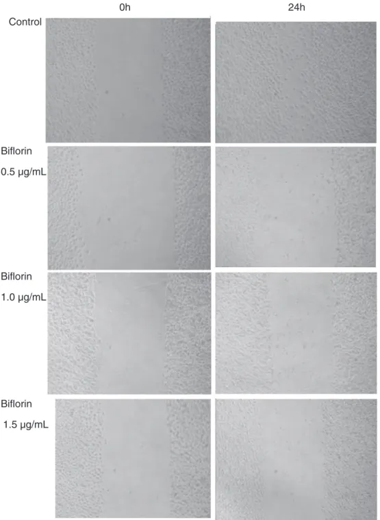
In migration assay, B16-F10 cells rapidly moved from the edge of the wound into the open area (Fig. 6). The biflorin-induced inhibition of cell movement occurred at all doses (0.5, 1.0 and 1.5μg/mL). In addition, biflorin showed a dose-dependent inhibitory effect on cell invasion through the Matrigel chamber (Fig. 7).
Cytotoxic effect of biflorin on B16-F10 melanoma cells
The cytotoxicity of biflorin was determined using an MTT assay. This assay was performed to determine if the anti-adhesion and anti-migration effect of biflorin was caused by a false positive due to cytotoxicity.Fig. 8shows that biflorin had no inhibitory effect on the growth of B16-F10 cells after 24 h incubation at the concentra-tions used in this study (0.5, 1.0 and 1.5μg/mL).
Discussion
Initially, a previous study in our laboratory byVasconcellos et al. (2011) demonstrated in vitro and in vivo anticancer activities of biflorin against a murine melanoma cell line. At 25 mg/kg/day i.p., this quinone improved the lifespan and also reduced tumor growth in animals bearing B16 melanoma tumors (Vasconcellos et al., 2011). It is well known that B16-F10 melanoma cells are highly metastatic and form tumor nodules in the parenchyma of the lungs when
0 10 20 30
0 50 100
Control 25 mg/kg* 50 mg/kg*
Days
Percent survival
Fig. 3.Effect of biflorin on the lifespan of experimental metastasis. Mice (C57BL/6) were injected with B16-F10 cells (106cells/500 ml, i.v.). Biflorin was administered at 25 mg/kg and 50 mg/kg i.p., starting 1 day after tumor implantation, for 21 days (n = 08). Control mice received vehicle (10% dimethyl sulfoxide). Data are represen-tative of 08 animals per group. The analyses were performed using Kaplan–Meier curves, and the controls and treated groups were compared by theχ2 test. *Pb0.01 compared with the control.
Fig. 4.Mice (C57BL/6) were inoculated with B16-F10 cells (106cells/500 ml, i.v.). Biflorin was administered intraperitoneally at a dose of 25 mg/kg and 50 mg/kg, starting 1 day after tumor implant, for 21 days (n = 08). Control mice (n = 08) received vehicle (10% DMSO). After the treatment, the lungs were dissected and observed for any metastases. Black arrows: hemosiderin, white arrows: melanoma cells. Magnification = 400×.
Control BSA 0.5 1.0 1.5
0 50 100 150
biflorin
(µg/mL)
*
*
*
Relative Adhesion
(% of Control)
administered through the tail vein (Fidler, 1970). Since biflorin can en-hance the lifespan of melanoma tumor-bearing animals, it was decided to determine the effect of biflorin on the metastatic process using in vitro and in vivo approaches.
First, in the present study, the anti-metastatic activity of biflorin was assessed using the experimental metastasis assay. The lungs with meta-static melanoma nodules produced after B16-F10 cell infection can be seen inFig. 1. Melanoma nodules occurred in untreated control but the presence of these nodules was reduced on the lungs of the animals treat-ed with biflorin (25 and 50 mg/kg/day). As seen inFig. 2, the number of lung metastases was significantly reduced after administration of biflorin (57 and 71% for 25 and 50 mg/kg/day, respectively) with an increase of
lifespan (22.3 ± 1.9, 24.4 ± 0.5 and 25.3 ± 0.42 days for control and 25 and 50 mg/kg/day, respectively) (Fig. 3).
Some drugs, in the literature, have demonstrated similar anti-metastatic activity as biflorin using this experimental melanoma me-tastasis assay. Lapachol, for example, is also a naphthoquinone and also has antitumor activity. When this drug was tested by serial oral administration of low non-toxic doses (5–20 mg/kg), it produced a weak but significant suppression of metastasis after injection of mu-rine B16-BL6 melanoma cells into the tail vein (Lee et al., 2006). The mechanism of action of this drug probably involves an alteration in protein profile and inhibition of cellular invasiveness, thus representing an important antimetastatic activity (Almeida, 2009).
0h
24h
Control
Biflorin
0.5 µg/mL
Biflorin
1.0 µg/mL
Biflorin
1.5 µg/mL
However, the histological analyses of the lungs of the tumor-bearing animals after administration of biflorin (25 and 50 mg/kg/day), showed the presence of hemosiderin and erythrocytes in the lung parenchyma, which is evidence of hemorrhage and probably an adverse effect. Nev-ertheless, more experiments are necessary to confirm this untoward effect.
It is well known that the metastasis occurs through several steps, including dissociation of cancer cells from the primary site, change in adhesion capability between cells and extracellular matrix (ECM), migration and invasion through the extracellular matrix (ECM) and circulation in the blood and lymph (Han et al., 2009; Lee et al., 2006; Chu et al., 2007; Zhao et al., 2008; Kurschat and Mauch, 2000). The invasion of tumor cells into adjacent tissues, a crucial event in metastasis, involves cell–cell and cell–ECM interactions (Zhao et al., 2008). These interactions involve a number of adhesive molecules on the cell surface, which have been described in detail (Kurschat and Mauch, 2000). Alterations in these interactions are one of the ini-tial events in cancer invasion, permitting cellular detachment from the primary tumor. Drugs that can inhibit the adhesion of the cells to ECM may have antimetastatic potential.
It is well been known that B16-F10 cells can adhere to a wide variety of cell-adhesive protein, including collagen, the most common
component of the ECM, and typical integrin ligands (Hatai et al., 1993; Zhou et al., 2008). In the present study, biflorin was found capable of inhibiting the adhesion of B16-F10 cells to type I collagen in a concentration-dependent manner (Fig. 5).
Salvicine, a diterpenoid quinone, identified as a non-intercalative topoisomerase II poison, has a broad range of antitumor and antimetastatic activity. Zhou et al. (2008) showed that salvicine down-regulatesβ1 integrin function and inhibits cell-ECM interaction, thereby providing further clues to the mechanism underlying the antimetastatic efficacy of salvicine.
Integrins are transmembrane glycoproteins composed of non-covalently linkedαandβsubunits. Integrins are essential for cell mi-gration and invasion because they mediate the adhesion of cells to the ECM and regulate intracellular signaling pathways that control cyto-skeletal organization, force generation, and survival (Zhou et al., 2008). Since these molecules are so important to cell adhesion, we can hypothesize that biflorin inhibits cell adhesion by blocking integrin function, like salvicine. However, this mechanism was not clear, and more experiments are necessary to confirm this hypothesis. After adhesion, the migration process is another characteristic of cell invasion. The invasion of cells to ECM may be due to growth pressure, but it may be due to cell migration. Since this process is so important for the study of metastasis, it was decided to evaluate the effect of biflorin on the cell migration of B16-F10 cells using the method devel-oped byBürk (1973), with modifications. In this assay, biflorin was able to inhibit the migration of B16-F10 cells in a concentration-dependent manner (Fig. 6). Furthermore, tumor cell invasion to the ECM is an important step in the process of tumor metastasis, which in-volves the adhesion, migration and degradation of the ECM [1, 4]. In order to investigate the effect of biflorin on invasiveness of B16-F10 cell line, we performed a cell invasion assay chamber coated with Matrigel (Lee et al., 2003, 2006). As expected, biflorin was able to inhibit the invasion of B16-F10 cells in a concentration-dependent manner (Fig. 7), which demonstrates the anti-invasive potential of this quinone. Taking together, these results showed that the antimetastatic ef-fect of biflorin may be related to anti-migration, anti-adhesion and anti-invasion activities.
It was also demonstrated that biflorin did not show any cytotoxicity at the concentrations used in the in vitro studies after 24 h of incubation (Fig. 8), which suggests that the anti-adhesion, anti-migration and anti-invasion effects of biflorin were not due to cytotoxicity.
Conclusion
We demonstrated that biflorin exhibits antimetastatic action in the mouse melanoma lung metastasis model. Furthermore, the anti-metastatic effect is suggested due to the inhibition of the adhesion, mi-gration and invasion of melanoma cells. These results indicate that biflorin is a promising candidate for an antimetastatic agent. However, more experiments are necessary to elucidate its mechanism of action.
Conflict of interest statement
The authors declare that there are no conflicts of interest.
Acknowledgments
We are grateful to the Brazilian agencies FINEP, CNPq, BNB/ FUNDECI, PRONEX, and CAPES for fellowships andfinancial support. We thank Silvana França dos Santos for providing excellent technical assistance. Dr. A. Leyva helped with English editing of the manuscript.
References
Acosta SL, Muro LV, Sacerio AL, Pena AR, Okwei SN.Analgesic properties ofCapraria bifloraleaves aqueous extract. Fitoterapia 2003;74:686–8.
Control 0,5 1,0 1,5
0 50 100
biflorin
(µg/mL)
*
*
*
Relative invasion (% of Control)
Fig. 7.Effects of biflorin on invasion in B16-F10 cells. The cells were treated with var-ious concentrations of biflorin for 16 h. After this treatment, B16-F10 cells were cul-tured with biflorin within a Matrigel invasion chamber. Quantitative analysis of the Matrigel chamber invasion assay is shown. Invasion was expressed as a percentage of control (DMSO). Each bar represents the mean ± S.D. calculated from three inde-pendent experiments. * pb0.001 compared with the negative control by ANOVA followed by the Student–Newman–Keuls test.
Control DOX 0.1 0.3 0.6 1.25 2.5 5.0 0 10 20 30 40 50 60 70 80 90 100 110 biflorin
(µg/mL)
* * *
Relative growth (% of Control)
Almeida ER.Preclinical and clinical studies of lapachol and beta-lapachone. Open Nat Prod J 2009;2:42–7.
Aquino TM, Amorim ELC, Feliciano GD, Lima EAC, Gomes ML, Lima CSA, et al.Influence of biflorin on the labelling of red blood cells, plasma protein, cell protein, and lympho-cytes with technetium-99 m: in vitro study. Braz J Pharmacogn 2007;17(2):181–5. Bürk RR.A factor from a transformed cell line that affects cell migration. Proc Natl Acad
Sci U S A 1973;70:369–72.
Cavallaro U, Christofori G.Cell adhesion in tumour invasion and metastasis: loss of the glue is not enough. Biochim Biophys Acta 2001;1552(1):39–45.
Chu SC, Yang SF, Liu SJ, Kuo WH, Chang YZ, Hsieh YS.In vitro and in vivo antimetastatic effects ofTerminalia catappaL. leaves on lung cancer cells. Food Chem Toxicol 2007;45: 1194–201.
Fidler IJ.Metastasis: quantitative analysis of distribution and fate of tumour emboli labeled with 125I-5-iodo-2_-deoxyuridine. J Natl Cancer Inst 1970;45:773–82. Fonseca AM, Pessoa ODL, Silveira ER, Lemos TLG, Monte FJQ, Braz-Filho R.Total
assign-ments of 1Hand 13C spectra of biflorin and bis-biflorin fromCapraria biflora. Magn Reson Chem 2003;41:1038–40.
Han EH, Choi JH, Hwang YP, Park HJ, Choi CY, Chung YC, et al.Immunostimulatory activity of aqueous extract isolated fromPrunella vulgaris. Food Chem Toxicol 2009;47:62–9.
Hatai M, Hashi H, Kato I, Yaoi Y.Inhibition of cell adhesion by proteolytic fragments of Type V collagen. Cell Struct Funct 1993;18:53–60.
Huey-Chun Huang, Guey-Yueh Shi, Shinn-Jong Jiang, Shi Chung-Sheng Wu, Chun-Mei Yang Hsi-Yuan, Hua-Lin Wu.Thrombomodulin-mediated cell adhesion: involve-ment of its lectin-like domain. J Biol Chem 2003;278:46750–9.
Kurschat P, Mauch C.Mechanisms of metastasis. Clin Exp Dermatol 2000;25:482–9. Lee H, Sakuraia K, Oshimac SH, Kimb I, Saikia A.Anti-metastatic and anti-angiogenic
activities of a new matrix metalloproteinase inhibitor, TN-6b S.J. Eur J Cancer 2003;39:1632–41.
Lee KJ, Kim JY, Choi JH, Kim HG, Chung YC, Roh SH, et al.Inhibition of tumor invasion and metastasis by aqueous extract of the radix ofPlatycodon grandiflorum. Food Chem Toxicol 2006;44:1890–6.
Mosmann T.Rapid colorimetric assay for the cellular growth and survival: application to proliferation and cytotoxicity assays. J Immunol Methods 1983;65:55–63. Vasconcellos MC, Bezerra DP, Fonseca AM, Pereira MRP, Lemos TLG, Pessoa ODL, et al.
Antitumor activity of biflorin, an o-naphthoquinone isolated fromCapraria biflora. Biol Pharm Bull 2007;30(8):1416–21.
Vasconcellos MC, Bezerra DP, Fonseca AM, Araujo AJ, Pessoa C, Lemos TLG, et al.The in vitro and in vivo inhibitory activity of biflorin in melanoma. Melanoma Res 2011;21(2): 106–14.
Weiss L.Metastatic inefficiency. Adv Cancer Res 1990;54:159–211.
Yang SF, Chub SC, Liu SJ, Chend C, Change YZ, Hsieh YS.Antimetastatic activities ofSelaginella tamariscina(Beauv.) on lung cancer cells in vitro and in vivo. J Ethnopharmacol 2007;110:483–9.
Zhao YY, Sato Y, Isaji T, Fukuda T, Matsumoto A, Miyoshi E, et al.Branched N-glycans regulate the biological functions of integrins and cadherins. FEBS J 2008;275: 1939–48.
Zhou J, Chen Y, Lang J-Y, Lu J-J, Ding J.Salvicine inactivatesβ1integrin and inhibits